A Multiplex Real-Time PCR Assay to Diagnose
and Separate
Helicoverpa armigera
and
H
.
zea
(Lepidoptera: Noctuidae) in the New
World
Todd M. Gilligan1*, Luke R. Tembrock2, Roxanne E. Farris3, Norman B. Barr3, Marja J. van der Straten4, Bart T. L. H. van de Vossenberg4, Eveline Metz-Verschure4
1USDA-APHIS-PPQ-Science & Technology, Identification Technology Program, Fort Collins, Colorado, United States of America,2Department of Biology, Colorado State University, Fort Collins, Colorado, United States of America,3USDA-APHIS-PPQ-Science & Technology, Mission Laboratory, Edinburg, Texas, United States of America,4National Plant Protection Organization, Netherlands Food and Consumers Product Safety Authority, Ministry of Economic Affairs, Wageningen, The Netherlands
Abstract
The Old World bollworm,Helicoverpa armigera(Hübner), and the corn earworm,H.zea (Boddie), are two of the most important agricultural pests in the world. Diagnosing these two species is difficult—adults can only be separated with a complex dissection, and larvae
can-not be identified to species using morphology, necessitating the use of geographic origin for identification in most instances. With the discovery ofH.armigerain the New World, identifi-cation of immatureHelicoverpabased on origin is no longer possible becauseH.zeaalso occurs in all of the geographic regions whereH.armigerahas been discovered. DNA bar-coding and restriction fragment length polymorphism (RFLP) analyses have been reported in publications to distinguish these species, but these methods both require post-PCR pro-cessing (i.e., DNA sequencing or restriction digestion) to complete. We report the first real-time PCR assay to distinguish these pests based on two hydrolysis probes that bind to a segment of the internal transcribed spacer region 2 (ITS2) amplified using a single primer pair. One probe targetsH.armigera, the second probe targetsH.zea, and a third probe that targets a conserved segment of 18S rDNA is used as a control of DNA quality. The assay can be completed in 50 minutes when using isolated DNA and is successfully tested on lar-vae intercepted at ports of entry and adults captured during domestic surveys. We demon-strate that the assay can be run in triplex with no negative effects on sensitivity, can be run using alternative real-time PCR reagents and instruments, and does not cross react with other New World Heliothinae.
OPEN ACCESS
Citation:Gilligan TM, Tembrock LR, Farris RE, Barr NB, van der Straten MJ, van de Vossenberg BTLH, et al. (2015) A Multiplex Real-Time PCR Assay to Diagnose and SeparateHelicoverpa armigeraandH. zea(Lepidoptera: Noctuidae) in the New World. PLoS ONE 10(11): e0142912. doi:10.1371/journal. pone.0142912
Editor:Daniel Doucet, Natural Resources Canada, CANADA
Received:June 19, 2015
Accepted:October 27, 2015
Published:November 11, 2015
Copyright:This is an open access article, free of all copyright, and may be freely reproduced, distributed, transmitted, modified, built upon, or otherwise used by anyone for any lawful purpose. The work is made available under theCreative Commons CC0public domain dedication.
Data Availability Statement:All DNA sequences generated for this study were submitted to GenBank
under accession numbers KT945996–KT946005
(18S), KT946006–KT946021 (ITS2), and KT946022– KT946127 (COI). RFLP sequence data was submitted to GenBank under accession numbers
KJ460240–KJ460246.
Introduction
The Old World bollworm,Helicoverpa armigera(Hübner), and the corn earworm,H.zea
(Boddie), are two of the most important agricultural pests in the world. A native of the Old World,H.armigerais the widest distributed species in the genusHelicoverpa[1–2], occurring from the Canary Islands east across much of Europe, Africa, Asia, and Australasia to the islands of Tonga in the southern Pacific Ocean. In the New World,H.zeais distributed across much of North and South America and the Caribbean [1]. Both species are highly polypha-gous, withH.armigerafeeding on hosts in 68 plant families andH.zeafeeding on hosts in 36 plant families [3]. Preferred hosts for both species include important agricultural crops, such as corn, cotton, soybean, tobacco, tomato, and many others [3–4].
In late 2012/early 2013, an outbreak ofHelicoverpalarvae was observed damaging soybean and cotton in the Cerrado region of central Brazil [4–6]. Adults reared from larvae were identi-fied using morphology [5], and later DNA sequence data [6–7], asH.armigera. Subsequent reports confirmedH.armigeraas present throughout much of Brazil [4,7–9], and this species was also reported from Argentina, Bolivia, Paraguay, and Uruguay [10–11]. In September, 2014, authors of this study from the United States Department of Agriculture (USDA) Mission Lab confirmed the first U.S. detection ofH.armigerain San Germán, Puerto Rico using DNA sequence data [12]. Between April, 2014 and February, 2015 authors of this study from the National Plant Protection Organization (NPPO) of the Netherlands confirmed interceptions of
H.armigerafrom the Dominican Republic and Peru using DNA sequence data. Netherlands NPPO also identified two larvae intercepted from Surinam in 2011 and 2012 asH.armigera
using sequence data; however, at that time presence ofH.armigerain the New World was not known, and therefore the reported origin of these consignments was assumed to be incorrect. These findings corroborate analysis ofH.armigeramitochondrial DNA (mtDNA) sequence data from Brazil that implies multiple invasion events or invasion several years prior to first discovery [9]. In June and July, 2015, three individuals ofH.armigerawere discovered in Flor-ida; these are the first reports ofH.armigerafrom the Continental U.S. [13–14].
Adults of bothH.armigeraandH.zeaare morphologically variable and cannot be identified reliably without genitalic dissection [15]. Brambila [16] provides step-by-step instructions for dissecting and diagnosingH.armigeraandH.zeausing male genitalia. This process is very tedious and time consuming, even for a lepidopteran specialist, especially when dealing with potentially hundreds of moths per trap. Identification ofHelicoverpalarvae is much more problematic than the identification of adults. Gilligan and Passoa [17] compared the head chaetotaxy, mandibles, hypopharyngeal complex, body coloration and markings, body chaeto-taxy, pinacula size and shape, setal color, cuticle texture, and crochet counts and arrangement for various instars ofH.armigeraandH.zeaand could not identify any morphological charac-ters that would reliably separate larvae of these two species.
For efficient diagnosis of adults captured during domestic surveys and larvae (and other immature stages) intercepted at ports of entry, means of identification other than morphology are necessary. Molecular techniques have been used to diagnosis these two pests based on vari-ation in the mitochondrial genome. For example, Mastrangelo et al. [4] used DNA barcoding to distinguish betweenH.armigeraandH.zeain Brazil. Behere et al. [18] developed a restric-tion fragment length polymorphism (RFLP) assay using two regions of mitochondrial DNA to distinguish betweenH.armigera,H.assulta,H.punctigera, as well asH.zea. These techniques, however, require post-PCR analysis steps that increase the processing time for samples. Restriction digestion and sequencing of PCR products add several hours or days to the time required to complete an analysis.
and analysis, decision to publish, or preparation of the manuscript.
Real-time PCR [19–20] is a molecular method that can be used for the detection and diag-nosis of biological organisms. The benefits of using real-time PCR versus conventional PCR include: reduced assay time; elimination of post-PCR electrophoresis; potential of scaling for high throughput testing; and increased sensitivity and specificity when using a quenched dye system (such as dual-labeled hydrolysis probes) [21]. In addition, real-time PCR eliminates the need to process and sequence the final PCR product, the lengthiest step in DNA barcod-ing. We have previously developed a real-time PCR assay to diagnose economically impor-tant Tortricidae using the internal transcribed spacer region 2 (ITS2) locus as a diagnostic marker and the 18S rDNA locus as an internal control [22]. Here we apply a similar method to develop a real-time PCR assay for diagnosing and separatingH.armigeraandH.zeain the New World.
Materials and Methods
Heliothinae collection and identification
Specimens used in this study are summarized inTable 1. A total of 452 Heliothinae represent-ing 18 species were used to develop the real-time PCR assay. One hundred and thirty-nineH.
armigeraadults and larvae were obtained from: port interceptions in the Netherlands and U.S.; fresh collections in South Africa; and colleagues in Australia, Brazil, Spain, and South Africa. Two hundred and fifty-eightH.zeaadults and larvae were obtained from: port interceptions in the U.S.; fresh collections in various locations in the U.S.; a lab colony at Mississippi State Uni-versity; and various USDA Cooperative Agricultural Survey (CAPS) programs, primarily those in Colorado, Florida, and Mississippi. Fifty-five other Heliothinae were obtained from: port interceptions in the U.S.; fresh collections in various locations in the U.S.; colleagues in Spain and South Africa; the C. P. Gillette Museum of Arthropod Diversity at Colorado State Univer-sity; and the Smithsonian Institution. Field collections in the U.S. were on private land (with permission of the land owner) or public land not requiring a collecting permit (National Forest, National Grassland, or BLM public land). Field collections in Gauteng Province, South Africa did not require a collecting permit under the Nature Conservation Ordinance of 1983 or the South African National Environmental Management: Biodiversity Act of 2004. Specimens pro-vided by colleagues from other foreign countries were collected on private land where no col-lecting permit was required, or were sourced from experimental colonies. Specimens from port interceptions and USDA CAPS surveys were obtained under the authority of the USDA and Netherlands NPPO. No endangered or protected species were collected for this study. The intercepted and CAPS specimens are representative of insect material expected from sampling at ports of entry and during surveys. The majority of larvae and fresh collected specimens were stored in>95% alcohol in microcentrifuge tubes, pinned adult specimens were stored dry, and
adults from CAPS traps were stored dry in plastic bags at−50°C.
Identification of specimens was performed using genitalic dissection (adults only) [15] and/ or sequencing of cytochrome c oxidase1 (COI) DNA barcodes (adults and larvae) [23]. Ampli-fications of COI were performed using the primers LepF1/LepR1 [24]. PCR conditions and sequencing steps were identical to those described below for 18s rDNA and ITS2. Edited DNA barcodes were identified using the“BOLD Identification System”ofwww.boldsystems.org [25]. In all cases, the DNA barcode identifications agreed with the morphological identification and/or real-time PCR ITS2 diagnosis.
DNA extraction, conventional PCR, and sequencing
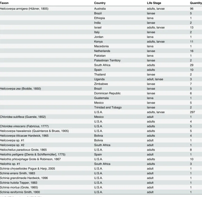
Total genomic DNA was extracted using a Qiagen DNeasy Blood and Tissue Kit (Qiagen, Valencia, Calif.). Tissue used for the extraction varied by specimen type: one to three legs from dried pinned adults; a portion of the thorax for adults in alcohol; two to three abdominal seg-ments for late instar larvae; and the entire larva for early instar larvae. For single column Table 1. Taxa sampled for study with country of origin, life stage, and number of specimens.
Taxon Country Life Stage Quantity
Helicoverpa armigera(Hübner, 1805) Australia adults, larvae 96
Brazil larvae 5
Ethiopia larva 1
India larvae 2
Israel adults, larvae 13
Italy larvae 2
Jordan larva 1
Kenya adults, larvae 11
Macedonia larva 1
Netherlands larvae 16
Pakistan larva 1
Palestinian Territory larvae 2
South Africa adults 29
Spain adults 10
Thailand larvae 2
Uganda adult, larvae 3
Zimbabwe larvae 7
Helicoverpa zea(Boddie, 1850) Brazil larvae 5
Dominican Republic larvae 6
Guatemala larva 1
Mexico larvae 5
Trinidad and Tobago larvae 2
U.S.A. adults, larvae 297
Chloridea subflexa(Guenée, 1852) Mexico adult 1
U.S.A. adults 4
Chloridea virescens(Fabricius, 1777) U.S.A. adults 5 Helicoverpa hawaiiensis(Quaintance & Brues, 1905) U.S.A. adults 5 Helicoverpa titicacaeHardwick, 1965 Bolivia adults 4
Helicoverpasp. #1 Bolivia adult 1
Helicoverpasp. #2 South Africa adult 1
Heliocheilus paradoxusGrote, 1865 U.S.A. adults 8
Heliothis peltigera([Denis & Schiffermüller], 1775) Spain adult 1 Heliothis phloxiphagaGrote & Robinson, 1867 U.S.A. adults 10
Heliothissp. #1 South Africa adults 3
Schinia chryselloidesPogue & Harp, 2005 U.S.A. adult 1
Schinia erransSmith, 1883 U.S.A. adult 1
Schinia grandimediaHardwick, 1996 U.S.A. adult 1
Schinia hulstiaTepper, 1883 U.S.A. adult 1
Schinia mortua(Grote, 1865) U.S.A. adult 1
Schinia reniformisSmith, 1900 U.S.A. adult 1
extractions, tissue samples were crushed dry in a 1.5 ml microcentrifuge tube, incubated in a solution of 180μl Buffer ATL and 20μl Proteinase K overnight at 56°C (in a dry bath), and eluted in 100μl of AE buffer after following the manufacturer’s recommended protocol. For plate extractions, tissue samples were chopped and placed in 1.2 ml tubes in deep 96-well plates and incubated overnight at 80°C (56°C dry bath equivalent) in an Eppendorf ThermoMixer (Eppendorf AG, Hamburg, Germany). The rest of the extraction followed the manufacturer’s recommended protocol with 96-well filter plates (Epoch Life Science, Missouri City, Texas) substituted for individual columns. To avoid possible contamination, all equipment and mate-rials were sanitized between specimens and filter tips were used to handle any liquids contain-ing DNA. No-tissue extraction controls were used for each extraction batch/plate when possible. DNA concentration and absorbance for a representative number of samples was esti-mated with a NanoDrop 1000 Ver. 3.7.0 spectrophotometer (Thermo Scientific/NanoDrop Wilmington, Delaware). Two readings were taken for each sample.
Conventional PCR used a Biometra T3000 (Biometra GmbH, Goettingen, Germany) or Bio-Rad C1000 Touch (Bio-Bio-Rad Laboratories, Inc., Hercules, Calif.) thermal cycler. PCR reactions were performed with TaKaRa Ex Taq HS polymerase (Takara Bio, Shiga, Japan) in total volumes of 50μl using the manufacturer’s recommended volumes of 10X Ex Taq buffer, dNTP mixture, and water. PCR conditions included an initial denaturation step of 94°C (3 min), 32 cycles of 94°C (20 sec) / 50°C (20 sec) / 72°C (30 sec), and an extension step of 72°C (5 min). PCR products were purified with a Qiagen QIAquick PCR Purification Kit (Qiagen, Valencia, Calif.). Purified PCR products were sequenced by the University of Chicago Cancer Research Center DNA Sequencing Facility using an Applied Biosystems 3730XL DNA sequencer (Applied Biosystems, Foster City, Calif.). The same primers used for PCR were also used for sequencing. Individual contigs were assembled, trimmed, and aligned using Geneious Pro 6.1.6 (Biomatters, Auckland, New Zealand) [26]. Following the methods described in Barr et al. [27], select ITS2 fragments were amplified, cloned, and purified prior to sequencing to generate multiple copies from a single moth.
Primer and probe design
Following Barr et al. [22,27], the ITS2 locus was selected as a potential diagnostic marker and the 18S rDNA locus was selected as a control. Conventional PCR was used to test the Barr et al. [22] 18S rDNA real-time PCR primers on four samples ofH.armigera, four samples ofH.zea, and twoHeliothissamples. Seven samples ofH.armigera, five samples ofH.zea, and two
Heliothissamples were used to sequence a region of 5.8S-ITS2-28S using the ITSF/ITSR prim-ers [22,27], and sequences generated using these primers were loaded into Geneious. Primer3 [28] was used to design internal primers to amplify a smaller region of the ITS2 locus that would maximize differences betweenH.armigeraandH.zea. Primers were designed to avoid regions of intragenomic variation detected in ITS2 for several individuals. After testing several primer combinations, the region amplified by primers 425F/568R (this study) was selected as appropriate for use in real-time PCR. The 425F/568R primers were tested with conventional PCR and sequenced for an additional 19 individuals ofH.armigera, 11 individuals ofH.zea, and threeHeliothisspecies (four individuals total).
All primers and probes used in this study are listed inTable 2. Melting temperatures (Tm; salt adjusted) were calculated with OligoCalc [31]. Primers were ordered from Integrated DNA Technologies (IDT; Coralville, Iowa) and stored in 1 × TE buffer or sterile water at−50°C;
working stocks were diluted to 10μM in sterile water and stored at−20°C. The 18S rDNA and
ITS2 primers generate amplicons of 68 and 140–143 bp, respectively. LNA probes were ordered from IDT and non-LNA probes were ordered from Biosearch Technologies (Petaluma, Calif.) Probes were diluted to 8μM in sterile water and stored at−20°C. The ITS2H.armigeraprobe
was labeled with FAM and theH.zeaprobe was labeled with HEX (both with BHQ-1 as quencher). The 18S rDNA control probe was designed identical to that of Barr et al. [22] with the exception of Quasar 670 (= Cy5; BHQ-2 as quencher) replacing CAL Red 610 for greater compatibility with real-time PCR machines that use the Red 610 channel for calibration. All probes were HPLC purified.
Real-time PCR multiplex protocol
Initial real-time PCR experiments were performed on a Roche LightCycler 480 Real-time PCR System (Roche Diagnostics Corp., Indianapolis, Indiana). Unless stated otherwise, all other assay development and testing was performed on a Bio-Rad CFX96 Touch Real-time PCR Detection System (Bio-Rad Laboratories, Inc., Hercules, Calif.). Development of real-time PCR protocols followed the MIQE Guidelines whenever possible [20]. All real-time PCR reactions used Roche LightCycler 480 Probes Master 2× hot start master mix (Roche Diagnostics Corp.) although master mixes from other vendors were tested in limited quantities (see below). The ITS2 (FAM or HEX) and 18S rDNA (Quasar 670) probe systems were first optimized as duplex assays and then combined into a triplex assay. Assay conditions were optimized so that the 18S rDNA control probe was less sensitive than the diagnostic ITS2 probes in order to limit the probability of false negatives [22,27] and the control/diagnostic probe combinations generated consistent quantification cycle (Cq) differences of less than seven cycles.
Bio-Rad CFX Manager 3.1 (Bio-Rad Laboratories, Inc.) was used to manage all real-time PCR analyses on the CFX96 Touch instrument. Quantification cycle determination mode was set to“single threshold”and baseline setting was set to“baseline subtracted curve fit.”Baseline cycles and single threshold were set to“auto calculated”for initial testing. However, both diag-nostic probes often exhibited a low level of amplification on the alternate species (e.g., theH.
armigeraprobe withH.zea) that exceeded the automatically calculated baseline threshold. This resulted in“false positive”Cq values with end relative fluorescence unit (RFU) values that Table 2. Primers and probes used in this study (Tm = melting temperature in °C).
Name Description Sequence Tm (°C) Source
RT-18S-F2 18S forward primer for real-time PCR 5'-ACCGCCCTAGTTCTAACCGTAAA 62.9 Barr et al. [22,27] RT-18S-R2 18S reverse primer for real-time PCR 5'-CCGCCGAGCCATTGTAGTAA 60.5 Barr et al. [22] RT-18S-P2 18S real-time PCR probe 5'-Quasar
670-TGTCATCTAGCGATCCGCCGA-BHQ-2
63.2 Barr et al. [22]/This study
ITSF 5.8S-ITS2-28S 5'-TTGAACATCGACATTTCGAACGCAC 64.1 Barr et al. [22,27]
ITSR 5.8S-ITS2-28S 5'-TCCTCCGCTTATTGATATGC 56.4 Barr et al. [22,27]
425F ITS2 forward primer for real-time PCR 5'-ACAAYACCAGAGGGGGTYGC 60.5–
64.6 This study 568R ITS2 reverse primer for real-time PCR 5'-CGTCGATGCGCTCTTCGG 60.8 This study
QP-Harm-ITS2-P8 ITS2 real-time PCR probe for H. armigera
5'-FAM-TGTCGTCCGYTTTAGCGTGAGAC-BHQ-1 64.6–
66.6 This study RT-ITS-zea ITS2 real-time PCR probe forH.zea 5'-HEX-CAACGCCATTAGTAGGCGGACTC-BHQ-1 66.6 This study
never exceeded 1,000 (usually<500). Because it would be difficult to replicate standardized
samples across different platforms and locations to set a baseline threshold, we changed the threshold setting from“auto calculated”to“user defined”with the value set to“1000.00”for all runs.
The optimized triplex real-time PCR assay was performed in 20μl reactions consisting of the following (concentrations listed are final): 10μl 2× master mix; 1μl each of the forward and reverse ITS2 and 18S rDNA primers (0.5μM); 0.5μl each of theH.armigeraandH.zea
probes (0.2μM); 1μl of the 18S rDNA control probe (0.4μM); 3μl of sterile water; and 1μl of DNA template. Real-time PCR conditions were as follows: 95°C for 7 min 30 sec (4.4°C/sec ramp rate); 40 cycles of 95°C for 10 sec (4.4°C/sec ramp rate), 62°C for 20 sec (2.2°C/sec ramp rate); and 40°C for 10 sec (1.5°C/sec ramp rate). A plate read was set to occur at the end of each 62°C cycle. All real-time PCR reactions were performed in Roche LightCycler 480 white 96 multi-well plates (Roche Diagnostics Corp.). The triplex real-time PCR assay was tested in two independent runs on all 452 samples.
Sensitivity analyses
Sensitivity of the real-time PCR assay was evaluated using a series of serial dilutions of bothH.
armigeraandH.zeasamples. Three samples of each species with known DNA concentrations (as estimated using a NanoDrop 1000) were diluted to a 100 ng/μl stock solution with sterile water and then a 10-fold dilution series was created using the previous solution and sterile water in a 1:9 ratio at each step. Each concentration of the dilution series for each species was tested in triplex (Quasar 670, FAM, HEX), duplex (Quasar 670 and FAM or HEX), and simplex (only FAM or HEX) to observe effects of DNA concentration on the assay and any possible complications created by multiplexing the three probes. Real-time PCR conditions were the same as those listed above with sterile water replacing the unused probe volumes in duplex and simplex reactions. The results from each of the three samples of each species were averaged and the Cq values plotted against DNA concentration on a logarithmic scale. Slopes, y-inter-cepts, and correlation coefficients were calculated in Excel for the triplex reactions.
Master mix testing
The effect of different real-time PCR master mix solutions on assay performance was evaluated using kits from three manufacturers. Three 96-well plates with identical samples (94 moth samples + 2 no template controls) were processed on the Bio-Rad CFX96 using the following kits: Roche LightCycler 480 Probes Master 2× hot start master mix (Roche Diagnostics Corp.); Takara Premix Ex Taq (Perfect Real Time) DNA Polymerase (Clontech Laboratories, Inc., Mountain View, Calif.); and Bio-Rad iTaq Universal Probes Supermix (Bio-Rad Laboratories, Inc.).
Mixed template experiments
Spike experiments were performed to determine the sensitivity of the real-time PCR assay to contamination (e.g., mixed samples). Template DNA (100 ng/μl) fromH.armigeraorH.zea
was spiked with DNA of the other species (100 ng/μl starting concentration) across a 10-fold dilution series in ratios of 1:1, 1:10, 1:100, and 1:1000 and tested using the triplex real-time PCR assay. Three replicates of each ratio were tested for each species.
microcentrifuge tubes. Legs were crushed using a pestle and DNA extracted following the sin-gle column protocol listed above. Five replicates of each ratio were extracted and tested using the triplex real-time PCR assay.
Assay precision testing
Precision, specifically intermediate precision and reproducibility, of the real-time PCR multi-plex protocol was evaluated by testing at Mission Lab using a Cepheid SmartCycler II (Cepheid, Sunnyvale, Calif.). All real-time PCR reactions used Takara Premix Ex Taq (Perfect Real Time) DNA Polymerase (Clontech Laboratories, Inc., Mountain View, Calif.). Primer and probe sequences were identical to those inTable 2. Probes were ordered with different fluoro-phores for better compatibility with the Cepheid system: CAL Fluor Red 610 (Texas Red) for the 18S rDNA control probe, TET for theH.zeaprobe, and FAM for theH.armigeraprobe. Reactions were performed in triplex with probes and primers at 10μM working stock (1μl of each primer, 1μl of the control probe, and 0.5μl of each ITS2 probe). Real-time PCR condi-tions were identical to those already reported (62°C annealing temperature). The Cepheid SmartCycler Software used the default threshold (30 fluorescent units) and growth curve (pri-mary curve) settings. Aliquots of 32H.armigeraextracts from the Fort Collins lab were used to test the triplex assay. Sensitivity tests using serial dilutions and spike experiments using leg ratios (described above) were also repeated on the Cepheid system.
Results
DNA extraction, primer and probe development
DNA concentrations ranged from 3.0 to 878.5 ng/μl (mean 125.4 ng/μl; average of two Nano-drop readings) for the subset of samples measured (approximately 60H.armigeraand 140H.
zea). Freshly collected specimens generally had higher DNA concentrations than those obtained from CAPS traps, most of which remained in the field for several weeks.
The 18S rDNA control primers and probe used by Barr et al. [22] produced positive results for all Heliothinae specimens tested. With the exception of replacing CAL Red 610 with Quasar 670 as the fluorophore for the probe, no other sequence modifications to the 18S rDNA mark-ers were made for this study.
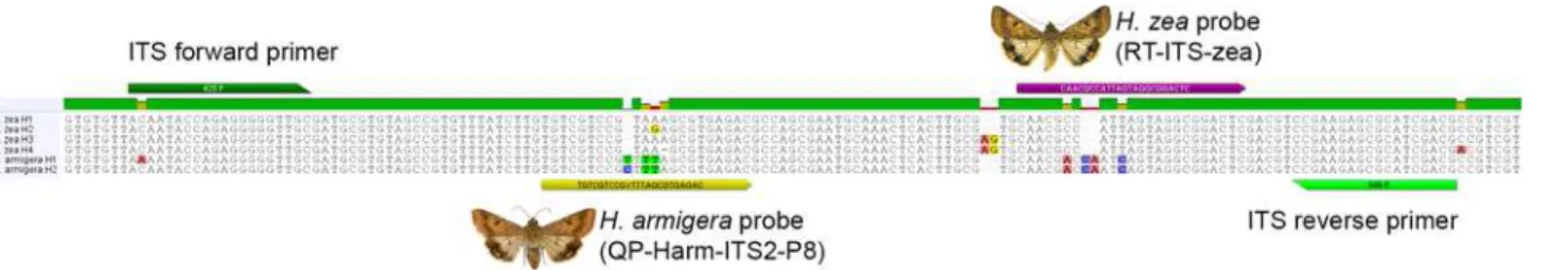
Direct sequencing and cloning of PCR products resulted in multiple distinct copies of ITS2 per species along with multiple copies in single individuals. The primers 425F and 568R were selected to maximize differences betweenH.armigeraandH.zeawhile minimizing intraspe-cific and intra-individual variation. TwoH.armigeraand fourH.zeahaplotypes were identi-fied within the ampliidenti-fied region of ITS2—these are illustrated in the alignment inFig 1along with primer and probe locations.
Helicoverpa armigeraprobe development focused on a 5-bp region that was variable inH.
zeaand consistently CTTTA or TTTTA forH.armigera. Initial 15-bp probes incorporating
Fig 1. Alignment of ITS2 forH.armigeraandH.zeashowing sequence variation along with primer and probe locations.
LNAs targeted at binding to the middle of this region were successful but all also bound toH.
zeawith varying levels of efficiently. Moving the target region (and associated LNAs) to the 5’ or 3’end of the probe reduced binding efficiency for both species. Because non-LNA probes with additional bases added to maintain a similar Tm (total 23–28 bp) produced similar results, subsequent probes were developed without LNAs. The probe“QP-Harm-ITS2-P8”was selected because it produced consistent binding (low Cq values) withH.armigeraand only low levels of binding (high Cq values and low RFUs) withH.zea. This probe was designed with a “Y”as the 10thbase to account for C/T variability in the twoH.armigerahaplotypes.
Helicoverpa zeaprobe development focused on a region further 3’of theH.armigeraprobe with a 2-bp indel flanked by two base pair differences from theH.armigerahaplotypes (Fig 1). A 23-bp probe“RT-ITS-zea”was designed that bound consistently toH.zea.
All probes exhibited low levels of binding with the alternate species (H.armigeraprobe with
H.zeaorH.zeaprobe withH.armigera); however, end RFU values in these instances never exceeded 1,000 (usually<500). Higher annealing temperatures were tested to attempt to
reduce incorrect probe binding, but temperatures>62°C interfered with performance of the
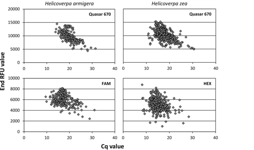
18S rDNA control probe when multiplexed. The problem of positive Cq values with low end RFUs was eliminated by manually adjusting the threshold level to“1000.00”in the CFX Man-ager software. When binding to the correct species, all three probes returned Cq values with end RFUs in excess of 2,000 for most samples; seeFig 2for plots of these values.
Fig 2. Cq values plotted against end RFU values for each probe in the triplex real-time PCR assay.
Performance of triplex assay on
H
.
armigera
All 139 of theH.armigerasamples tested with the triplex real-time PCR assay generated Cq values for the control (Quasar 670) and target (FAM) probes. Quasar 670 Cq values ranged from 13.87 to 31.45 (mean 20.15 ± SD 2.80) and FAM Cq values ranged from 9.55 to 31.01 (mean 17.58 ± SD 3.37). Only oneH.armigeraspecimen (HELICOV-595) had a FAM Cq value>30 on one run (Cq = 31.01); all other specimens had Cq values28.89.
As demonstrated by the standard curve of Cq values plotted versus DNA template concen-trations (Fig 3), Cq values decrease as DNA template concentration increases in a linear fash-ion. The FAM probe generated the same Cq values for each DNA concentration regardless if run in simplex, duplex (with Quasar 670), or triplex (with Quasar 670 and HEX). Similarly, the Quasar 670 probe generated the same Cq values for each DNA concentration if run in duplex (with FAM) or triplex (with FAM and HEX). No interactive or negative effects of multiplexing the three probe systems were observed. The difference between Quasar 670 and FAM probe Cq values for an individual is reported asΔCq (i.e., Quasar 670 Cq−FAM Cq) and used to detect
an association between Cq values of the target and control probes. TheΔCq values ranged from 0.09 to 6.68 (mean 3.41 ± SD 1.07). In all cases the control (Quasar 670) was higher than the diagnostic (FAM) probe Cq values.
Performance of triplex assay on
H
.
zea
All 258 of theH.zeasamples tested with the triplex real-time PCR assay generated Cq values for the control (Quasar 670) and target (HEX) probes. Quasar 670 Cq values ranged from 10.74 to 30.62 (mean 17.58 ± SD 2.89) and HEX Cq values ranged from 8.24 to 28.40 (mean 14.94 ± SD 2.48). OneH.zea(HELICOV-207) failed for both probes on one of the two runs, while an adjacent sample (HELICOV-208) failed for only theH.zeaprobe on one run; it is unknown if these failures were due to pipetting error. Two other samples produced consistently high or low Cq values relative to the other samples. Sample HELICOV-026 produced consis-tently low Cq values for HEX (8.24–8.65) and consisconsis-tently high Cq values for Quasar 670 (27.10–30.62), with the Cq difference between the two probes well outside of the mean for all other samples. Sample HELICOV-333 produced consistently high Cq values for HEX (26.02– 28.40) and similar but slightly lower values for Quasar 670 (24.86–27.95). Although the identi-fication of both samples was verified using morphology, their molecular identity would remain “inconclusive”using the interpretation rules developed here (see below).
Similar to results forH.armigera, Cq values decrease as DNA template concentration increases in a linear fashion (Fig 3), and no negative effects of multiplexing the three probe sys-tems were observed. The Quasar 670 probe Cq values were consistently higher than those of the HEX probe for all specimens except the two listed above and two additional specimens (HELICOV-373, 374) that had HEX Cq values exceeding the Quasar 670 Cq values for only the first run; these two samples were within the range of other specimens on the second run. TheΔCq values ranged from 0.04 to 6.16 (mean 2.64 ± SD 0.86) with the Quasar 670 Cq values consistently higher than the diagnostic (HEX) probe Cq values.
Performance of triplex assay on non-target Heliothinae
Master mix testing
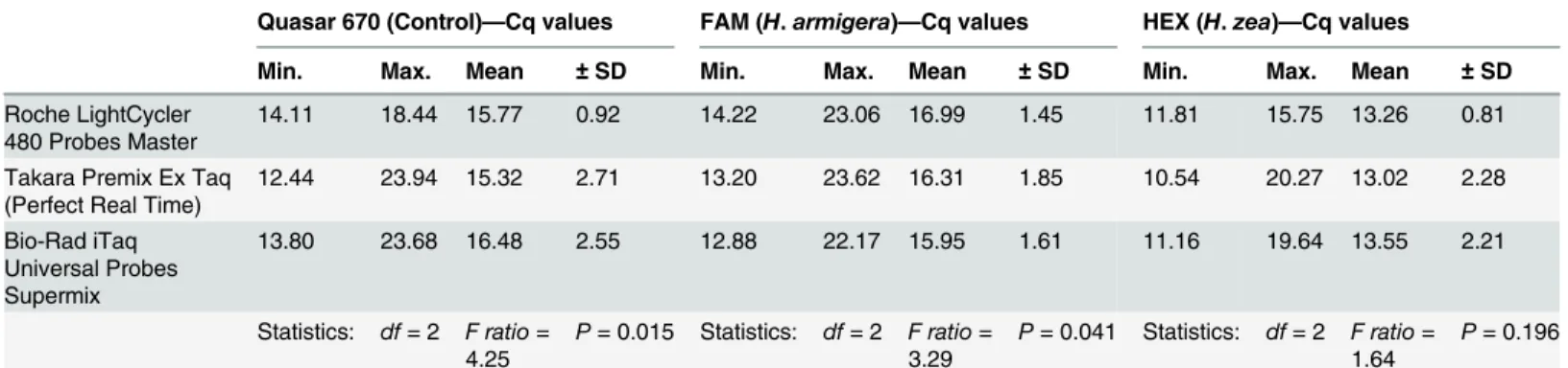
Results of the master mix testing are listed inTable 3. The Roche LightCycler 480 Probes Mas-ter performed more consistently (lowest SD) across all three probes. A one-way ANOVA revealed statistical significance between kits for the control (Quasar 670) probe (P = 0.015) and
H.armigera(FAM) probe (P = 0.041). No significant difference was found for theH.zea
(HEX) probe (P = 0.196). None of the kits produced Cq values outside the range of those speci-fied in the interpretation rules, and none of the samples would have been scored differently using different master mixes.
Mixed template experiments
WhenH.armigeraDNA was spiked withH.zeaDNA in ratios (armigera:zea) of 1:1, 1:10, 1:100, and 1:1000, and tested with the triplex real-time PCR assay, only the first ratio (1:1) pro-duced Cq values above the threshold level for theH.armigera(FAM) probe (mean
Cq = 19.35). WhenH.zeaDNA was spiked withH.armigeraDNA in ratios (zea:armigera) of 1:1, 1:10, 1:100, and 1:1000, theH.zea(HEX) probe returned Cq values for the ratios 1:1 (mean Cq = 16.70), 1:10 (mean Cq = 19.85), and 1:100 (mean Cq = 22.77).
Bulk leg extractions produced similar results for theH.armigera(FAM) probe. Only the 1:1 ratio extraction (1 leg ofH.armigera+ 1 leg ofH.zea) returned Cq values (mean Cq = 21.02). All other ratios (1:4, 1:9, and 1:19) did not produce Cq values above the threshold level.
Assay precision
All 32 of theH.armigerasamples tested with the triplex real-time PCR assay on the Cepheid system generated Cq values for the control (Texas Red) probe that ranged from 18.21 to 24.17 (mean 20.40 ± SD 1.38). All but one of the samples generated a Cq>0 for theH.armigera
(FAM) probe, with values ranging from 16.71–20.79 (mean 18.58 ± SD 0.99). This sample (HELICOV-107) generated a similar result after repeating the experiment on all 32 samples. However, analysis of an independent DNA extraction of that same sample (larva) generated a Cq for theH.armigeraprobe. It is not clear if the initial failure was due to degradation of nucleic acids in the first extraction sample, reduced sensitivity of the Cepheid protocol, and/or another factor. Consequently, this sample is recorded as a failure in the intermediate precision Fig 3. Standard curve of Cq values for serial dilutions ofH.armigeraandH.zeaDNA run with the real-time PCR assay in triplex, duplex, and simplex for the ITS2 probe (FAM or HEX) and in triplex and duplex for the control probe (Quasar 670).
doi:10.1371/journal.pone.0142912.g003
Table 3. Cq values for each probe resulting from testing different real-time PCR master mixes with the triplex assay.ANOVA statistics for compari-sons of each data set are provided in the last row of the table.
Quasar 670 (Control)—Cq values FAM (H.armigera)—Cq values HEX (H.zea)—Cq values
Min. Max. Mean ±SD Min. Max. Mean ±SD Min. Max. Mean ±SD
Roche LightCycler
480 Probes Master 14.11 18.44 15.77 0.92 14.22 23.06 16.99 1.45 11.81 15.75 13.26 0.81 Takara Premix Ex Taq
(Perfect Real Time) 12.44 23.94 15.32 2.71 13.20 23.62 16.31 1.85 10.54 20.27 13.02 2.28 Bio-Rad iTaq
Universal Probes Supermix
13.80 23.68 16.48 2.55 12.88 22.17 15.95 1.61 11.16 19.64 13.55 2.21
Statistics: df= 2 F ratio= 4.25
P= 0.015 Statistics: df= 2 F ratio= 3.29
P= 0.041 Statistics: df= 2 F ratio= 1.64
P= 0.196
assay (i.e., how the assay performs on a different platform using identical samples). None of the samples produced a Cq value for theH.zea(TET) probe. The control (Texas Red) Cq val-ues were higher than those of the target (FAM) for all samples (meanΔCq = 1.78 ± SD 0.65).
Based on serial dilution data, the Cepheid system is less sensitive than the Bio-Rad CFX96. Samples ofH.armigerafailed to register a Cq>0 for DNA concentrations less than 1.0 ng/μl
for theH.armigera(FAM) probe. Samples ofH.zeaproduced Cq values>0 for
concentra-tions down to 0. 1 ng/μl for theH.zea(TET) probe; however, the control probe (Texas Red) failed to produce Cq values>0 below DNA concentrations of 1.0 ng/μl for these same samples.
The control (Texas Red) Cq values were higher than those of the targets (FAM, TET) for all samples with DNA concentrations1.0 ng/μl (meanΔCq = 3.68 ± SD 1.44).
Aliquots of the bulk leg extractions produced results similar to those of the Bio-Rad CFX96. Only the 1:1 ratio extraction (1 leg ofH.armigera+ 1 leg ofH.zea) returned Cq values>0
(mean Cq = 20.47 ± SD 0.45). All other ratios (1:4, 1:9, and 1:19) did not produce Cq values>0.
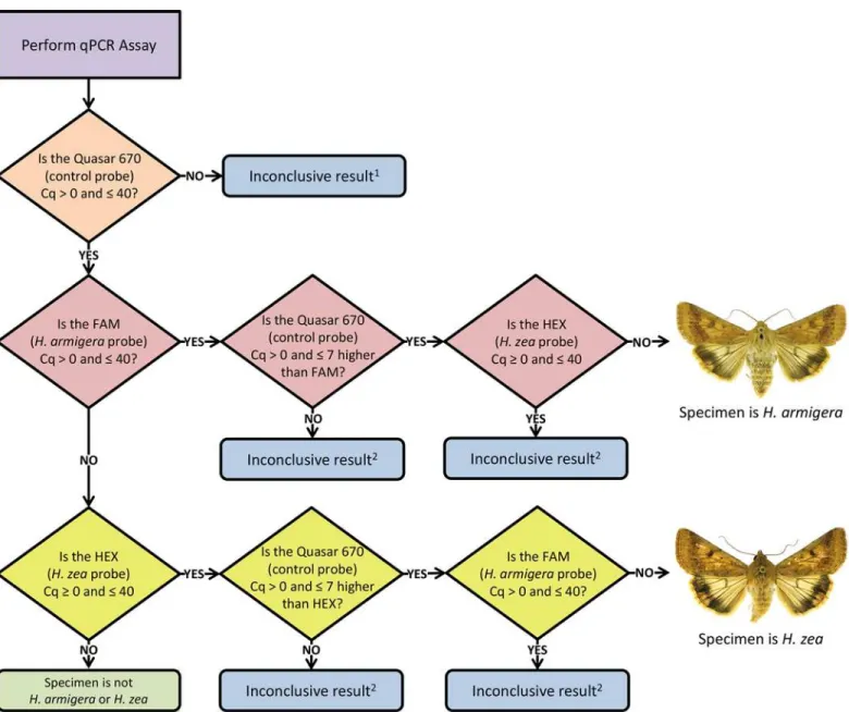
Development of interpretation rules
The interpretation rules developed here are based on the Cq values obtained from the 452 sam-ples tested with the real-time PCR assay on a Bio-Rad CFX96 Touch Real-time PCR Detection System. As demonstrated by testing on a Cepheid SmartCycler II, modification of the rules pre-sented here may be desirable if the assay is performed on other real-time PCR systems. Simply detecting fluorescence in a reaction (e.g., DNA is present) is not sufficient because the source of the DNA is not always known [22]. Abnormally low or high Cq values could be the result of contamination, equipment malfunction, incorrect assay preparation, non-specific probe bind-ing, or low quality DNA extract. The rules presented here are designed to function with target DNA concentrations as low as 0.01 ng/μl.
The following interpretation rules were developed: 1) the control (Quasar 670) probe Cq value must be>0 and40 to ensure DNA is present in concentrations sufficient for analysis; 2) the target (FAM or HEX) probe Cq value must be>0 and40; 3) the difference between the control (Quasar 670) probe and target (FAM or HEX) probe Cq values must be>0
and7 (the control Cq value must be higher); and 4) both target probes (FAM and HEX) can-not have Cq values>0 and40 at the same time. These interpretation rules are the basis for the decision tree illustrated inFig 4.
Discussion
We have developed a real-time PCR assay using the internal transcribed spacer region 2 (ITS2) locus as a diagnostic marker and the 18S rDNA locus as an internal control that is capable of diagnosingH.armigeraandH.zealarvae and adults intercepted at ports of entry and captured during domestic surveys. The assay can be run in triplex with no negative effects on sensitivity. Total analysis time is approximately 50 minutes for isolated DNA. This is a significant time savings over other methods, such as DNA barcoding, where sequencing of the final PCR prod-uct is required. The assay is capable of providing a correct diagnosis in the presence of other New World Heliothinae such asC.virescens.
correct products) but at values outside of the range defined in the interpretation rules for an identification. A singleH.zea(on one run) failed to generate a Cq value for theH.zeaprobe— this is the only result that would be considered a false negative. No false negatives were observed for theH.armigeraprobe.
No false positives were generated from any samples once the baseline threshold was estab-lished. Standard curves demonstrate that the assay is applicable to DNA concentrations of0.01 ng/μl. The coefficient of determination (r2) for both ITS2 probes was 0.998 (Fig 3); an r2>0.980 is recommended to maintain amplification efficiency across different starting
template copy numbers [32].
Although the assay was tested with a variety of other Helothinae, including other Helicov-erpa, caution should be used when applying these protocols outside of the New World. We Fig 4. Decision tree flowchart developed using the interpretation rules for the triplex real-time PCR assay run on a Bio-Rad CFX96 system.
were unable to obtain specimens of two other important Old WorldHelicoverpapests:H. punc-tigeraandH.assulta.Helicoverpa punctigerais an important polyphagous agricultural pest in Australia and New Zealand [2].Helicoverpa assultais an important pest of Solanaceae in East Asia; it also occurs in Africa, Australasia, and the Pacific [2]. We aligned a sequence of ITS2 for
H.punctigeradownloaded from Genbank (accession number AF047759.1) [33] with those of
H.armigeraandH.zea, and observed a 12 bp insertion inH.punctigerathat would prevent the
H.armigera(FAM) probe from binding. If this insert is present in allH.punctigera, the assay will be able to positively separateH.armigerafromH.punctigera. We were unable to obtain ITS2 sequence data forH.assulta. Preliminary testing of the triplex real-time PCR assay by Netherlands NPPO on a variety of Heliothinae, including otherHeliothis,Protoschinia, and
Pyrrhia, resulted in no false positives or false negatives using the interpretation rules developed here (data not shown).
The decision tree inFig 4outlines the process used to diagnose a specimen using the inter-pretation rules developed for the Bio-Rad CFX96 real-time PCR system. The overall process is applicable to any real-time PCR system capable of multiplexing several probes; however base-line threshold levels may need to be adjusted for other systems. We evaluated the intermediate precision of the assay using a Cepheid SmartCycler II in a different laboratory. The Cepheid produced results consistent with the Bio-Rad system, although it was less sensitive, requiring DNA concentrations1.0 ng/μl.
Sequencing of ITS2 forH.armigera,H.zea, and other Heliothinae revealed multiple distinct copies per species (four inH.zeaand two inH.armigera) and evidence of intra-individual vari-ation (multiple, distinct copies within a single individual). Direct sequencing of ITS2 was diffi-cult because of intra-individual variability and multiple copies of ITS2 per individual were confirmed in two individuals by cloning PCR products (GenBank accession numbers KT946006–KT946009).
Concerted evolution across rDNA copies is expected to result in intra-genomic uniformity [34], although intra-genomic variation in ITS2 has been documented in several studies [35– 36]. We observed similar variability when attempting to sequence ITS2 for Tortricidae [22]. Although intra-individual variation in our moths could be due to the presence of multiple, divergent copies within the genome of each cell (intra-genomic), our data do not distinguish this state from the presence of multiple, divergent copies within the tissue (i.e., cell lineages in the insect have different gene copies [37]). None of the sequences generated fromH.zea speci-mens were identical to sequences fromH.armigeraspecimens. We did not attempt to quantify the amount of intra-individual variation within ITS2 for each species.
Mixed template testing demonstrates that the real-time assay developed here is not applica-ble for diagnosing bulk samples. Detecting a single specimen ofH.armigerain a trap sample predominately composed ofH.zeais desirable for processing pheromone traps. WhenH.
armigeratemplate DNA was spiked withH.zeaDNA in different ratios and processed as a sin-gle sample, theH.armigera(FAM) probe only produced acceptable Cq values for a 1:1 (100 ng/μl of each) ratio. Based on the standard curve produced forH.armigera, the assay should be capable of detecting DNA concentrations as low as 0.01 ng/μl. When the experiment was reversed andH.zeatemplate DNA was spiked withH.armigeraDNA, the assay was capable of detectingH.zeaDNA down to 0.01 ng/μl (1:100 ratio ofzea:armigera), which is towards the lower limit of detection according to the standard curve. Thus, it appears that includingH.zea
results. However, increasing the FAM probe concentration to the same level did produce Cq values>0 for a small number of samples down to 0.01 ng/μl (1:100 ratio ofarmigera:zea).
Fur-ther testing is required to determine if increasing FAM probe concentration and optimization of PCR conditions can be used to improve results with mixed DNA templates. More problem-atic for bulk sample processing is the DNA extraction itself. When legs were extracted from individuals ofH.armigeraandH.zeain the same reaction in ratios above 1:1, the assay failed to produce Cq values above the threshold level. Alternate extraction methods may also be nec-essary to improve results with bulk samples.
Detection of hybrid individuals is also potentially problematic. It has been estimated thatH.
zeasplit from a common ancestor withH.armigeraby a founder event approximately 1.5 mil-lion years ago [15]. This hypothesis is supported by DNA sequence data as well as very similar morphology and mating compatibility between the two species. Under laboratory conditions,
H.armigeraandH.zeahave been demonstrated to be able to mate and produce fertile off-spring [1,6,38,39–40]. We were able to obtain severalarmigera×zeahybrids produced dur-ing sterility studies performed in Mississippi in the mid-1980s; however DNA extraction for these individuals failed. Thus, we have not tested the real-time assay on any hybrid individuals and cannot hypothesize on how it would function on hybrids. Although noarmigera×zea
hybrids have yet to be identified in the wild, recognition of hybrids is potentially problematic for any identification method [4].
Other rapid molecular methods that do not require sequencing have proven successful in separatingH.armigerafrom other species ofHelicoverpapresent in the Old World [41–42]. Behere et al. [18] developed a PCR-RFLP assay using two regions of mtDNA, COI, and cyto-chromeb(Cytb), to distinguish betweenH.armigera,H.assulta,H.punctigera, as well asH.
zea. We tested the Behere et al. [18] assay and found it separatedH.armigerafromH.zea, as reported. However, the method is less useful when additional species present in the New World are included in the assay. The PCR protocol for Cytbdoes not work well for many of these species precluding diagnosis because failure to amplify is not a diagnostic characteristic (data not shown). More troubling is that we found some species such asChloridea virescens
generate RFLP patterns identical to those forH.armigera(GenBank accession numbers KJ460240–KJ460246).Chloridea virescensis a common heliothine that is distributed across much of the New World, and is commonly intercepted at ports of entry in the U.S. and the EU (Netherlands).
Acknowledgments
We thank Joaquin Baixeras, Julieta Brambila, Richard Brown, John Gilligan, Janet Hardin, Boris Kondratieff, Paul Opler, O. P. Perera, Michael Pogue, Ricardo Polanczyk, Hermann Staude, Wee Tek Tay, and private collectors in the Netherlands for providing specimens of Heliothinae used in this study. Donald Mykles and Daniel Sloan provided access to real-time PCR machines and software. Laura Hartmann assisted with DNA extractions and Terrance Todd assisted with cloning experiments. Geoff Dennis and two anonymous reviewers provided helpful comments that greatly increased the quality of the manuscript. The use or mention of a trademark or proprietary product does not constitute an endorsement, guarantee, or warranty of the product and does not imply its approval to the exclusion of other suitable products by the U.S. Department of Agriculture, an equal opportunity employer.
Author Contributions
reagents/materials/analysis tools: TMG LRT REF NBB MJvS BTLHvV. Wrote the paper: TMG LRT REF NBB MJvS BTLHvV.
References
1. Hardwick DF. The corn earworm complex. Memoirs Entomol Soc Can. 1965; 40: 1–247. 2. Matthews M. Heliothine moths of Australia: a guide to pest bollworms and related noctuid groups.
Monographs on Australian Lepidoptera, Volume 7. CSIRO Publishing, Collingwood, Victoria, Austra-lia; 1999.
3. Cunningham JP, Zalucki MP. Understanding heliothine (Lepidoptera: Heliothinae) pests: What is a host plant? J Econ Entomol. 2014; 107: 881–896. PMID:25026644
4. Mastrangelo T, Paulo DF, Bergamo LW, Morais EGF, Silva M, Bezerra-Silva G, et al. Detection and genetic diversity of a heliothine invader (Lepidoptera: Noctuidae) from North and Northeast of Brazil. J Econ Entomol. 2014; 107: 970–980. PMID:25026655
5. Czepak C, Albernaz KC, Vivan LM, Guimarães HO, Carvalhais T. Research note. First reported occur-rence ofHelicoverpa armigera(Hübner) (Lepidoptera: Noctuidae) in Brazil. Pesq Agropec Trop. 2013; 43: 110–113.
6. Tay WT, Soria MF, Walsh T, Thomazoni D, Silvie P, Behere GT, et al. A brave new world for an Old World pest:Helicoverpa armigera(Lepidoptera: Noctuidae) in Brazil. PLoS One. 2013; 8(11): e80134. doi:10.1371/journal.pone.0080134PMID:24260345
7. Specht A, Sosa-Gómez DR, de Paula-Moraes SV, Cavaguchi Yano SA. Identificação morfológica e molecular deHelicoverpa armigera(Lepidoptera: Noctuidae) e ampliação de seu registro de ocorrência no Brasil. Pesq Agropec Brasil. 2013; 48: 689–692.
8. Bueno RCOF, Yamamoto PT, Carvalho MM, Bueno NM. Occurrence ofHelicoverpa armigera(Hübner, 1808) on citrus in the state of São Paulo, Brazil. Rev Bras Frutic. 2014; 36: 520–523.
9. Leite NA, Alves-Pereira A, Corrêa AS, Zucchi MI, Omoto C. Demographics and genetic variability of the New World bollworm (Helicoverpa zea) and the Old World bollworm (Helicoverpa armigera) in Brazil. PLoS One. 2014; 9(11): e113286. doi:10.1371/journal.pone.0113286PMID:25409452
10. Murúa MG, Scalora FS, Navarro FR, Cazado LE, Casmuz A, Villagrán ME, et al. First record of Helicov-erpa armigera(Lepidoptera: Noctuidae) in Argentina. Fla Entomol. 2014; 97: 854–856.
11. Kriticos DJ, Ota N, Hutchison WD, Beddow J, Walsh T, Tay WT, et al. The potential distribution of
invadingHelicoverpa armigerain North America: Is it just a matter of time? PLoS One. 2015; 10(3): e0119618. doi:10.1371/journal.pone.0119618PMID:25786260
12. Smith E. Detection of old world bollworm (Helicoverpa armigera) in Puerto Rico. North American Plant Protection Organization, Phytosanitary Alert System Bulletin. 2014 Oct 28. Available:http://www. pestalert.org/oprDetail.cfm?oprID=600.
13. Hayden, J, Brambila, J. Pest alert:Helicoverpa armigera(Lepidoptera: Noctuidae), the Old World boll-worm. Florida Department of Agriculture and Consumer Services. 2015 Jun 17. Available:http://www. freshfromflorida.com/Divisions-Offices/Plant-Industry/Plant-Industry-Publications/Pest-Alerts/Pest-Alert-The-Old-World-Bollworm.
14. El-Lissy, O. Detection of Old World bollworm (Helicoverpa armigera) in Florida. 2015 Jul 30. Available: http://www.aphis.usda.gov/plant_health/plant_pest_info/owb/downloads/DA-2015-43.pdf.
15. Pogue MG. A new synonym ofHelicoverpa zea(Boddie) and differentiation of adult males ofH.zeaand H.armigera(Hübner) (Lepidoptera: Noctuidae: Heliothinae). Ann Entomol Soc Am. 2004; 97: 1222–
1226.
16. Brambila J. 2009. Instructions for dissecting male genitalia ofHelicoverpa(Lepidoptera: Noctuidae) to separateH.zeafromH.armigera. USDA-APHIS-PPQ. Available:http://www.aphis.usda.gov/plant_ health/plant_pest_info/owb/downloads/owb-screeningaids2.pdf.
17. Gilligan TM, Passoa SC. LepIntercept, An identification resource for intercepted Lepidoptera larvae.
Identification Technology Program, USDA, Fort Collins. 2014. Available:www.lepintercept.org.
18. Behere GT, Tay WT, Russell DA, Batterham P. Molecular markers to discriminate among four pest spe-cies ofHelicoverpa(Lepidoptera: Noctuidae). Bull Entomol Res. 2008; 98: 599–603. doi:10.1017/
S0007485308005956PMID:18631420
19. Heid CA, Stevens J, Livak KJ, Williams PM. Real time quantitative PCR. Genome Res. 1996; 6: 986–
994. PMID:8908518
20. Bustin SA, Benes V, Garson JA, Hellemans J, Huggett J, Kubista M, et al. The MIQE Guidelines: Mini-mum information for publication of quantitative real-time PCR experiments. Clin Chem. 2009; 55: 611–
21. Logan JMJ, Edwards KJ. An overview of real-time PCR platforms. In: Edwards K, Logan J, Saunders N, editors. Real-time PCR an essential guide. Horizon Bioscience, Norfolk, United Kingdom; 2004. pp. 13–29.
22. Barr NB, Ledezma LA, Farris RE, Epstein ME, Gilligan TM. A multiplex real-time polymerase chain reaction assay to diagnoseEpiphyas postvittana(Lepidoptera: Tortricidae). J Econ Entomol. 2011; 104: 1706–1719. PMID:22066202
23. Hebert PDN, Cywinska A, Ball SL, deWaard JR. Biological identifications through DNA barcodes. Proc R Soc Lond B Biol Sci. 2003; 270: 313–322.
24. Hebert PDN, Penton EH, Burns JM, Janzen DH, Hallwachs W. Ten species in one: DNA barcoding reveals cryptic species in the neotropical skipper butterflyAstraptes fulgerator. Proc Natl Acad Sci USA. 2004; 101: 14812–14817. PMID:15465915
25. Ratnasingham S, Hebert PDN. BOLD: The Barcode of Life Data System (Available:http://www. barcodinglife.org). Mol Ecol Notes. 2007; 7: 355–364. doi:10.1111/j.1471-8286.2007.01678.xPMID:
18784790
26. Kearse M, Moir R, Wilson A, Stones-Havas S, Cheung M, Sturrock S, et al. Geneious Basic: an inte-grated and extendable desktop software platform for the organization and analysis of sequence data. Bioinformatics. 2012; 28: 1647–1649. doi:10.1093/bioinformatics/bts199PMID:22543367 27. Barr NB, Ledezma LA, Vasquez JD, Epstein ME, Kerr PH, Kinnee S, et al. Molecular identification of
the light brown apple moth (Lepidoptera: Tortricidae) in California using a polymerase chain reaction assay of the internal transcribed spacer 2 locus. J Econ Entomol. 2009; 102: 2333–2342. PMID:
20069865
28. Untergrasser A, Cutcutache I, Koressaar T, Ye J, Faircloth BC, Remm M, et al. Primer3—new
capabili-ties and interfaces. Nucleic Acids Res. 2012; 40(15): e115. PMID:22730293
29. Braasch DA, Corey DR. Locked nucleic acid (LNA): fine-tuning the recognition of DNA and RNA. Chem Biol. 2001; 8: 1–7. PMID:11182314
30. Owczarzy R, You Y, Groth CL, Tataurov AV. Stability and mismatch discrimination of locked nucleic
acid-DNA duplexes. Biochemistry. 2011; 50: 9352–9367. doi:10.1021/bi200904ePMID:21928795 31. Kibbe WA. OligoCalc: an online oligonucleotide properties calculator. Nucleic Acids Res. 2007; 35:
W43–W46. doi:10.1093/nar/gkm234PMID:17452344
32. Taylor S, Wakem M, Dijkman G, Alsarraj M, Nguyen M. A practical approach to RT-qPCR–Publishing
data that conform to the MIQE guidelines. Methods. 2010; 50: S1–S5. doi:10.1016/j.ymeth.2010.01.
005PMID:20215014
33. Amornsak W, Gordh G, Graham G. Detecting parasitised eggs with polymerase chain reaction and DNA sequence ofTrichogramma australicumGirault (Hymenoptera: Trichogrammatidae). Aust J Ento-mol. 1998; 37: 174–179.
34. Álvarez I, Wendel JF. Ribosomal ITS sequences and plant phylogenetic inference. Mol Phylogenet Evol. 2003; 29: 417–434. PMID:14615184
35. Harris DJ, Crandall KA. Intragenomic variation within ITS1 and ITS2 of freshwater crayfishes
(Deca-poda: Cambaridae): Implications for phylogenetic and microsatellite studies. Mol Biol Evol. 2000; 17: 284–291. PMID:10677851
36. Keller I, Chintauan-Marquier IC, Veltsos P, Nichols RA. Ribosomal DNA in the grasshopperPodisma pedestris: Escape from concerted evolution. Genetics. 2006; 174: 863–874. PMID:16951064 37. Magnacca KN, Brown MJF. Tissue segregation of mitochondrial haplotypes inheteroplasmic Hawaiian
bees: implications forDNA barcoding. Mol Ecol Res. 2010; 10: 60–68.
38. Behere GT, Tay WT, Russell DA, Heckel DG, Appleton BR, Kranthi KR, et al. Mitochondrial DNA analy-sis of field populations ofHelicoverpa armigera(Lepidoptera: Noctuidae) and of its relationship toH. zea. BMC Evol Biol. 2007; 7: 117. doi:10.1186/1471-2148-7-117PMID:17629927
39. Laster ML, Hardee DD. Intermating compatibility between North AmericanHelicoverpa zeaand Heliothis armigera(Lepidoptera: Noctuidae) from Russia. J Econ Entomol. 1995; 88: 77–80. 40. Laster ML, Sheng CF. Search for hybrid sterility forHelicoverpa zeain crosses between the North
AmericanH.zeaandH.armigera(Lepidoptera: Noctuidae) from China. J Econ Entomol. 1995; 88: 1288–1291.
41. Kranthi S, Kranthi KR, Bharose AA, Syed SN. A PCR-RFLP tool for differentiatingHelicoverpa armigera andH.assulta(Lepidoptera: Noctuidae). Curr Science. 2005; 89: 1322–1323.