Ü. İsaoğlu et al. Rupture of rudimentary horn 219
J Clin Exp Invest www.clinexpinvest.org Vol 2, No 2, June 2011
Yazışma Adresi /Correspondence: Dr. Ünal İsaoğlu,
Nenehatun Gynecology and Obstetrics Hospital, Erzurum/Turkey Email: [email protected] Geliş Tarihi / Received: 01.10.2010, Kabul Tarihi / Accepted: 13.10.2010
Copyright © Klinik ve Deneysel Araştırmalar Dergisi 2011, Her hakkı saklıdır / All rights reserved
Klinik ve Deneysel Araştırmalar Dergisi / 2011; 2 (2): 219-221
Journal of Clinical and Experimental Investigations
C A S E R E P O R T / O LG U S U N U M U
Rupture of a rudimentary horn at 16 weeks of gestation
Onaltıncı gebelik haftasında rudimenter horn rüptürü
İlhan Bahri Delibaş1, Ünal İsaoğlu1, Mehmet Yılmaz1, Sedat Kadanalı21Nenehatun Obstetrics and Gynecology Hospital, Erzurum, Turkey
2Department of Obstetrics and Gynecology, Ataturk University Aziziye Research Hospital, Erzurum, Turkey
ÖZET
Onaltıncı gebelik haftasında akut batın tablosu ile gelen hasta acil laparotomiye alındı. Laparotomide intraabdomi-nal iki litre ibrine ve deibrine kan ve ölü bir fetus ile bir-likte plasentası tesbit edildi. Sağ over ve tüp normal görü-nümde ve uterus normal boyutlardaydı. Uterusun solunda rupture ve aktif kanamalı uterin horn gözlendi. Rupture uterin horn sol over korunarak sol tüp ile birlikte eksize edildi. Hasta herhangi bir komplikasyon gelişmeden ta-burcu edildi. Klin Deney Ar Derg 2011;2(2):219-21
Anahtar kelimeler: Rudimenter horn, gebelik, yırtılma
ABSTRACT
A patient with acute abdomen at 16th week of gestation underwent an emergency laparotomy. At laparotomy two litres of ibrinated and free blood and a dead fetus with placenta was detected intraabdominally. The right ovary and tube was normal and the uterus was normal in size. There was a ruptured and actively bleeding uterine horn on the left side of the uterus. The ruptured uterine horn was excised with the left tube by preserving the left ovary. The patient recovered without any complications. J Clin Exp Invest 2011;2(2):219-21
Key words: Rudimentary horn, pregnancy, rupture
INTRODUCTION
Congenital uterine anomalies have been classiied
by the American Fertility Society.1 The prevalence
of uterine anomalies in general population is 0.5%.2
In a retrospective study conducted in our country, prevalence of uterine rupture in non-scarred uteri is reported as 1 per 2119 deliveries.3 Unicornuate
uterus is the second rarest seen Mullerian anomaly after uterus didelphys comprising 10% of all uter -ine anomalies.4 It is estimated to be due to failure
of migration of one of the Mullerian ducts to its normal destination. ASRM classiied this anomaly
as unicornuate uterus with communicating or
non-communicating horn, unicornuate uterus with no cavity and unicornuate uterus with no horn. Ninety percent of rudimentary uterine horns are non-com -municating.5
CASE
A 27 years-old multigravida who had a complaint of abdominal pain for 24 hours was referred to our
clinic from the emergency service of a local
mater-nity hospital with the general diagnosis of acute ab
-domen. She had a history of a caesarean delivery at term 3 years ago and a preterm vaginal delivery at 32 weeks 1 year ago. She attended no routine visits
in that pregnancy.
On admission she was somnolent and her blood pressure was 90/60 mmHg and her pulse was 96 bpm. On abdominal examination she had severe ab
-dominal tenderness and rebound on the left lower quadrant. Vaginal examination revealed severe cer
-vical motion tenderness. No vaginal bleeding or cervical dilatation was detected.
Her laboratory values were as follows: hemo
-globin 7.1 g/dl, hematocrit 21%, White blood cell count 18200 and platelets 322 x 10 9 /L. Her coagu
-lation proile was normal.
On abdominal ultrasound (USG) there was ex
-tensive abdominal luid, a normal-sized uterus with 16 mm endometrial thickness and a neighbouring extrauterine 16 week-fetus with no cardiac activity.
-Ü. İsaoğlu et al. Rupture of rudimentary horn 220
J Clin Exp Invest www.clinexpinvest.org Vol 2, No 2, June 2011
ferred to operation room for exploratory laparotomy with the suspected diagnosis of uterine rupture.
At laparotomy two litres of ibrinated and free blood and a dead fetus with placenta was detected intraabdominally. The right ovary and tube was nor
-mal and the uterus was nor-mal in size. There was a ruptured and actively bleeding uterine horn on the left side of the uterus. The left tube was short and connected to the uterine horn. There was no connec
-tion detected between the horn and the main uterine cavity. The ruptured uterine horn was excised with
the left tube by preserving the left ovary. The patient
was given 3 units of packed cells and two units of fresh frozen plasma.
The patient recovered without any complica
-tions. The histopathology report veriied the diag
-nosis of pregnancy and rupture of the uterine rudi -mentary horn.
Figure 1. Intra-operative photograph showing the poste-rior view of the uterus with ruptured rudimentary horn
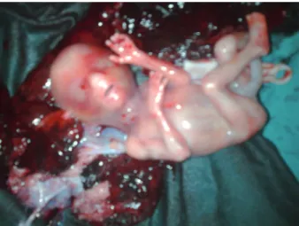
Figure 2. Fetus and Placenta
DISCUSSION
Congenital uterine anomalies have been classiied
by the American Fertility Society.1 The prevalence
of uterine anomalies in general population is 0.5%.2
In a retrospective study conducted in our country, prevalence of uterine rupture in non-scarred uteri is reported as 1 per 2119 deliveries.3 Unicornuate
uterus is the second rarest seen Mullerian anomaly after uterus didelphys comprising 10% of all uter -ine anomalies.4 It is estimated to be due to failure
of migration of one of the Mullerian ducts to its normal destination. ASRM classiied this anomaly
as unicornuate uterus with communicating or
non-communicating horn, unicornuate uterus with no cavity and unicornuate uterus with no horn. Ninety percent of rudimentary uterine horns are non-com -municating.5
Ultrasonography may be helpful in diagnos -ing such anomalies before rupture. It is helpful in
decreasing the morbidity and mortality associated with rapid and massive haemoperitoneum due to
rupture.
Pregnancy in a non-communicating rudimen -tary horn occurs through transperitoneal migration
of sperm or fertilized ovum.6 It is associated with spontaneous abortion, preterm labor, intrauterine growth retardation, intraabdominal haemorrhage and uterine rupture.7 Rupture is reported to occur
usually between 10-15 weeks.8 Time of rupture
depends on the structure of the rudimentary horn. Since myometrium is very thin and there is a high risk of uterine rupture, in rudimentary horn preg
-nancy prophylactic removal is recommended. To reduce the maternal mortality rate, removal of ru
-dimentary horn gestation is suggested in the irst
trimester before imminent rupture.9
In our case, rupture of the rudimentary horn oc
-cured at 16th week of gestation. Ambulatory
opera-tion and intensive care was life saving. Urinary sys
-tem malformations may also accompany Mullerian
anomalies in 38% of the cases.10 In this case there
was no urinary system anomaly in postoperative in-travenous pyelography.
REFERENCES
Ü. İsaoğlu et al. Rupture of rudimentary horn 221
J Clin Exp Invest www.clinexpinvest.org Vol 2, No 2, June 2011
pregnancies, müllerian anomalies and intrauterine adhe -sions. Fertil Steril 1988;49 (6):944-55.
2. Nahum GG. Uterine anomalies. How common are they, and what is their distribution among subtypes? J Reprod Med 1998;43 (10):877-87.
3. Evsen M, Sak M.E., Bozkurt Y, Kapan M, Bakır C. Uterine rupture: Regional incidence, causes and treatment. Dicle Tıp Dergisi 2008; 35(4):260-4.
4. Speroff L, Fritz M.A.Clinical Gynecologic Endocrinology and Infertility.7th edn. Philadelphia: Lippincott Williams & Wilkins, 2005:133.
5. Goel P, Aggarwal A, Devi K, Takkar N, Saha PK, Huria A. Unicornuate uterus with non-communicating rudimentary horn. Different clinical presentations. J Obstet Gynecol In -dia 2005;55 (2):155-8.
6. Panayotidis C, Abdel-Fattah M, Leggott M. Rupture of rudi -mentary uterine horn of a unicornuate uterus at 15 weeks’ gestation. J Obstet Gynaecol 2004; 24 (3):323-4.
7. Jin Woo Shin, Hai Joong Kim. Case of live birth in a non-communicating rudimentary horn pregnancy. J Obstet Gy -naecol Res. 2005,31(4):329–31.
8. O’Leary J. L. and O’Leary J. A Rudimentary horn pregnancy. Obstet Gynecol 1963; 22(3): 371-5.
9. Kriplani A, Relan S, Mittal S, Buckshee K. Pre-rupture di -agnosis and management of rudimentary horn pregnancy in the irst trimester. Eur J Obstet Gynecol Reprod Biol 1995;58 (2):203-5.