Prevalence of oral hairy leukoplakia and epithelial infection by
Epstein-Barr virus in pregnant women and diabetes mellitus patients –
cytopathologic and molecular study
Adrianna Milagres, Eliane Pedra Dias/
+, Débora dos Santos Tavares,
Roberta Miranda Cavalcante, Vivian Antunes Dantas, Silvia Paula de Oliveira,
José Paulo Gagliardi Leite*
Programa de Pós-graduação em Patologia, Hospital Universitário Antônio Pedro, Universidade Federal Fluminense Rua Marquês de Paraná, 303 – 4º andar, sala 1, 24033-900 Niterói, RJ, Brasil *Laboratório de Virologia,
Instituto Oswaldo Cruz-Fiocruz, Rio de Janeiro, RJ, Brasil
Oral hairy leukoplakia (OHL) is generally reported in patients with severe immunosuppression, except for a few cases in individuals with moderate degree of immunodeficiency. It is a white lesion that appears mainly in the lateral border of the tongue, caused by Epstein-Barr virus (EBV). The nuclear changes caused by EBV (Cowdry A inclusion, ground glass and nuclear beading), observed in cytopathology, are specific and enough for the definitive diagnosis of OHL, independent of the identification of the virus. Here we investigated the prevalence of OHL and the presence of EBV-DNA in the lateral borders of the tongue from 90 pregnant women, 90 diabetes mellitus (DM) patients, 30 healthy individuals (negative group) and 30 HIV+ with OHL (positive group). Smears were analyzed by cytopathology and polymerase chain reaction (PCR). A case of subclinical OHL and candidiasis was identificated in a DM patient by cytopathologic analysis. PCR results demonstrated EBV-DNA in 65% of the pregnant women, in 35% of DM patients, and in 20% of the healthy individuals. We concluded that DM patients can develop OHL with a low prevalence. Furthermore, the prevalence of the EBV in lateral border of the tongue is larger in pregnant women than in healthy individuals.
Key words: oral hairy leukoplakia - pregnant - diabetes mellitus - Epstein-Barr virus - cytopathology - polymerase chain reaction
Oral hairy leukoplakia (OHL) is a white lesion caused by Epstein Barr virus (EBV) that occurs mainly in the lateral border of the tongue and it is often associated with severe immunodeficiency as in HIV infection, ma-lignant tumor, and organ transplant recipients (Blomgren & Bäck 1996, Ammatuna et al. 2001, Casiglia & Woo 2002). However, according to the literature (Table I) there are cases of OHL in patients with moderate or mi-nor immunosuppression (self-immune diseases) and also in immunocompetent individuals (Eisenberg et al. 1992, Felix et al. 1992,Lozada-Nur et al. 1994, Schiodt et al. 1995, Zakrzewska et al. 1995). Besides molecular meth-ods to detect the presence of EBV in OHL suspect le-sions,the diagnosis might be done through the identifi-cation of nuclear alterations that represent the cytopathic effect of EBV on keratinocytes: Cowdry A inclusion, ground glass and nuclear beading. Although these alter-ations are observed in both histopathological and cytopathological exams, the later one has been consid-ered the best method to diagnose clinical and subclini-cal OHL (Fraga-Fernández & Vicandi-Plaza 1992, Migliorati et al. 1993, Dias et al. 2000).
Financial support: Capes, CNPq
+Corresponding author: mptepd@vm.uff.br Received 3 October 2006
Accepted 22 February 2007
There is only one disease associated with EBV repli-cative infection in immunocompetent patients: infec-tious mononucleosis. Moreover, latentinfection has been associated with several malignant and benign tumorsin which EBV identification appears only when the disease is already installed (Gulley 2001, Sand et al. 2002). Re-ports on the seroprevalence of EBV infection are usu-ally taken in healthy subjects, with the seropositivity in 70-90% (Ferres et al. 1995, Pancharoen et al. 2001, Ozkan et al. 2003). Nevertheless, the presence of EBV in oral epithelium has been investigated only by some authors: HIV-1+ subjects, prevalence of 11 to 67% (Mabruck et al. 1995, Scully et al. 1998, Triantos et al. 1998, Ammatuna et al. 2001), organ transplant recipi-ents, 4-58% (Schimidt-westhausen et al. 1993, Am-matuna et al. 2001) and on healthy individuals, 7 to 90% (Mabruk et al. 1994, Scully et al. 1998, Triantos et al. 1998, Ammatuna et al. 2001, Sand et al. 2002).
MATERIALS AND METHODS
This work was approved by ethics committee, and comprised 90 DM patients (group A) and 90 pregnant women (group B) attending, respectively, the Endocri-nology and Gynecology and Obstetrics Departments at Hospital Universitário Antonio Pedro (HUAP) and Hos-pital Maternidade Fernando Magalhães. It has also been established a negative control group composed of 30 clinically healthy individuals and HIV seronegative (group C), and a positive control group of 30 patients (group D) HIV seropositive with OHL cytopatological diagnosis (data obtained from the files of the Pathologi-cal Anatomy Service at HUAP).
Every patient was submitted to clinical anamnesis, oral examination and a smear of the lateral border of the tongue was collected using a sterile endocervical brush. The smears were then fixed in 96oGL alcohol and stained by Papanicolaou technique. The following aspects were evaluated by cytopathology: (a) cellularity (good or in-sufficient); (b) predominant cellular disposition (loose, in plaques, overlapped); (c) presence or absence of bac-teria, polymorphonuclear (PMN), mononuclear (MN) cells and candidiasis; (d) presence or absence of Cowdry A inclusion, ground glass and nuclear beading.
After collecting the cytological samples, the brushes had their tips sectioned and conditioned into Eppendorf tubes containing a TBS solution stored at –80oC. The brushes from 20 patients from group A, 20 from group B, 10 from group C, and 2 from group D were randomly selected to be submitted to polymerase chain reaction (PCR) analysis and the human β-globin gene was ampli-fied in order to assess the adequacy of the specimen. EBV-DNA was extracted by means of DNAZOL® (Invitrogen) technique. NESTED-PCR was performed in all samples previously selected. Two primer sets that amplified a 297 bp or a 209 bp fragments from the EBNA-1 gene were used on these reactions (Cinque et al. 1993, Read et al. 1997). After initial denaturation for 5 min at 94oC followed by 2 min on ice, PCR mix-ture was added to 10 µl of the sample followed by the amplification program in the thermal cycler: 94oC for 2 min; 35 cycles (94oC for 30 s, 55oC for 30 s, 72oC for
30 s); one time (72oC for 10 min); 10oC forever; 5 µl of the PCR product was added to 45 µl of the NESTED mixture with the following cycle on the thermal cycler: 94oC for 45 s; 35 cycles (94oC for 20 s, 55oC for 30 s; 72oC for 30 s); one time (72oC for 10 min); 10oC for-ever. After this second amplification with inner primers, 10 µl of the amplified product was electrophoresed on a 1.5% agarose gel containing ethidium bromide. The re-sults were photographed under UV illumination and con-sidered positive when a band corresponding to the 209 bp DNA fragment was present. The PCR mixture used contained dNTP’s (2,5 mM - Invitrogen®) – 4 µl; 10X buffer (Invitrogen®) – 5 µl; TAQ Platinum DNA poly-merase 5UI (GIBCO®) – 0.3 µl; primers (20 µM) (pool) (Invitrogen®) – 2 µl; MgCl2 (50 mM - Invitrogen®) – 1.5 µl; H2O DNAse/RNAse free – 27.2 µl and the NESTED one: dNTP’s (2.5 mM - Invitrogen®) – 4 µl; 10X buffer (Invitrogen®) – 5 µl; MgCl2 (50mM -Invitrogen®) – 1.5 µl; TAQ Platinum DNA polymerase 5UI (GIBCO®) – 0.3 µl; primers (20 µM pool -Invitrogen®) – 2 µl; H2O DNAse/RNAse free – 32.2 µl. (Cinque et al. 1993).
RESULTS
The clinical examination of all subjects from groups A, B, and C showed that the tongue and oral mucosa were normal. In the positive control group (group D), 11 pati-ents (36.5%) presented a white lesion on borders of the tongue. Among these, 5 (16.5%) were bilateral, 3 (10%) on the right border and 3 (10%) on the left border.
All samples presented enough material to cytopa-thological analysis, and the majority of them were con-sidered of normal pattern (Table II). In groups A, B, and C only one case of OHL associated with candidiasis was identified (Fig.1) and two cases of candidiasis, both in the diabetic group.
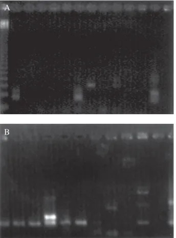
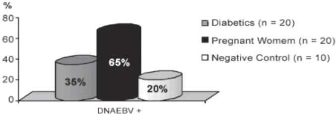
The PCR method (Fig. 2) used identified epithelial infection on the lateral border of the tongue in 7 (35%) of 20 selected diabetics; 13 (65%) of 20 pregnant wo-men and in 2 (20%) of 10 selected healthy individuals (Fig. 3).Comparing the percentage of EBV-DNA by PCR between pregnant women and diabetics, we found that
TABLE I
Hairy leukoplakia in immunocompetent patients and individuals with moderate or minor immunossupression Author Cases Sistemic condition Corticotherapy Diagnosis method Eisenberg (1992) 2 Immunocompetent - Histopathology in situ
Hibridization Felix (1992) 1 Immunocompetent - Histopathology in situ
Hibridization
Lozada-Nur (1994) 1 Pemphigus Topic Histopathology in situ 1 Pemphigoid Topic Hibridization 2 Immunocompetent
-Schiodt (1995) 1 Behçet’s syndrome Oral Histopathology in situ Hibridization Zakrzewska (1995) 1 Asthma Inalation Histopathology in situ
there is no representative difference in the proportions of patients with EBV-DNA in the two groups (Fisher Exact Test, p > 0.05). On the other hand, the proportion of pregnant women to healthy individuals showed that the former is statistically bigger (65%) than the latter (20%) (Fisher Exact Test, p value = 0.025). Likewise, comparing diabetic and healthy subjects, no significant difference was detected (Fisher Exact Test, p > 0.05). All EBV-DNA negative samples were tested for β-globin and presented a positive result.
DISCUSSION
Although OHL and candidiasis are not considered lesions associated with an important morbidity or mor-tality, they are important hallmarks of immunodefi-ciency, specially in HIV infection (Cherry-Peppers et
al. 2003, Greenspan et al. 2004, Chattopadhyay et al. 2005). The etiology of OHL has already been established, and EBV can be identified through electronic micros-copy, in situ hybridization, immunohistochemistry, and PCR (Fraga-Fernández & Vicandi-Plaza 1992, Mabruk et al. 1995, Greenspan et al.1998, Gulley 2001). How-ever, those techniques are very complex and expensive. The nuclear changes that represent EBV cytopathic ef-fects can be visualized by histopathology and cytopa-thology (Fraga-Fernández et al. 1990, Fraga-Fernández & Vicandi-Plaza 1992, Migliorati et al. 1993, Dias et al. 1999, 2000).
Considering that biopsy is an invasive procedure, we suggest that cytopathology should be the best choice to diagnose OHL. Among the advantages of the routine smears of the lateral border of the tongue for
cytopatho-TABLE II Cytopathologic diagnoses
Group NP (%) Inflammation (%) Candidiasis (%) OHL (%)
A 77 (86) 11 (12) 2 (2) 1 (1)
B 77 (86) 13 (14) -
-C 30 (87) - -
-D - - 9 (30) 30 (100)
A: diabetics; B: pregnant women; C: negative control; D: positive control; NP: normal pattern; OHL: oral hairy leukoplakia.
Fig. 1:oral hairy leukoplakia associated with candidiasis in diabetes mel-lituspatient. Smears (Papanicolaou, 1000x) showing: A: nuclei alterations - Cowdry type A inclusion and nuclear beading; B: candidiasis.
A
B
Fig. 2: polymerase chain reaction result. Gel electrophoresis showing EBV amplicons (on the left) and a positive control (on the right - samples of HIV+ patients); M = 123 bp DNA ladder marker; A: two positive samples of healthy individuals; B: seven positive samples of pregnant women.
A
logical exam are: (a) the diagnosis of inflammatory changes with the identification of the etiological agent (fungus and some bacteria) or cytopathic effects (HSV, CMV, EBV); (b) oncologic analysis of epithelial cells; (c) the possibility to use the material in different meth-ods, including molecular and electronic microscopy stud-ies.
The development of OHL and candidiasis in patients with severe immunodeficiency, as in HIV seropositive, organ-transplanted subjects and in individuals present-ing malignant neoplasias, is already well documented in the literature (Blomgren & Bäck 1996, Ammatuna et al. 2001, Casiglia & Woo 2002).Conversely, a relevant fact is that OHL findings in HIV seronegative patients, with low or moderate immunosuppression, or even in clini-cally healthy individuals are scarce (Eisenberg et al. 1992, Felix et al. 1992,Lozada-Nur et al. 1994, Schiodt et al. 1995, Zakrzewska et al. 1995). Considering the importance of candidiasis and OHL as markers and EBV association to several malignant neoplasias, it is perti-nent to identify the frequency of EBV infection and can-didiasis not only in healthy individuals, but also in those showing different degrees of immunosuppression.
Our research grouped an important number of HIV-1 seronegative individuals (n = 210), being the first study to investigate the presence of OHL in diabetics and preg-nant women. From this casuistic, 50 (24%) cases were submitted to EBV-DNA identification by PCR. OHL and candidiasis were identified only among diabetics and in a prevalence of 1 and 2%, respectively. The only case of OHL identified among all the samples studied, was re-lated to a subclinic lesion in a 75-year-old diabetic man, who presented a hyperglycemic condition and chronic obstructive pulmonary disease since 1995, taking oral and systemic steroid (40 mg/day), characterizing a sec-ond csec-ondition of immunosuppression. These results are in agreement to the literature that considers OHL and candidiasis hallmarks of severe immunosuppression (Cherry-Peppers et al. 2003, Greenspan et al. 2004, Chattopadhyay et al. 2005).
Studies reveal a high prevalence of oral candidiasis in diabetics. Amato and Pecora (1983) analyzing 50 DM type II patients, without any lesion on the oral cavity, identified Candida albicans hyphae in the saliva of 25 (50%) patients. Willis et al. (1999) demonstrated the presence of C. albicans in 77% of 414 DM patients us-ing insulin, from which 40% did not present any clinical manifestation of candidiasis. Redinova and Zlobina (2002) performedan oral exam in 102 DM types I and II patients with, attesting the incidence of C. albicans in
78.4%, among these only 35% presented lesion on oral mucosa. Surprisingly, we identified only two cases (2.2%) without clinical manifestation of the lesion. One possible explanation for that can be related to the fact that we performed the examinations in a group of stable patients that received regular outpatient medical care and psycho-nutritional orientation.
The prevalence of EBV on the tongue of healthy in-dividuals, by PCR, varies from 10 to 90%. Mabruck et al. (1994), collected material from the lateral border of the tongue of 10 healthy patients, where they identified EBV-DNA in 9 (90%) of 10 cases. In the same way, Scully et al. (1998), identified in cytological smears of the tongue EBV-DNA in 4 (20%) out of 20 healthy sub-jects. Ammatuna et al. (2001), studying 30 healthy indi-viduals who presented normal tongue clinical aspect, detected by PCR, EBV-DNA in 3 (10%) individuals. The major sample studied was published by Triantos et al. (1998) and included 40 individuals, where EBV was iden-tified in the epithelial cells of the tongue in 4 (10%). Consequently, EBV identification in 20% of healthy in-dividuals of our study is in accordance with the few stud-ies already mentioned.
This is the first study that investigates the presence of EBV-DNA on smears of the lateral border of the tongue of diabetics (35%) and pregnant women (65%). Our results showed an increase in the percentage of la-tent EBV infected patients, on the lateral border of the tongue, when compared to the percentage identified among healthy individuals. In pregnant women, it is re-ported only serological analyses, which in large series identify an average prevalence of EBV of 98%(Icart et al. 1981, Fleisher & Bolognese 1982,Le et al. 1983). Notwithstanding, in diabetics, serological studies are rare. (Hyoty et al. 1991, Jaeckel et al. 2002).
The PCR analysis has called our attention because there were positive cases in only one of the borders of the tongue. All negative samples regarding the presence of EBV-DNA, were β-globin positive, that is, all of them presented enough material for analysis. This result may justify the unilateral identification of the OHL, clinical or subclinical, in several HIV+ patients.In addition, it suggests that EBV-DNA was not in the saliva; otherwise both samples should be positive.
The results showed that EBV infection on smears of the lateral border of the tongue of individuals with mi-nor immunodeficiency such as pregnant women and dia-betics is considerably higher than in healthy subjects. Considering OHL a probable marker of immunodefi-ciency, PCR analysis to diagnose EBV latent infection and cytopathology to follow a possible passage to repli-cative phase could be useful in patients with different degrees of immunodeficiency. We believe that the in-vestigation of the prevalence of EBV infection in the keratinocytes of the tongue, with proper identification of viral burden and respective phase of the infection, will determine the real contribution of OHL and EBV infec-tion as markers of immunodeficiency and it would be an alternative tool in patients’ clinical follow up.
REFERENCES
Amato R, Pecora A 1983. Incidence of oral candidiasis in a sample group of diabetics. Boll Soc Ital Biol Sper30: 532-534. Ammatuna P, Campisi G, Giovannelli L, Giambelluca D, Alaimo
C, Mancuso S, Margiotta V 2001. Presence of Epstein-Barr virus, cytomegalovirus and human papillomavirus in normal oral mucosa of HIV-infected and renal transplant patients.
Oral Dis7: 34-40.
Blomgren J, Bäck H 1996. Oral hairy leukoplakia in a patient with multiple mieloma. Oral Surg Oral Med Oral Pathol
Oral Radiol Endod 82: 408-410.
Casiglia J, Woo S-B 2002. Oral hairy leukoplakia as a nearly indicator of Epstein-Barr viru-associated post-transplant lymphoproliferative disorder. J Oral Maxillofac Surg60: 948-950.
Chattopadhyay A, Caplan D J, Slade G D, Shugars DC, Tien HC, Patton LL 2005. Incidence of oral candidiasis and hairy leu-koplakia in HIV-infected adults in North Carolina. Oral Surg
Oral Med Oral Pathol Oral Radiol Edond99: 39-47.
Cherry-Peppers G, Daniels CO, Meeks V, Sanders CF, Reznik D 2003. Oral manifestations in the era of HAART. J Natl
Med Assoc95: 21-32.
Cinque P, Brytting M, Vago L, Castagna A, Parravicini C, Zanchetta N, D’Arminio, Monfort A, Wahren B, Lazzarin A, Linde A 1993. Epstein-Barr virus DNA in cerebrospinal fluid from patients with AIDS-related primary lymphoma of the central nervous system. The Lancet342: 398-401. Dias EP, Rocha ML, Silva Júnior A, Spyrides KS, Ferreira SM,
Polignano GA, Feijo EC, Da Fonseca EC 2000. Oral hairy leukoplakia – Histophatologic and cytopathologic features of a subclinical phase. Am J Clin Pathol114: 394-400. Dias EP, Silva Junior A, Feijó EC, Polignano GAC 1999. Diagnóstico
citopatológico da leucoplasia pilosa. J Bras Patol Med Lab35: 23-28.
Eisenberg E, Krutchkof FD, Yamase H 1992. Incidental oral hairy leukoplakia in immunocompetent persons. Oral Surg
Oral Med Oral Pathol74: 332-333.
Felix DH, Watret K, Wray D, Southam JC 1992. Hairy leuko-plakia in a HIV-negative nonimmunosuppressed patient. Oral
Surg Oral Med Oral Pathol74: 563-566.
Ferres M, Prado P, Ovalle J, Fuentes R, Villarroel L, Ferreccio C, Vial P 1995. Seroprevalence of Epstein-Barr virus infec-tion in a healthy populainfec-tion of Santiago de Chile. Rev Med Chil123: 1447-1452.
Fleisher GR, Bolognese R 1982. Seroepidemiology of Epstein-Barr virus in pregnant women. J Infect Dis145: 537-541. Fraga-Fernández J, Vicandi-Plaza B 1992. Diagnosis of hairy
leukoplakia by exfoliative cytologic methods. Am J Clin
Pathol97: 262-266.
Fraga-Fernández J, Benito C, Lizaldez EB 1990. Oral hairy leu-koplakia: a histopathologic study of 32 cases. Am J
Dermatopathol12: 571-578.
Greenspan D, Gange SJ, Phelan JA, Navazesh M, Alves ME, MacPhail LA, Mulligan R, Greenspan JS 2004. Incidence of oral lesions in HIV-1-infected women: reduction with HAART. J Dent Res83: 145-150.
Greenspan JS, De Souza YG, Regezi JA, Daniels TE, Greenspan D, MacPhail LA, Hilton JF 1998. Comparison of cytopathic
changes in oral hairy leukoplakia with in situ hybridization for EBV DNA. Oral Dis4: 95-99.
Gulley ML 2001. Molecular diagnosis of Epstein-Barr virus – Related diseases. J Mol Diag3: 1-10.
Hyoty H, Rasanen L, Hiltunen M, Lehtinen M, Huupponen T, Leinikk P 1991. Decreased antibody reactivity to Epstein-Barr virus capsid antigen in type 1 (insulin-dependent) diabe-tes mellitus. APMIS99: 359-363.
Icart J, Didier J, Dalens M, Chabanon G, Boucays A 1981. Pro-spective study of Epstein-Barr virus (EBV) infection during pregnancy. Biomedicine34: 160-163.
Jaeckel E, Manns M, Von Herrat M 2002. Viruses and diabetes.
Ann NY Acad Sci 958: 7-25.
Le CT, Chang RS, Lipson MH 1983. Epstein-Barr virus infec-tions during pregnancy. Am J Dis Child137: 466-468. Lozada-Nur F, Robinson J, Regezi JA 1994. Oral hairy
leuko-plakia in nonimmunosuppressed patiens – Report of four cases. Oral Surg Oral Med Oral Pathol78: 599-602. Mabruk MJ, Flint SR, Torner M, Balluz I Coleman DC, Sullivan
D, Atkins GJ 1994. In situ hybridization and the polimerase chain reaction (PCR) in the analysis of biopsies and exfolia-tive cytology specimens for definiexfolia-tive diagnosis of oral hairy leukoplakia (OHL). J Oral Pathol Med23: 302-308. Mabruk MJ, Flint SR, Torner M, Leonard N, Sheils O, Coleman
DC, Atkins GJ 1995. Detection of Epstein-Barr virus DNA in tongue tissues from AIDS autopsies without clinical evi-dence of oral hairy leukoplakia. J Oral Pathol Med24: 109-112.
Migliorat CA, Jones AC, Baughman P 1993. Use of exfoliative cytology in the diagnosis of oral hairy leukoplakia. Oral Surg76: 704-710.
Nakamura N, Miyazaki K, Kitano Y, Fujisaki S, Okamura H 1993. Suppression of cytotoxic T-lymphocyte activity during pregnancy. J Reprod Immuno23: 119-130.
Ozkan A, Kilic SS, Kalkan A, Ozden M, Demirdag K, Ozdarendeli A 2003. Seropositivity of Epstein-Barr virus in Eastern Anatolian Region of Turkey. Asian Pac J Allergy Immunol 21: 49-53.
Pancharoen C, MekmullicA J, Chinratanapisit S, Bhattarakosol P, Thisyakom U 2001. Seroprevalence of Epstein-Barr vi-rus antibody among children in various age groups in Bangkok, Thailand. Asian Pac J Allergy Immunol19: 135-137. Priddy KD 1997. Immunologic adaptations during pregnancy.
J Obstet Gynecol Neonatal Nurs26: 388-394.
Read SJ, Jeffery KJM, Bangham CRM 1997. Aseptic meningitis and encephalitis: the role of PCR in diagnostic laboratory.
J Cli Microbiol35: 691-695.
Redinova TL, Zlobina AO 2002. Incidence of candidiasis of the buccal mucosa and efficiency of its treatment in diabetics.
Stomatologiia (Mosk)80: 20-22.
Sand LP, Jalouli J, Larsson PA, Hirsch JM 2002. Prevalence of Epstein-Barr virus in oral squamous cell carcinoma, oral li-chen planus, and normal oral mucosa. Oral Surg Oral Med
Oral Pathol Oral Radiol Edond93: 586-592.
Schiodt M, Norgaard T, Greenspan JS 1995. Oral hairy leuko-plakia in an HIV-negative woman with Behçet’s syndrome.
Oral Surg Oral Med Oral Pathol 79: 53-56.
Scully C, Porter SR, Di Albert L, Jalal M, Maitland N 1998. Detection of Epstein-Barr virus in oral scrapes in HIV infec-tion, in hairy leukoplakia, and in healthy non-HIV-infected people. J Oral Pathol Med 27: 480-482.
Stiehm R, Fulginiti V 1980. Immunologic Disorders in Infants
and Children, Sauders, Philadelphia.
Triantos D, Leao JC, Porter SR, Scully CM, Teo CG 1998. Tis-sue distribtion of Epstein- Barr vírus genotypes in hosts coinfection by HIV. AIDS12: 2141-2146.
Willis AM, Coulter WA, Fulton CR, Hayes JR, Bell PM, Lamey PJ 1999. Oral candidal carriage and infection in insulin-treated diabetic patients. Diabet Med16: 675-679.
Zakrzewska JM, Aly Z, Speight PM 1995. Oral hairy leukoplakia in a HIV-negative asthmatic patient on systemic steroids.