INTRODUCTION
The antibiotic novobiocin (NB), a nonspecific in-hibitor of DNA topoisomerase II (DNA topo II) enzyme, can interfere with the action of many antitumoral agents, including topo II inhibitors, such as amsacrine (m-AMSA), mitoxantrone, adriamycin and ellipticine (Utsumi et al., 1990; Stetina and Veselá; 1991; Lee et al., 1992; Sakamoto-Hojo et al., 1994). NB increases the radiation-induced ab-errations at G2-phase of the cell cycle in Chinese hamster V79 cells (Takahashi et al., 1985), inhibits the repair of DNA single-strand breaks and causes partial DNA degra-dation in irradiated HeLa cells (Seregina et al., 1996).
NB cannot be regarded as a specific inhibitor of DNA topo II in mammalian cells since it can disturb many biological processes, including eukaryotic DNA replica-tion, transcription and repair (Mattern and Scudiero, 1981; Mattern et al., 1982; Seregina et al., 1996), as well as chro-mosome condensation (Aijiro and Nishimoto, 1985). Even though many studies have shown that treatment of cells with inhibitors of DNA synthesis may increase the fre-quency of chromatid-type aberrations in human lympho-cyte cultures which were previously exposed to radiation or chemical mutagens (Kihlman and Natarajan, 1984; Kihlman and Anderson, 1985; Sakamoto-Hojo and Takahashi, 1991; Traganos et al., 1991), there is evidence that NB suppresses the induction of chromosomal aberra-tions (CA) by camptothecin (Palitti et al., 1994), mitoxan-trone (Medeiros and Takahashi, 1994) and ellipticine (Sakamoto-Hojo et al., 1994) under certain cell treatment protocols.
Although the induction of CA by γ-irradiation is quite well known, the mechanisms underlying the repair of DNA lesions in cycling cells leading to chromosome damage still need to be clarified. As it is known that NB interferes with the activity of many enzymes, the objec-tive of the present study was to determine the influence of NB posttreatment on the induction of micronuclei (MN) and CA in γ-irradiated G0-lymphocytes.
MATERIAL AND METHODS
Cell culture and irradiation
Human peripheral blood lymphocytes from nor-mal, healthy male and female donors aged 20-30 years were grown in RPMI 1640 medium supplemented with 20% fetal calf serum plus penicillin and streptomycin. Cells were stimulated with 4% phytohemagglutinin (PHA) prepared in our laboratory from Phaseolus vul-garis. In all experiments, cells were irradiated before PHA stimulation (at the G0-phase of the cell cycle) using a
60-cobalt (60-Co) γ-radiation source (Department of Genet-ics, Faculty of Medicine of Ribeirão Preto-USP) at a dose rate of 0.1 Gy/min.
Chemicals
Novobiocin (Sigma) was freshly prepared in deion-ized sterile distilled water and used at concentrations of 25 or 50 µg/ml of culture medium.
NB treatment at the G0-phase of the cell cycle
Unstimulated lymphocytes from three donors (a fe-male and two fe-males) were irradiated with 60-Co γ-rays (0.75, 1.5 and 3.0 Gy) and posttreated with 50 µg/ml NB for 30 min in serum and in phytohemagglutinin-free me-dium. After treatment with NB, the cells were washed twice
INFLUENCE OF NOVOBIOCIN ON
γ
-
IRRADIATED G
0-LYMPHOCYTES AS
ANALYZED BY CYTOGENETIC ENDPOINTS
Marcela Savoldi-Barbosa1, Elza T. Sakamoto-Hojo1,2 and Catarina S. Takahashi1,2
A
BSTRACTExperiments with novobiocin (NB) post-treatment were performed to verify its effect on the frequencies of micronuclei (MN) and chromosomal aberrations (CA) induced byγ-irradiation (0.75, 1.5 and 3.0 Gy) in human lymphocytes at G0-phase. The frequencies of MN significantly decreased by 44 and 50%, for the treatment with NB 50 µg/ml (30-min pulse) after radiation doses of 1.5 and 3.0 Gy, respectively. However, CA frequencies were not significantly affected. No significant effect on CA was observed when lymphocyte cultures were exposed to a single dose of 2.0 Gy at the G0-phase and posttreated with 25 µg/ml NB for three hours either immediately after irradiation (G0-phase) or after 24 h (S-phase). The significant suppressive effect of NB on MN frequencies supports the hypothesis that NB interaction with chromatin increases access to DNA repair enzymes.
1Departamento de Genética, Faculdade de Medicina de Ribeirão Preto, USP, Ribeirão Preto, SP, Brasil.
with NB without NB
1500
1200
900
600
300
0
0 0.75 1.5 3
Radiation dose (Gy)
Micr
onuclei/1000 binucleated cells
with RPMI medium and incubated at 37ºC. For CA analysis, lymphocyte cultures were fixed 52 h after cul-ture initiation and exposed to 0.4 µg/ml colchicine (Sigma) during the last 90 min of incubation. For MN analysis, the cytokinesis-block (CB) method (Fenech and Morley, 1985) was applied; 3 µg/ml cytochalasin B (Cyt-B, Sigma) was added to the cultures after 44 h of incubation, and cells were fixed after 72 h. MN were scored in 2000 binucleated CB cells per treatment per individual, using the criteria of Fenech and Morley (1985). The results were analyzed statistically by the Fisher exact test.
NB treatment at S-phase
Lymphocytes from three donors (all female) were
γ-irradiated with 2.0 Gy at the G0-phase of the cell cycle
and posttreated with 25 µg/ml NB for 3 h, immediately after irradiation (G0-phase) and 24 h after culture
initia-tion (when most cells were in the S-phase). Lymphocytes were washed twice in serum-free medium, and reincubated for 52 h in complete medium at 37ºC. The Student t-test was applied to the results.
Chromosome preparation and analysis
Metaphase preparations for CA analysis were ob-tained by the conventional technique of Moorhead et al. (1960). For mitotic index (MI) calculation, mitotic cells were scored in 2000 cells per treatment per individual. CA were analyzed in a blind test and classified according to Savage (1975) and IAEA (1986). One hundred metaphases per treatment were analyzed in all experiments.
RESULTS
Effect of NB posttreatment in irradiated G0-lymphocytes
a) Micronucleus analysis
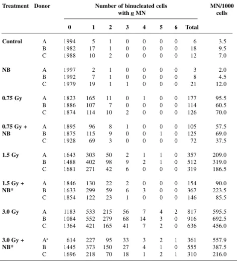
The effects of NB posttreatment (30-min pulse) on γ-irradiated unstimulated lymphocytes were analyzed in three experiments with different donors (Table I). As expected, MN frequencies increased proportionally to the radiation doses (0.75 to 3.0 Gy). The induction of chro-mosome damage was significant (P < 0.01) and dose de-pendent, as can be seen in the distribution of cells with increased numbers of MN. There was a clear interaction effect between the radiation treatment and 50 µg/ml NB posttreatment, since significant reductions of 44 and 50% in the frequencies of MN (P < 0.05) were observed for the doses of 1.5 and 3.0 Gy, respectively (Figure 1), despite a considerable degree of variability among individuals.
b) Chromosomal aberrations
Similarly to MN analysis, three experiments were carried out in parallel to verify the effects of NB post-treatment on the induction of CA in γ-irradiated unstimu-lated-lymphocytes. A significant (P < 0.01) induction of CA was obtained for all lymphocyte cultures treated with radiation at all doses (Table II). There was a lack of in-teraction effects between the radiation and NB treat-ments. Differences in mitotic indices were not significant for all treatments (P > 0.05). The aberrations detected were predominantly dicentrics and double fragments.
NB posttreatment at different phases of the cell cycle in irradiated G0-lymphocytes
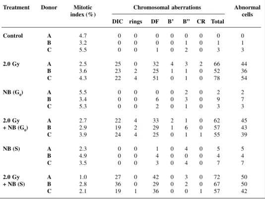
The influence of NB treatment at different phases of the cell cycle was evaluated in G0-lymphocytes irradi-ated with 2.0 Gy. There were no significant differences (P > 0.05) among treatments (Table III), indicating an ab-sence of interaction effect between radiation and NB treat-ment at the G0- and S-phase.
DISCUSSION
The influence of NB on radiation-induced chro-mosome damage in human lymphocytes was investigated. Posttreatment of cells with NB immediately after irra-diation (G0-phase) significantly reduced frequencies of MN. There was a decreasing trend in radiation-induced CA frequencies with NB, but it was not significant.
Combinations of NB with other genotoxic
com-pounds have been studied by many authors. Lazutka and Rudaitiene (1992) reported that NB suppressed SCE induc-tion by tumor necrosis factor (TNF-α) in human lympho-cytes. Furthermore, Sakamoto-Hojo et al. (1994) showed that pretreatment of lymphocyte cultures with NB dur-ing the S/G2 transition can reduce CA frequencies
in-duced by the antitumoral drug ellipticine. Medeiros and Takahashi (1994) observed a similar result by combin-ing NB with mitoxantrone durcombin-ing the G2-phase of the cell
cycle. However, the cell response to chemical and/or physical agents may be influenced by several factors, such as cell line or organism, drug concentration or dose, pro-tocol of cell treatment, etc.
It is well known that the firm compactation of chro-matin may impair the accessibility of DNA repair enzymes to the damaged sites, since variations in chromatin con-formation and organization can affect the extent of DNA damage and repair (Wheeler and Wierowski, 1983; Yasui et al., 1987). Chiu et al. (1986) showed that chromatin
Table I - Frequencies of micronuclei (MN) in 0.75 to 3.0 Gy gamma-irradiated cytokinesis-blocked G0-lymphocytes posttreated with 50 µg/ml novobiocin (NB) (30-min pulse). Three experiments (A, B, C) were performed with different donors and 2000 binucleated cells/treatment/
donor were analyzed.
Treatment Donor Number of binucleated cells MN/1000
with n MN cells
0 1 2 3 4 5 6 Total
Control A 1994 5 1 0 0 0 0 6 3.5
B 1982 17 1 0 0 0 0 18 9.5
C 1988 10 2 0 0 0 0 12 7.0
NB A 1997 2 1 0 0 0 0 3 2.0
B 1992 7 1 0 0 0 0 8 4.5
C 1979 19 1 1 0 0 0 21 12.0
0.75 Gy A 1823 165 11 0 1 0 0 177 95.5
B 1886 107 7 0 0 0 0 114 60.5
C 1874 114 10 2 0 0 0 126 70.0
0.75 Gy + A 1895 96 8 1 0 0 0 105 57.5
NB B 1875 115 9 0 0 1 0 125 69.0
C 1928 69 3 0 0 0 0 72 37.5
1.5 Gy A 1643 303 50 2 1 1 0 357 209.0
B 1488 402 98 9 2 1 0 512 319.0
C 1681 271 42 6 0 0 0 319 186.5
1.5 Gy + A 1846 130 22 2 0 0 0 154 90.0
NB* B 1633 299 59 6 3 0 0 367 223.5
C 1854 122 23 1 0 0 0 146 85.5
3.0 Gy A 1183 533 215 56 7 4 2 817 595.5
B 1084 552 279 68 14 3 0 916 692.5
C 1364 421 165 41 7 2 0 636 456.0
3.0 Gy + Aa 0614 227 95 33 3 2 1 361 557.9
NB* B 1445 373 150 27 4 1 0 555 387.5
C 1696 218 70 18 1 2 1 310 216.0
structure plays an important role in the formation and repair of DNA lesions in Chinese hamster cells exposed to γ-irradiation. Likewise, Udvardy and Schedl (1991) showed that chromatin structure, not sequence speci-ficity, is the primary determinant in DNA topo II site selection, and that chromatin organization may provide a general mechanism for generating specificity in a wide range of DNA-protein interactions. Kapizewska and Lange (1991) observed that NB applied before and dur-ing irradiation prevents the induction of alterations in DNA supercoiling in two mouse leukemia cell lines dif-fering in radiosensitivity, by modifying the DNA relax-ation in the chromatin structure. Consequently, these alterations influence the X-ray-induced DNA damage and its repair.
Therefore, many lines of evidence in the literature are showing that the repair mechanisms may be influenced by the state of chromatin relaxation, supporting the possi-bility that in the present study, NB may have acted by changing the DNA structure in irradiated G0-lymphocytes, and consequently the DNA strands became more
acces-sible to the enzymes responacces-sible for DNA repair. Thus, NB interference in the processing of the DNA lesions, caused by ionizing radiation at the G0-phase, may lead to a modulation of cell response by reducing the degree of chro-mosome damage.
No complete correlation between the CA and MN frequencies was verified when G0-lymphocytes were ir-radiated and posttreated with NB. It is well known that radiation-induced MN arise not only from acentric chro-mosomes or fragments, but also from whole chromo-somes which were not incorporated into the main nu-clei during mitosis (Fenech and Morley, 1985); thus, the presence of MN indicates not only structural chro-mosome damage, but also an alteration in chrochro-mosome number (aneugenic effect). With the conventional mi-cronucleus test it is not possible to distinguish among micronuclei arising from acentric fragments induced by clastogens and those arising from whole chromosomes induced by spindle poisons (aneugenic agents). Attempts to overcome this limitation of the micronucleus test have included measurement of micronucleus size and DNA
Table II -Frequencies of chromosomal aberrations in gamma-irradiated G0-lymphocytes posttreated with 50
µg/ml novobiocin (NB) (30-min pulse). Three experiments (A, B, C) were performed with different donors and 100 metaphaseswere analyzed per treatment for each donor.
Treatment Donor Mitotic Chromosomal aberrations Abnormal
index (%) cells
DIC rings DF B’ B” CR Total
Control A 3.9 0 0 2 4 1 0 7 7
B 7.5 0 0 4 4 0 0 8 8
C 3.2 0 0 3 1 0 0 4 3
NB A 1.0 1 0 3 3 0 0 7 7
B 6.1 0 0 8 1 0 0 9 8
C 3.1 0 0 1 2 0 0 3 3
0.75 Gy A 1.7 7 0 9 2 0 0 18 16
B 4.1 0 0 8 3 1 0 12 12
C 1.8 2 0 9 0 0 0 11 9
0.75 Gy A 2.5 1 0 12 6 0 0 19 15
+ NB B 4.6 3 0 17 0 1 0 21 18
C 1.8 3 1 4 1 0 0 9 9
1.5 Gy A 3.4 25 3 45 3 6 0 82 46
B 3.1 5 0 17 0 0 0 22 18
C 4.7 11 2 18 4 3 2 40 32
1.5 Gy A 2.2 25 4 37 11 6 1 84 55
+ NB B 4.1 12 0 40 5 0 0 57 39
C 4.1 15 3 23 3 3 1 48 38
3.0 Gy A 3.2 51 5 86 2 5 0 149 71
B 9.4 49 3 84 6 3 0 145 69
C 2.8 56 1 58 3 2 2 122 66
3.0 Gy A 1.5 79 2 62 4 4 4 155 84
+ NB B 4.2 34 2 63 2 0 2 103 62
C 2.5 50 4 66 4 4 1 129 77
content, C-banding and, more recently, in situ hybrid-ization with DNA probes (Miller et al., 1992).
Cornforth and Goodwin (1991) demonstrated that about 50% of all radiation-induced chromosome fragments in metaphase cells were found as MN in the subsequent interphase. Fragments which were unable to form MN could be incorporated into the daughter nuclei during di-vision, however some fragments could coalesce and form micronuclei containing more than one acentric fragment (Savage, 1988; Kramer et al., 1990; Cornforth and Goodwin, 1991). Miller et al. (1992) used simultaneous fluorescence in situ hybridization with telomere- and cen-tromere-specific probes, to show that in nearly all MN containing a centromeric signal (spontaneous and radia-tion induced) the centromeric signal was accompanied by two to four telomeric signals in the same MN, indicating the presence of a whole chromosome. Similarly, the use of antibodies against centromeres has revealed that usually 30 to 50% of the total MN include whole chromosomes in untreated established cell lines and in biopsies from hu-man rectal carcinomas (Weissenborn and Streffer, 1991). Therefore, the absence of correlation between CA and MN frequencies observed in our study is in accor-dance with the following evidence: some specific types of CA may form MN in preference to other aberrations (Wakata and Sasaki, 1987), multiple fragments may re-side within a single MN (Jonhston et al., 1997) and a con-siderable percentage of MN may have originated from
whole chromosomes due to disturbance of the cell spindle (Walker et al., 1996). According to Johnston et al. (1997), the interactions between the initial damage, repair pro-cesses, cell cycle-dependent variations in sensitivity and changes in cell kinetics can produce complex patterns of MN expression. In the case of CA analysis, the cell cycle kinetics of lymphocyte cultures should be considered as an important factor influencing the cell response when colchicine-arrested metaphases are scored for aberrations. Although cultures were fixed after 52 h, most of the aber-rations were observed in 1st-cycle cells, since the chro-mosome-type ones were most frequent, resulting in a low percentage of 2nd-cycle cells.
Boei et al. (1996) showed that most chromosome breaks are repaired within a few hours after the induction of the damage in human lymphocytes, since the frequencies of radiation-induced aberrations decrease between the first and the second cycle, and are reduced drastically between the second and third cell cycle. Similarly, Darroudi et al. (1998) observed that in irradiated human lymphocytes (1, 2, 4 and 6 Gy) almost 50% of breaks were rejoined after 1 h, and about 20% of breaks remained after 18 h. In addition, these authors showed that at low doses of 1 and 2 Gy most of the exchanges were formed immediately, and, at higher doses, the frequency of exchanges increased at rates similar to those observed for the rejoining of breaks.
Thus, it seems that an efficient mechanism of re-pair process occurs immediately after cell irradiation,
in-Table III - Frequencies of chromosomal aberrations in gamma-irradiated G0-lymphocytes posttreated with 25
µg/ml novobiocin (NB) for three hours at G0- and S-phases of the cell cycle. Three experiments (A, B, C) were performed with different donors and 100 metaphases were analyzed per treatment for each donor.
Treatment Donor Mitotic Chromosomal aberrations Abnormal
index (%) cells
DIC rings DF B’ B” CR Total
Control A 4.7 0 0 0 0 0 0 0 0
B 3.2 0 0 0 0 1 0 1 1
C 5.5 0 0 1 0 2 0 3 3
2.0 Gy A 2.5 25 0 32 4 3 2 66 44
B 3.6 23 2 25 1 1 0 52 36
C 4.3 22 4 51 0 1 0 78 54
NB (G0) A 5.5 0 0 0 0 2 0 2 2
B 3.4 0 0 6 0 3 0 9 7
C 5.3 0 0 2 0 1 0 3 3
2.0 Gy A 2.7 22 4 33 2 1 0 62 45
+ NB (G0) B 2.9 19 2 29 1 6 0 57 43
C 3.9 24 4 25 0 1 1 55 39
NB (S) A 2.3 0 0 1 0 4 0 5 5
B 4.9 0 0 4 0 0 0 4 4
C 3.5 0 0 3 0 4 0 7 7
2.0 Gy A 1.0 27 0 42 0 3 0 72 50
+ NB (S) B 2.8 36 0 29 0 2 0 67 50
C 2.1 19 1 36 0 0 1 57 42
dicating that in the present work, NB may have acted in a critical period after the induction of DNA damage, reduc-ing the number of lesions which would be converted into chromosome damage. NB reduced the MN but not the CA frequencies induced by γ-irradiation during the G0-phase of the cell cycle. Even though NB acts on different cell targets and by different pathways, our results support the evidence (Villeponteau et al., 1984; Kapizewska and Lange, 1991) that NB interaction increases the accessibil-ity of chromatin to DNA repair enzymes during the G0 -phase of the cell cycle by affecting its structure.
ACKNOWLEDGMENTS
We thank Luiz A. Costa Jr., Silvio A. dos Santos and Sueli A. Neves for valuable technical assistance, and Prof. Dr. Flávio R. Gorini for the statistical analysis. This research was supported by FAPESP, CAPES and CNPq (Brazil). Publication supported by FAPESP.
RESUMO
Com o objetivo de estudar o mecanismo de indução do dano cromossômico, foi testada a influência do pós-tratamento com a novobiocina (NB) sobre as freqüências de micronúcleos (MN) e aberrações cromossômicas (CA) induzidas pela radiação gama (0,75, 1,5 e 3,0 Gy) em linfócitos humanos na fase G0. Os experimentos realizados com 4 doadores demonstraram que as freqüências de MN sofreram um decréscimo estatisticamente significativo, 44 e 50%, para o tratamento com 50 µg/ml de NB (pulso de 30 min) após as doses de 1,5 e 3,0 Gy de radiação, respectivamente. Entretanto, o mesmo grau de interação não foi observado para as freqüências de AC e reduções não significativas foram obtidas para todos os tratamentos combinados em protocolos semelhantes. A ausência de um efeito de interação foi também observada quando os linfócitos em cultura foram expostos à dose única de 2,0 Gy na fase G0 e pós-tratados com 25 µg/ml de NB por 3 horas logo após a irradiação (fase G0) e após 24 horas (fase S) de incubação. O efeito supressivo da NB sobre as freqüências de MNs na fase G0 do ciclo celular é discutido em termos de indução de modificações na estrutura da cromatina, aumentando o acesso às enzimas de reparo do DNA, e podendo, assim, ser observada uma modulação da resposta celular nos linfócitos irradiados em G0 e pós-tratados com NB.
REFERENCES
Aijiro, K. and Nishimoto, T. (1985). Specific site of histone H3 phospho-rylation related to the maintenance of premature chromosome con-densation. J. Biol. Chem. 260: 15379-15381.
Boei, J.J.W.A., Vermeulen, S. and Natarajan, A.T. (1996). Detection of chromosomal aberrations by fluorescence in situ hybridization in the first three postirradiation divisions of human lymphocytes. Mutat. Res. 349: 127-135.
Chiu, S.M., Friedman, L.R., Sokany, N.M., Xue, L.Y. and Oleinick, N.L.
(1986). Nuclear matrix proteins are crosslinked to transcriptionally active gene sequences by ionizing radiation. Radiat. Res. 107: 24-38.
Cornforth, M.N. and Goodwin, E.H. (1991). Transmission of radiation-induced acentric chromosomal fragments to micronuclei in normal human fibroblasts. Radiat. Res. 126: 210-217.
Darroudi, F., Fomina, J., Meijers, M. and Natarajan, A.T. (1998). Kinet-ics of the formation of chromosome aberrations in X-irradiated hu-man lymphocytes, using PCC and FISH. Mutat. Res. 404: 55-65.
Fenech, M. and Morley, A.A. (1985). Measurement of micronuclei in hu-man lymphocytes. Mutat. Res. 147: 29-36.
IAEA (1986). Biological Dosimetry: Chromosomal Aberration Analysis for Dose Assessment. Technical Report No. 260, International Atomic Energy Agency, Vienna.
Johnston, P.J., Stoppard, E. and Bryant, P.E. (1997). Induction and dis-tribution of damage in CHO-K1 and the X-ray-sensitive hamster cell line xrs-5, measured by the cytochalasin-B-cytokinesis block micro-nucleus assay. Mutat. Res. 385: 1-12.
Kapizewska, M. and Lange, C.S. (1991). Novobiocin treatment reverses radiation-induced alterations in higher-order DNA structure in L5178Y nucleoids. Radiat. Res. 127: 285-291.
Kihlman, B.A. and Anderson, H.C. (1985). Synergistic enhancement of the frequency of chromatid aberrations in cultured human lympho-cytes by combinations of inhibitors of DNA repair. Mutat. Res. 150: 313-325.
Kihlman, B.A. and Natarajan, A.T. (1984). Potentiation of chromosomal alterations by inhibitors of DNA repair. In: DNA Repair and its Inhi-bition: Towards an Analysis of Mechanism (Collins, A., Johnson, R.T. and Downes, S., eds.). Nuclei Acids Symposium Ser. 13, IRL Press, Oxford, pp. 319-339.
Kramer, J., Schaich-Walch, G. and Nüsse, M. (1990). DNA synthesis in radiation-induced micronuclei studied by bromodeoxyuridine (BrdUrd) labelling and anti-BrdUrd antibodies. Mutagenesis 5: 491-495.
Lazutka, J.R. and Rudaitiene, S. (1992). Modulation by novobiocin of sister-chromatid exchanges induced by tumor necrosis factor in hu-man lymphocytes. Mutat. Res. 268: 217-221.
Lee, F.Y.F., Flannery, D.J. and Siemann, D.W. (1992). Modulation of the cell cycle-dependent cytotoxicity of adriamycin and 4-hydroperoxy-cyclophosphamide by novobiocin, an inhibitor of mammalian topoisomerase II. Cancer Res. 52: 3515-3520.
Mattern, M.R. and Scudiero, D.A. (1981). Dependence of mammalian DNA synthesis on DNA supercoiling. III. Characterization of the inhibi-tion of replicative and repair type DNA synthesis by Novobiocin and nalidixic acid. Bioch. Bioph. Acta 653: 248-258.
Mattern, M.R., Paone, R.F. and Day, R.S. (1982). Eukaryotic DNA repair is blocked at different steps by inhibitors of DNA topoisomerases and of DNA polymerases alpha and beta. Biochim. Biophys. Acta 697: 6-13.
Medeiros, M.G. and Takahashi,C.S. (1994). Effects of treatment with mitoxantrone in combination with novobiocin, caffeine and ara-C on human lymphocytes in culture. Mutat. Res. 307: 285-292.
Miller, B.M., Werner, T., Weier, H.-U. and Nüsse, M. (1992). Analysis of radiation-induced micronuclei by fluorescence in situ hybridization (FISH) simultaneously using telomeric and centromeric DNA probes.
Radiat. Res. 131: 177-185.
Moorhead, P.S., Nowell, P.C., Mellman, W.J., Battipps, D.M. and
Hungerford, D.A. (1960). Chromosome preparation of leukocytes cultured from human peripheral blood. Exp. Cell Res. 20: 613-616.
Palitti, F., Mosesso, P., Dichiara, D., Schinoppi, A., Fiore, M. and Bassi, L. (1994). Use of antitopoisomerase drugs to study the mechanism of induction of chromosomal damage. In: Chromosomal Alterations - Origin and Significance (Obe, G. and Natarajan, A.T., eds.). Springer-Verlag, Berlin, pp. 103-115.
Sakamoto-Hojo, E.T. and Takahashi, C.S. (1991). Clastogenic action of ellipticine over the cell cycle of human lymphocytes and influence of posttreatments with caffeine and ara-C at G2. Mutat. Res. 248:
195-202.
Sakamoto-Hojo, E.T., Dias, F.L. and Takahashi, C.S. (1994). Mecanismos de indução de aberrações cromossômicas: interação de compostos inibidores de topoisomerases II em linfócitos humanos. Rev. Bras. de Genét. 17: (Suppl. 3): 244.
Savage, J.R.K. (1975). Classification and relationships of induced chro-mosomal structural changes. J. Med. Genet. 12: 103-122.
Savage, J.R.K. (1988). A comment on the quantitative relationship between micronuclei and chromosome aberrations. Mutat. Res. 207: 33-36.
of DNA single-stranded breaks and degradation under the action of novobiocin in gamma-irradiated HeLa cells. Tsitologiia 38: 57-65.
Stetina, R. and Veselá, D. (1991). The influence of DNA-topoisomerase II inhibitors novobiocin and fostriecin on the induction and repair of DNA damage in Chinese hamster ovary (CHO) cells treated with mitoxantrone. Neoplasma 38: 109-117.
Takahashi, K., Kaneko, I., Nishiyama, C. and Nakano, K. (1985). Effect of novobiocin on the frequencies of chromatid-type aberrations and sister-chromatid exchanges following γ-iradiation. Mutat. Res. 144: 265-270.
Traganos, F., Kaminska-Eddy, B. and Darzynkiewicz, Z. (1991). Caf-feine reverses the cytotoxic and cell kinetic effects of novantrone (mitoxantrone). Cell Prolif. 24: 305-319.
Udvardy, A. and Schedl, P. (1991). Chromatin structure, not DNA sequence specificity, is the primary determinant of topoisomerase II sites of action in vivo. Mol. Cell Biol. 11: 4973-4984.
Utsumi, H., Shibuya, M.L., Kosaka, T., Buddeenbaum, W.E. and Elkind, M.M. (1990). Abrogation by novobiocin of cytotoxicity due to the topoisomerase II inhibitor amsacrine in Chinese hamster cells.
Can-cer Res. 50: 2577-2581.
Villeponteau, B., Lundell, M. and Martinson, H. (1984). Torsional stress promotes the DNase I sensitivity of active genes. Cell 39: 469-478.
Wakata, A. and Sasaki, M.S. (1987). Measurement of micronuclei by cytoki-nesis-block method in cultured Chinese hamster cells: comparison with types and rates of chromosome aberrations. Mutat. Res. 190: 51-57.
Walker, J.A., Boreham, D.R., Unrau, P. and Duncan, A.M.V. (1996). Chromosome content and ultrastructure of radiation-induced micro-nuclei. Mutagenesis 11: 419-424.
Weissenborn, U. and Streffer, C. (1991). Micronuclei with kinetochores in human melanoma cells and rectal carcinomas. Int. J. Radiat. Biol. 59: 373-383.
Wheeler, K. and Wierowski, J. (1983). DNA accessibility: a determinant of mammalian cells differention? Radiat. Res. 93: 312-318.
Yasui, L.S., Higashikubo, R. and Warters, R.L. (1987). The effect of chro-matin decondensation DNA damage and repair. Radiat. Res. 112: 318-330.


