Pro life rative signaling initiate d in
ACTH re ce pto rs
1Departamento de Bioquímica, Instituto de Q uímica, Universidade de São Paulo,
São Paulo, SP, Brasil
2Departamento de Biofísica, Escola Paulista de Medicina,
Universidade Federal do Estado de São Paulo, São Paulo, SP, Brasil C.F.P. Lotfi1, A.P. Lepique1,
F.L. Forti1, T.T. Schwindt1,
C.B. Eichler1, M.O . Santos1,
I.T. Rebustini1,
G.N.M. Hajj1, L. Juliano2
and H.A. Armelin1
Abstract
This article reviews recent results of studies aiming to elucidate modes of integrating signals initiated in ACTH receptors and FGF2 receptors, within the network system of signal transduction found in Y1 adreno-cortical cells. These modes of signal integration should be central to the mechanisms underlying the regulation of the G0®G1®S
transi-tion in the adrenal cell cycle. FGF2 elicits a strong mitogenic response in G0/G1-arrested Y1 adrenocortical cells, that includes a) rapid and transient activation of extracellular signal-regulated kinases-mitogen-activated protein kinases (ERK-MAPK) (2 to 10 min), b) transcription activation of c-fos, c-jun and c-myc genes (10 to 30 min), c) induction of c-Fos and c-Myc proteins by 1 h and cyclin D1 protein by 5 h, and d) onset of DNA synthesis stimulation within 8 h. ACTH, itself a weak mitogen, interacts with FGF2 in a complex manner, blocking the FGF2 mitogenic response during the early and middle G1 phase, keeping
ERK-MAPK activation and c-Fos and cyclin D1 induction at maximal levels, but post-transcriptionally inhibiting c-Myc expression. c-Fos and c-Jun proteins are mediators in both the strong and the weak mitogenic responses respectively triggered by FGF2 and ACTH. Induction of c-Fos and stimulation of DNA synthesis by ACTH are independent of PKA and are inhibited by the PKC inhibitor GF109203X. In addition, ACTH is a poor activator of ERK-MAPK, but c-Fos induction and DNA synthesis stimulation by ACTH are strongly inhibited by the inhibitor of MEK1 PD98059.
Co rre spo nde nce
H.A. Armelin
Departamento de Bioquímica Instituto de Q uímica, USP 05508-990 São Paulo, SP Brasil
E-mail: haarmeli@ quim.iq.usp.br Presented at the First
International Meeting on Adrenal Disease: Basic and Clinical Aspects, Ribeirão Preto, SP, Brazil, August 31-September 2, 1999. Research supported by FAPESP and CNPq to, respectively, H.A. Armelin and L. Juliano. C.F.P. Lotfi is a post-doctoral and A.P. Lepique, F.L. Forti, T.T. Schwindt, C.B. Eichler, M.O . Santos, I.T. Rebustini and G.N.M. Hajj are recipients of pre-doctoral fellowships from FAPESP.
Received December 20, 1999 Accepted March 22, 2000
Ke y wo rds ·ACTH ·FGF2
·Signal transduction ·MAP kinases ·Early response genes
Intro ductio n
Elucidation of the molecular mechanisms by which ACTH exerts its growth effects on adrenal cortex has defied investigators for decades. In the intact organism ACTH seems to be a mitogen, but in cultures of isolated adrenocortical cells ACTH behaves like a growth inhibitory hormone (1).
ACTH growth effects on adrenal cortex. Considerable attention has been recently given to mitogenic effects triggered by pep-tide hormones that activate G protein-coupled receptors. The majority of these studies has centered on determining whether, and how, signals initiated in G protein-coupled recep-tors can activate the mitogen-activated pro-tein kinase (MAPK) cascades, particularly the one including the class of the extracellu-lar signal-regulated kinases (ERK-MAPK) (4).
In Y1 adrenocortical cells, we started by showing that ACTH induces transcription of the fos and jun gene families (5,6), a charac-teristic of peptide growth factors that bind to tyrosine kinase receptors initiating signals to activate the ERK-MAPK pathway. The Fos and Jun protein families consist of four (c-Fos, FosB, Fra1 and Fra2) and three gene products (c-Jun, JunB and JunD), respec-tively, that dimerize via a leucine zipper domain to form hetero- (Fos-Jun) and homo-dimers (Jun-Jun), that comprise the series of transcription factors denominated AP1 (7). AP1 factors seem to play a critical role in the control of G0®G1®S transition of the cell cycle. Quiescent G0-arrested fibroblasts ex-hibit very low levels of AP1 factors and upon stimulation by peptide growth factors, like fibroblast growth factor 2 (FGF2), rapidly induce the fos and jun genes in an ordered fashion (7). Blocking the induction of c-Fos and c-Jun proteins is sufficient to block the G0®G1®S transition in G0-arrested fibro-blasts stimulated with FGF2 (8-10). Thus, ACTH, inducing the fos and jun genes in Y1 adrenocortical cells, resembles FGF2 trig-gering the G0®G1®S transition in G0 -ar-rested 3T3 fibroblasts.
FGF2 is the prototype of the large family of fibroblast growth factors (11), presently consisting of 18 members (12) that are widely expressed in embryo, fetus and adult tissues of vertebrates and invertebrates. It has been recognized for years that forms of FGF play key roles in mitogenesis, cellular
differentia-tion, angiogenesis and repair of tissue injury in the adult organism (11). In addition, re-ports of the last few years have involved molecular pathways commanded by FGFs in fundamental steps of embryo and fetal velopment such as left-right asymmetry de-termination in chick and mouse embryo (13), hepatogenesis in mouse embryo (14), devel-opment of the Drosophila tracheal system and the mouse lung (15), and development of the rat exocrine pancreas (16). In fact, it is becoming more and more clear that the FGF family of proteins and their respective set of receptors are major players governing the classical epithelial-mesenchymal interactions that underlie differentiation and organogen-esis in the embryo. In the adult organism FGF forms persist throughout a number of different tissues as local paracrine and auto-crine regulators of very basic biological pro-cesses like mitogenesis and cellular differ-entiation. The interaction between classical trophic hormones and FGFs, locally pro-duced at the target cell level, is likely to be a major part of the molecular mechanisms by which trophic hormones regulate functions of target cells.
promising. Our aim is to elucidate modes of integrating signals initiated in ACTH recep-tors and FGF2 receprecep-tors within the network system of signal transduction found in Y1 adrenocortical cells. These modes of signal integration should be central to the mechan-isms underlying the regulation of the G0®G1®S transition in the adrenal cell cycle.
FGF2 is a stro ng mito ge n fo r Y1 adre no co rtical ce lls
In serum-free medium, G0/G1-arrested Y1 adrenocortical cells stimulated with bovine recombinant FGF2 (5-200 pM; short pulse, 30 min to 2 h, or sustained treatment) display a mitogenic response consisting of the fol-lowing sequential events: a) rapid and tran-sient activation of p42 and p44 ERK-MAPK, peaking between 2 and 5 min; b) transient transcription activation of the c-fos, c-jun, junB and c-myc genes, between 10 and 30 min; c) induction of c-Fos protein: 80% of the nuclei labeled with anti-c-Fos antibody by 1 h (control under 0.1%); d) induction of c-Myc protein: 70% labeled nuclei with anti-c-Myc antibody by 2 h (control levels at 35%); e) induction of the cyclin D1 protein: a sharp increase from negligible control lev-els detected by Western blot at 5 h, and f) the onset of DNA synthesis stimulation by 8 h, showing 90% of BrdU-labeling index by 20 h, whereas control levels increased linearly from 10% at zero time to 25% by 20 h.
It is still unknown whether endogenous growth factors play an autocrine role in the growth control of Y1 adrenocortical cells. Nonetheless, total RNA of Y1 cells, probed with RT-PCR and specific primers, has yielded cDNA sequences of FGF2, FG5 and PDGF A, indicating that these peptide fac-tors are produced by this adrenocortical cell line. In addition, serum-free medium condi-tioned with Y1 cells is rich in PDGF-like factors, but recombinant forms of PDGF AB and BB do not trigger a mitogenic response
in G0/G1-arrested Y1 cells. Furthermore, Y1 cell lysates, but not serum-free conditioned medium, possess active FGF-like factors, suggesting that Y1 cells do not liberate this kind of factors into the culture medium.
Sho rt pulse s o f ACTH can e licit a we ak mito ge nic re spo nse in Y1 adre no co rtical ce lls
Short pulses (up to 2 h) of synthetic ACTH39 and natural porcine corticotropin A, at physiological concentrations, trigger DNA synthesis stimulation in G0/G1-arrested Y1 adrenocortical cells. In a 24-h experi-ment, a 2-h pulse of 1 nM ACTH39 or 0.1 mU/ml corticotropin A respectively leads to a 2.6- and 2.5-fold increase in BrdU-labeling index; under the same conditions, 0.2 nM FGF2 causes a 7-fold increase in BrdU-labeling index. This mitogenic activity of ACTH is not dependent on the cAMP/PKA pathway, since it is observed in PKA-defi-cient mutants of the Y1 cell line. In addition, ACTH mitogenic activity is inhibited by the PKC inhibitor GF109203X. Thus, according to this mitogenic assay, ACTH resembles FGF2, except that ACTH is a weak mitogen (2,3).
The synthetic ACTH fragment 7-38, a well-known competitive inhibitor of ACTH39 (21), at 100 nM inhibits by 60% and 80% the DNA synthesis respectively stimulated by 1 nM ACTH39 and 1 mU/ml corticotropin A, indicating that this mitogenic effect of ACTH39 and corticotropin A is mediated by specific ACTH receptors.
activ-ity decreases sharply beyond 2 h of pulse length, whereas for FGF2, mitogenic activ-ity is the same for short pulses or sustained treatment.
ACTH induce s transcriptio n o f the
fos and jun ge ne s, but no t o f the c-m yc ge ne
ACTH, like FGF2, is a strong inducer of the fos and jun families of proto-oncogenes in Y1 adrenocortical cells, independently of protein synthesis (5,6). Experiments of run-off transcription have shown that ACTH transiently induces transcription of c-fos, fosB, c-jun and junB between 10 and 30 min. Similar results were obtained with FGF2 and PMA (phorbol-12-myristate-13-acetate) in identical experiments of run-off transcrip-tion. To further probe into fos and jun induc-tion by signals initiated in ACTH receptors, we took the entire ACTH receptor cDNA, inserted it in the pSVK3 mammalian vector, and transfected it into Balb3T3 fibroblasts (22). Stable 3T3 transfectants, expressing active ACTH receptors, respond to ACTH displaying induction of Fos, Fra1, Fra2, c-Jun and c-JunB proteins; circumstantial evi-dence suggests that this induction is inde-pendent of the cAMP/PKA pathway. Taken together, these results indicate that ACTH receptors can potentially initiate signals suf-ficient to induce the whole set of fos and jun genes.
On the other hand, ACTH, in contrast to FGF2, does not have any effect on transcrip-tion of the c-myc oncogene in Y1 adrenocor-tical cells. Thus, with respect to transcrip-tion inductranscrip-tion of the early response genes, required for G0®G1®S transition, namely
the fos, jun and c-myc genes, ACTH effects
are not equal to FGF2 effects. In fact, ACTH post-transcriptionally inhibits the expression of c-myc induced by FGF2, by mechanisms dependent on cAMP/PKA (see below).
We should keep in mind that Y1 adreno-cortical cells are tumor cells whose control
of c-myc transcription is partially relaxed, due to amplification and over-expression of the c-Ki-ras proto-oncogene. For these rea-sons, Y1 cells serum starved to arrest the cell cycle show significantly high basal levels of c-myc expression, i.e., 20 to 30% nuclear labeling with anti-c-Myc antibodies. This conclusion is supported by transfections of the N17 dominant negative mutant of c-ras into Y1 adrenocortical cells that yields stable transfectants displaying a reconstituted tight control of c-myc expression.
c-Fo s and c-Jun pro te ins are m e diato rs o f the m ito ge nic re spo nse to bo th ACTH and FGF2
To test whether fos and jun induction is necessary for cell entry into the S phase stimulated by ACTH and FGF2, we blocked c-Fos and c-Jun protein synthesis with anti-sense phosphorotioate oligodeoxynucleo-tides (23).
The pattern of antisense inhibition is dif-ferent for FGF2 and ACTH. Combination of both c-fos and c-jun antisense oligonucle-otides is required to completely inhibit DNA synthesis stimulation by FGF2; fos or c-jun oligonucleotide alone causes only about 50% inhibition. On the other hand, DNA synthesis stimulated by ACTH is essentially abolished by either c-fos or c-jun antisense oligonucleotides. In spite of these differ-ences in patterns of antisense inhibition, al-together these results support the notion that the induction of c-Fos and c-Jun proteins is a necessary step in the triggering mechanisms by which both ACTH and FGF2 drive G0/ G1-arrested Y1 adrenal cells to enter the S phase.
ACTH antago nize s the mito ge nic actio n o f FGF2
but the phenomenology of this interaction is complex (3). A 30-min pulse of 1 nM FGF2 causes a 12-fold increase in BrdU-labeling index of G0/G1-arrested Y1 cells, after 2 h incorporation of 1 µM BrdU between 12 and 14 h. A 30-min pulse of 100 mU/ml ACTH, under the same conditions, yields an in-crease in BrdU-labeling index of only 3-fold. But combination of FGF2 with ACTH in the 30-min pulse reduces the mitogenic activity of FGF2 by 50%. On the other hand, a 2-h pulse of ACTH, between 6 and 8 h, after the 30-min initial pulse of FGF2 com-pletely abolishes the mitogenic response of G0/G1-arrested Y1 cells. This ACTH inhibi-tion of FGF2 mitogenic activity is dependent on PKA, but its mechanisms are still ob-scure.
ACTH post-transcriptionally inhibits the expression of c-Myc protein in Y1 adreno-cortical cells by a process dependent on PKA. This inhibitory effect is likely to be part of the mechanisms by which ACTH inhibits the mitogenic activity of FGF2. In exponentially growing Y1 cells, 0.1 nM ACTH39 causes a 3-fold reduction in % nuclear labeling with anti-c-Myc antibodies within 15 min. In G0/G1-arrested Y1 cells, c-Myc levels stimulated by FGF2 (1 nM) de-crease anti-c-Myc antibody nuclear labeling from 70 to 30% within 30 min of ACTH39 (10 nM) treatment. In PKA-deficient mu-tants of Y1 cells, c-Myc protein expression is completely resistant to ACTH39.
On the other hand, ACTH39 has no effect whatsoever on the induction of cyclin D1 protein by FGF2 in Y1 adrenocortical cells. Intriguingly, commercially available prepa-rations of natural porcine corticotropin A (100 mU/ml; Sigma Chemical Co., St. Louis, MO, USA) block cyclin D1 induction when added together with FGF2 (1 nM) in a pulse of 1 h. But addition of the same preparation of corticotropin A (100 mU/ml) after the 1-h pulse of FGF2 (1 nM) has no effect on the induction of cyclin D1, detected by Western blot. Apparently, these commercial
prepara-tions of corticotropin A contain as a con-taminant an unknown small peptide (or pep-tides) of pituitary origin and distinct from ACTH that blocks cyclin D1 induction by FGF2 in Y1 adrenocortical cells (see be-low).
ACTH is a po o r activato r o f ERK-MAPK, but a MEK1 inhibito r blo cks ACTH mito ge nic activity o n Y1 adre no co rtical ce lls
Several lines of evidence suggest that ACTH induces the fos and jun genes and stimulates DNA synthesis in G0/G1-arrested
ERK-2 42
ERK-1 44 ERK-2 42 ERK-1 44
0 5 10 20 40 5 10 20 40 5 5 10 20 40 min
Corticotropin A FGF2
dcAM P + FGF2 M W
kDa A
B FGF Cort A Fr4 Fr3 Fr2 Fr1
1 2
1 2 1 2
1 2
1 2 3
FGF ACTH Fraction 2
5 4
2 1 3 2
4 3
39 24 5
Fraction 1 Corticotropin A
2
5 4 3 1
1 ERK-1 44
ERK-2 42 46.5
d
c
A
M
P
C
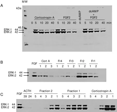
Figure 1 - ERK-M APK activation in Y1 adrenocortical cells assayed by Western blot. A, Kinetics of activation; 100 mU/ml corticotropin A (Cort A), 1 nM FGF2, 1 mM dibutyryl cAM P (dcAM P). B, Screening of activities of fractions (Fr) 1 to 4 of porcine corticotropin A batch 96H0687 (Sigma) fractionated in an HPLC reverse phase column. C, Dose-response curves for the tw o most active fractions, namely 1 and 2. Dilution series from 1 to 5, respectively. Corticotropin A: 100, 1, 0.01 and 0.001 mU/ml; fractions 1 and 2: 10, 0.1, 0.01, 0.001 and 0.0001 µg/ml. Cell lysates w ere boiled for 5 min, submitted to 10% SDS-PAGE and electroblotted onto nitrocellulose Hybond-C extramembranes (Amersham Life Science Inc., Cleveland, OH, USA). M embranes w ere incubated w ith anti-phospho-M APK rabbit antibody (New England Biolabs Inc., Beverly, M A, USA) and developed by chemiluminescence w ith HRP-linked anti-rabbit antibody.
U U
Y1 cells through pathways other than the classical cAMP/PKA pathway. A likely can-didate to transduce mitogenic signals initi-ated in ACTH receptors would be the ERK-MAPK pathway.
The phosphorylated forms of ERK1 and ERK2 are not detected in lysates of G0/G1 -arrested Y1 cells by Western blots, devel-oped with specific antibodies to both ERK-phosphorylated forms. FGF2 and corticotro-pin A rapidly and transiently promote activa-tion of both ERK-MAPK forms with similar kinetics: peaking between 2 and 5 min and sharply decreasing after 10 min (Figure 1A). cAMP analogs (0.5 to 3 mM) have no effect on ERK activation promoted by FGF2 (Fig-ure 1A). The synthetic forms of ACTH, ACTH39 and ACTH24, do not duplicate the ERK-activating effect of corticotropin A (Fig-ure 1C), whereas PMA (10 ng/ml) and fetal
calf serum (10%) promote maximal activa-tion of both ERK1 and ERK2. This observa-tion agrees with the report by Pestell and collaborators (24) that ACTH39 does not activate ERK-MAPK. Only lysates of Y1 cells treated with high, non-physiological concentrations (100 nM) of both ACTH39 and ACTH24 have shown faint bands of phos-phorylated ERK in Western blots (Figure 1C). These results imply that the most active chemical species in corticotropin A, that promotes ERK phosphorylation, is an un-known peptide different from ACTH39 and ACTH24. On the other hand, corticotropin A does not promote phosphorylation of ERK in Balb3T3 fibroblasts, suggesting that the active species of corticotropin A are specific for adrenal cells. As expected, FGF2 is equally active in Y1 adrenocortical cells and Balb3T3 fibroblasts in promoting ERK phos-phorylation.
One batch of a commercial preparation of porcine pituitary corticotropin A was frac-tionated on a reverse phase HPLC-column and analyzed by mass spectrometry and N-terminal sequencing. ACTH39, representing 40% of the total, is the major component and was isolated in a single homogeneous frac-tion (fracfrac-tion 4). Other identified major com-ponents were ßMSH (fraction 3) and gMSH (fraction 2), which are not active on Y1 cells. Fraction 1 displays 6 peaks that were not chemically identified. Fraction 1 shows the highest activity for promotion of ERK-MAPK phosphorylation (Figure 1B and C) and the lowest activity for steroidogenesis stimula-tion (Table 1).
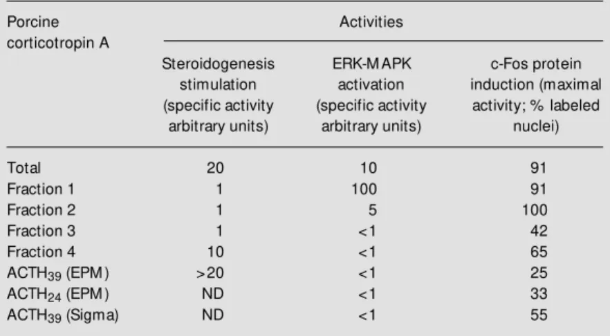
The MEK inhibitor, PD98059 (50 µM), strongly inhibits ERK phosphorylation pro-moted by corticotropin A and PMA in Y1 adrenal cells. In addition, as expected, 50 µM PD98059 inhibits at least 80% of the DNA synthesis stimulated by 100 mU/ml corticotropin A. Unexpectedly, 50 µM PD98059 turns out to be also a strong inhib-itor of both DNA synthesis stimulation and c-Fos induction by the synthetics ACTH39 Table 1 - Activities of ACTH39, ACTH24 and fractions of porcine corticotropin A
regulating functions of Y1 adrenocortical cells.
Steroidogenesis w as estimated by extracting and assaying the fluorescent steroids liberated by cells into serum-free medium; specific activities w ere determined from dose-response curves. The unit of activity w as arbitrarily defined as the ratio betw een fluorescence of stimulated cultures over fluorescence of untreated controls. Extracel-lular signal-regulated kinases-mitogen-activated protein kinases (ERK-M APK) activa-tion w as assayed by Western blot as described in Figure 1. c-Fos protein inducactiva-tion w as estimated by immunocytochemical staining w ith anti-c-Fos antibodies counting % labeled nuclei under the microscope. All fractions significantly activated adenylate cyclase but specific activities w ere not determined. ACTH39 (EPM ) and ACTH24 (EPM )
are peptides synthesized in the laboratory of L. Juliano. ACTH39 (Sigma) is a
commer-cial product. Porcine corticotropin A batch 96H0687 w as purchased from Sigma. All peptide fractions w ere chemically characterized by HPLC and mass spectrometry and also eventually by N-terminal sequencing. ND: Not determined.
Porcine Activities
corticotropin A
Steroidogenesis ERK-M APK c-Fos protein stimulation activation induction (maximal (specific activity (specific activity activity; % labeled
arbitrary units) arbitrary units) nuclei)
Total 20 10 91
Fraction 1 1 100 91
Fraction 2 1 5 100
Fraction 3 1 <1 42
Fraction 4 10 <1 65
ACTH39 (EPM ) >20 <1 25
ACTH24 (EPM ) ND <1 33
and ACTH24, in spite of the fact that these synthetic forms of ACTH are comparatively poor activators of ERK phosphorylation.
Co nclusio ns
FGF2 elicits a prototypic mitogenic re-sponse in Y1 adrenocortical cells that cannot be triggered by signals initiated in ACTH receptors.
Taken together, the results reviewed here suggest that the weak mitogenic response of Y1 adrenocortical cells to ACTH, whose unmasking we recently reported (3), con-sists of the following sequential steps: a) initiation of signals in ACTH receptors, b)
activation of PKC, c) activation of ERK-MAPK, d) induction of fos and jun genes, and e) stimulation of entry of G0/G1-arrested cells into the S phase of cell cycle.
Signals initiated in ACTH receptors and transduced by the cAMP/PKA pathway post-transcriptionally down-regulate c-Myc pro-tein expression, typifying an efficient mech-anism of growth inhibition.
We are still missing important links to explain the mechanisms that underlie the correlation between chronically elevated lev-els of circulating ACTH and, for instance, the adrenal cortex hyperplasia well docu-mented in classical inborn errors of steroido-genesis (25).
Re fe re nce s
1. Hornsby PJ (1985). Regulation of adreno-cortical cell proliferation in culture. Endo-crine Research, 10: 259-281.
2. Armelin HA, Lotfi CFP & Lepique AP (1996). Regulation of grow th by ACTH in the Y-1 line of mouse adrenocortical cells.
Endocrine Research, 22: 373-383. 3. Lotfi CFP, Todorovic Z, Armelin HA &
Schim m er BP (1997). Unm asking a grow th-promoting effect of the ACTH in Y1 mouse adrenocortical tumor cells.
Journal of Biological Chem istry, 272: 29886-29891.
4. Gutkind JS (1998). The pathw ays connect-ing G protein-coupled receptors to the nucleus through divergent mitogen-acti-vated protein kinase cascades. Journal of Biological Chemistry, 273: 1839-1842. 5. Kimura E & Armelin HA (1990). Phorbol
ester mimics ACTH action in corticoadre-nal cells stim ulating steroidogenesis, blocking cell cycle, changing cell shape and inducing c-fos proto-oncogene ex-pression. Journal of Biological Chemistry, 265: 3518-3521.
6. Kimura E, Sonobe M HH, Armelin M CS & Armelin HA (1993). Induction of FOS and JUN proteins by adrenocorticotropin and phorbol ester but not by 3' ,5' -cyclic a-denosine m onophosphate derivatives.
M olecular Endocrinology, 7: 1463-1471. 7. Angel P & Karin M (1991). The role of Jun,
Fos and the AP-1 complex in
cell-prolif-eration and transformation. Biochimica et Biophysica Acta, 1072: 129-157. 8. Holt JT, Gopal TV, M oulton AD & Nienhuis
AW (1986). Inducible production of c-fos
antisense RNA inhibits 3T3 cell prolifera-tion. Proceedings of the National Acade-my of Sciences, USA, 83: 4794-4798. 9. Riabow ol KT, Vosatka R, Ziff EB, Lamb NJ
& Feramisco JR (1988). M icroinjection of
fos-specific antibodies blocks DNA syn-thesis in fibroblast cells. M olecular and Cellular Biology, 8: 1670-1676.
10. Kovary K & Bravo R (1991). The Jun and Fos protein families are both required for cell cycle progression in fibroblasts. M o-lecular and Cellular Biology, 11: 4466-4472.
11. Burgess WH & M aciag T (1989). The hep-arin-binding fibroblast grow th factor fam-ily of proteins. Annual Review of Bio-chemistry, 58: 575-606.
12. Nishimura T, Utsunomiya Y, Hoshikaw a M , Ohuchi H & Itoh N (1999). Structure and expression of a novel human FGF, FGF19, expressed in fetal brain. Biochimi-ca et BiophysiBiochimi-ca Acta, 1444: 148-151. 13. M eyers EN & M artin GR (1999).
Differ-ences in left-right axis pathw ays in mouse and chick: functions of FGF8 and SHH.
Science, 285: 403-406.
14. Jung J, Zheng M , Goldfarb M & Zaret KS (1999). Initiation of mammalian liver de-velopment from endoderm by fibroblast
grow th factors. Science, 284: 1998-2003. 15. M etzger RJ & Krasnow M A (1999). Ge-netic control of branching morphogen-esis. Science, 284: 1635-1639.
16. M iralles F, Czernichow P, Ozaki K, Itoh N & Scharfman R (1999). Signaling through FGF receptor 2b plays a key role in the development of the exocrine pancreas.
Proceedings of the National Academy of Sciences, USA, 96: 6267-6272.
17. M esiano S, M ellon SH, Gospodarow icz D, DiBlasio AM & Jaffe RB (1991). Basic FGF expression is regulated by corticotropin in the human fetal adrenal - a model for adrenal grow th regulation. Proceedings of the National Academy of Sciences, USA, 88: 5428-5432.
18. Schimmer BP (1981). The adrenocortical tumor cell line, Y-1. In: Sato G (Editor),
Functionally Differentiated Cell Lines. Alan R. Liss, Inc., New York, 61-92. 19. Rae PA, Gutmann NS, Tsao J & Schimmer
BP (1979). M utations in cyclic AM P-de-pendent protein kinase and corticotropin (ACTH)-sensitive adenylate cyclase affect adrenal steroidogenesis. Proceedings of the National Academy of Sciences, USA, 76: 1896-1900.
M olecular Biology, 43: 937-950. 21. Li CH, Chung D, Yamashiro D & Lee CY
(1978). Isolation, characterization and syn-thesis of a corticotropin-inhibiting peptide from human pituitary glands. Proceedings of the National Academy of Sciences, USA, 75: 4306-4309.
22. Forti FL & Armelin HA (1998). ACTH
in-duces c-fos proto-oncogene in fibroblasts expressing the ACTH receptor. Endocrine Research, 24: 433-437.
23. Lotfi CFP & Armelin HA (1998). c-Fos pro-tein is a mediator in mitogenic response to ACTH. Endocrine Research, 24: 421-424.
24. W at anabe G, Pena P, Albanese C,
Wilsbacher LD, Young JB & Pestell RG (1997). Adrenocorticotropin induction of stress-activated protein kinase in the ad-renal cortex in vivo. Journal of Biological Chemistry, 272: 20063-20069.
25. New M I (1998). Diagnosis and manage-ment of congenital adrenal hyperplasia.