INTRODUCTION
Ionizing radiation induces chromosome aberrations in G0 human peripheral lymphocytes which can be classified as unstable (dicentrics, centric rings and acentric fragments) or stable (various kinds of translocations) (Natarajan et al., 1992, 1994). Unstable aberrations are lost during succes-sive cell divisions whereas the stable ones can persist for a long time (Natarajan et al., 1991; Lucas et al., 1992; Boei et al., 1994). The frequencies of structurally aberrant chro-mosomes in peripheral lymphocytes of persons acciden-tally exposed to ionizing radiation have been used since the 1960s to estimate absorbed radiation dose (Bender and Gooch, 1966; Sasaki and Miyata, 1968; Natarajan, 1984). According to classical models of formation of chro-mosome exchange aberrations, the ratio of radiation-in-duced symmetrical exchanges (reciprocal translocations, inversions) and asymmetrical exchanges (dicentrics, rings) should be 1:1 (Evans, 1962). A change in this ratio in favor of translocation frequencies has been observed (Natarajan
et al., 1992; Schmid et al.,1992; Nakano et al.,1993), and it has been suggested that the higher estimate of transloca-tions in these studies may be due to the misclassification of exchange aberrations. This problem could be overcome by combination of whole chromosome marking with cen-tromere specific DNA probes (Weier et al., 1991). Straume and Lucas (1993) found a close correlation between di-centrics and reciprocal translocations even after the appli-cation of these techniques. Finnon et al. (1995) also ob-served that the yield of radiation-induced translocations was
not significantly higher than that of dicentrics. Natarajan
et al. (1996) explained this by suggesting that if one con-fines scoring only to reciprocal translocations, ignoring other types such as terminal and interstitial translocations, then the frequencies of dicentrics and translocations are almost equal. In general, the frequency of translocations has been found to be higher than the frequency of dicen-trics. This shift may be dependent upon the structure of the chromosomes, position of the centromere (Natarajan et al., 1996), and the nature of the chromatin at the time of irra-diation (Vyas et al., 1991). The formation of these two types of aberrations (translocations and dicentrics) may be the result of different mechanisms (Natarajan et al., 1996).
Fanconi anemia (FA) and xeroderma pigmentosum (XP) are rare repair-deficient, mutagen-sensitive, autoso-mal recessive human disorders with chromosome instabil-ity, which are cancer prone. Cells derived from FA patients have more spontaneous chromosomal damage than those of other instability syndromes (Schroeder et al., 1964, 1989; Natarajan et al., 1989). There exist inter- and intra-individual variations specifically with regard to suscepti-bility to cross-linking agents and X-rays (Sasaki and Tonomura, 1973).
XP is characterized by high sensitivity to sun expo-sure, susceptibility to skin cancer, cutaneous pigmentation, impaired DNA repair and, in some patients, neurological degeneration (Kraemer and Slor, 1985). These patients have a greater than 1000-fold-increased frequency of UV-in-duced skin cancer (Cleaver and Kraemer, 1989) when com-pared with the normal population.
Frequencies of X-ray induced chromosome aberrations in lymphocytes of
xeroderma pigmentosum and Fanconi anemia patients estimated by Giemsa
and fluorescence
in situ
hybridization staining techniques
Radha Saraswathy and A.T. Natarajan
Abstract
Blood lymphocytes from xeroderma pigmentosum (XP) and Fanconi anemia (FA) patients were assessed for their sensitivity to ionizing radiation by estimating the frequency of X-ray (1 and 2 Gy)-induced chromosome aberrations (CA). The frequencies of aberrations in the whole genome were estimated in Giemsa-stained preparations of lymphocytes irradiated at G0 or G2 stages. The frequencies of
transloca-tions and dicentrics involving chromosomes 1 and 3 as well as the X-chromosome were determinedin slides stained by fluorescence in situ hybridization (FISH) technique. An increase in all types of CA was observed in XP and FA lymphocytes irradiated at G0 when compared
to controls. The frequency of dicentrics and rings was 6 to 27% higher (at 1 and 2 Gy) in XP lymphocytes and 37% higher (at 2 Gy) in FA lymphocytes than in controls, while chromosome deletions were higher in irradiated (30% in 1 Gy and 72% in 2 Gy) than in control XP lymphocytes and 28 to 102% higher in FA lymphocytes. In G2-irradiated lymphocytes the frequency of CA was 24 to 55% higher in XP
lymphocytes than in controls. In most cases the translocation frequencies were higher than the frequencies of dicentrics (21/19).
Several studies (Bigelow et al., 1979; Arlett and Harcourt, 1980; Parshad et al., 1983) have shown increased radiosensitivity of XP and FA cells in the G2 phase of the cell cycle, but in other studies an increase in chromosomal aberrations was not observed in FA cells (Evans et al., 1978; Sasaki, 1978) and XP fibroblasts (Darroudi et al., 1995).
The present study was designed to estimate the fre-quencies of X-ray-induced chromosome aberrations in lym-phocytes of XP and FA patients.
MATERIAL AND METHODS
Culture conditions
Venous blood drawn in lithium heparin tubes was set up for whole blood cultures in Ham’s F10 medium supple-mented with 15% heat inactivated fetal calf serum (Gibco), phytohemagglutinin and antibiotics. The cultures were in-cubated at 37°C in a 5% CO2 atmosphere. The FA blood sample was obtained from the Academic Hospital, Leiden, and XP from the Institute of Human Genetics, Budapest, Hungary; the complementation groups of these patients are not known.
X-Rays were generated by an ENRAF machine, oper-ating at 200 kV, 6 mA at a dose rate of 2 Gy/min.
Treatment
G0 irradiation
Normal, XP and FA blood lymphocyte cultures were irradiated with a dose of 1 or 2 Gy and harvested 48 h after initiation of the cultures. Colcemid (Sigma; 0.3 µg/ml) was added to the cultures 2-3 h before harvest. 5-Bromo-2-deoxyuridine (BrdU, Sigma; 10 µM) was added to all the cultures after irradiation for identification of first and sec-ond division cells. Scoring of chromosomal aberrations was restricted to cells at first mitosis.
G2 irradiation
Lymphocyte cultures were set up and grown for 69 h and then irradiated with doses of 0.5 or 1 Gy. Colcemid was added to these cultures 0.5 h after irradiation and the lymphocytes were harvested and fixed after 2.5 h. For har-vesting the cultures, cells were subjected to hypotonic shock (0.075 mM KCl) for 25 min and fixed in acetic acid:methanol (1:3). Appropriate controls were prepared. For G0 irradiation studies, air-dried preparations were stained by the fluorescence plus Giemsa (FPG) technique (Perry and Wolff, 1974). Two hundred metaphases were analyzed for the presence of dicentrics, rings and chromo-some fragments for each radiation dose. For G2 irradiation studies, the slides were stained with 5% Giemsa solution. Chromatid gaps, chromatid breaks, isochromatid breaks and chromatid exchanges were scored in 200 cells at each dose.
Fluorescence in situ hybridization
Slides to be processed for in situ hybridization were stored dry at -20°C. Chromosome specific libraries were obtained from blue-scribe plasmids. A mixture of three dif-ferent chromosomes (1, 3 and X) was used. The in situ
hybridization method routinely used in our laboratory (Natarajan et al., 1992) was adapted. In order to analyze the aberrations in the first mitosis, the slides were stained with Hoechst 33258 (Sigma), exposed to UV light and pro-cessed for in situ hybridization (Boei et al., 1994).
Triple-color hybridization was performed using bi-otin 11-dUTP (chromosome number 1; Sigma), digoxigenin 11-dUTP (chromosome number 3; Boehringer Manheim, Germany) and fluorescein 12-dUTP (X-chromosome; Boehringer). The labelled DNA representing the library was combined with competitive (human cot 1 DNA) DNA fol-lowed by denaturation, then hybridized in situ (overnight at 37°C) with metaphase preparation.
For immunological detection, after hybridization the slides were washed successively in 50% formamide/2X SSC (42°C), 0.1X SSC (60°C) and 4X SSC/0.05% Tween 20, pH 7, at room temperature. The first incubation was carried out with immunological buffer (NFDM) for 30 min at room temperature. After four washes with SSC, 0.05% Tween 20 and TNT (0.1 M Tris-HCl, 0.15 M NaCl and 0.05% Tween 20), the second incubation with anti-bodies avidin-FITC and mouse anti-digoxigenin diluted in TNB (0.1 M Tris-HCl, 0.15 M NaCl and 0.5% Boehringer blocking agent) was performed at 37°C for 30 min. The third incubation was carried out with goat-anti-avidin D and sheep anti-mouse dig diluted in TNB for 30 min at 37°C. The fourth incubation was carried out with avidin-FITC and sheep anti-dig TRITC for 30 min at 37°C. Each incubation was followed by washing with TNT. After the final wash with PBS the slides were counter-stained with DAPI (4,6-diamidino-2-phenylindole; Sigma) diluted in an anti-fading agent, vectashield (Vector Laboratories) (Figure 1).
In order to detect translocations, dicentrics and com-plex exchanges involving chromosome numbers 1 and 3 and the X-chromosome, 300 to 500 metaphases were ana-lyzed for each radiation dose using a Zeiss fluorescence microscope equipped with triple filters. Due to variations in mitotic index the number of metaphases scored by FISH varied for each experimental point. The translocations and dicentrics were scored using the same slide. The back-ground frequencies were subtracted to obtain the induced frequency of translocations and dicentrics.
RESULTS AND DISCUSSION
Spontaneous chromosomal aberrations in normal, XP and FA lymphocytes
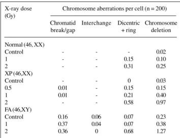
The baseline aberration frequencies in normal and XP lymphocytes were in the range of 2 to 3%, whereas in FA the frequency was 23% (Table I). Darroudi et al. (1995) also observed a similar spontaneous aberration frequency in the range of 2 to 3% in the normal and XP-C cell lines, and 16 to 22% in FA cells, which is known to be character-istic of FA patients (Schroeder et al., 1964; Natarajan et al., 1989). However, the frequency of spontaneous aberra-tions in XP lymphocytes was similar to that of normal lym-phocytes.
Figure 1 - A. Metaphase spread of an irradiated lympho-cyte painted with libraries for chromosomes 1, 3 and X showing translocations (a, b) involving chromosome 1 and dicentrics involving chromosome 3 (c) and chromo-some 1 (d). B. The same cell counter-stained with DAPI (4,6-diamidino-2-phenylindole).
Fp
2.5 x fp (1 - fp)
FG = G0 radiation induced CA in normal,XP and FA lymphocytes
chro-matid exchanges were only observed in FA lymphocytes, a known characteristic of these cells. G0 radiation produced chromatid breaks/gaps in FA lymphocytes (0.37 and 0.36 per cell in 1 Gy and 2 Gy, respectively) and were zero in normal lymphocytes and 0.01 in XP lymphocytes, which further confirms a positive defect in repair of radiation damage in FA cells.
G2 radiosensitivity in normal and XP lymphocytes
The frequency of X-ray-induced total CA aberrations in XP lymphocytes was 0.79 and 2.17 per cell for 0.5 Gy and 1 Gy, respectively, while in the normal lymphocytes it was 0.55 and 1.62, indicating an increase (24-55%) in XP lymphocytes (Table II). The frequency of chromatid and isochromatid breaks in XP lymphocytes was 0.72 and 2.03 for 0.5 and 1 Gy, respectively, while in normal
lympho-cytes the frequencies were 0.54 and 1.6, indicating an in-crease (18-43%) in the XP lymphocytes when compared to normal lymphocytes. The frequency of interchanges was seven times (6% at 0.5 Gy and 12% at 1 Gy) higher in the XP lymphocytes than in the controls. Isochromatid breaks induced by X-rays were observed only in XP lymphocytes and were dose related (0.15 and 0.22 per cell for 0.5 and 1 Gy, respectively). Even though Taylor (1978) observed an increase in chromatid aberrations in XP cells irradiated in G2, it was not as significant as it was in the case of ataxia telangiectasia cells. In XP-C lymphocytes an increase in ionizing radiation-induced chromosome aberrations has been observed in the G2 phase of the cell cycle (Parshad et
al., 1983).
Estimation of radiation-induced dicentrics and translocations in normal, XP and FA
lymphocytes using FISH
A. Genomic translocation frequency
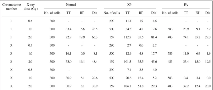
The genomic translocation frequency (including re-ciprocal and terminal ones) calculated for individual chro-mosomes (1, 3, X) represents about 20% of the total ge-nome. The translocation frequencies for chromosomes 1, 3 and the X-chromosome followed the order expected on the basis of their DNA contents in normal lymphocytes. The DNA contents of chromosomes 1, 3 and the X-chro-mosome are 8.15, 6.63 and 5.08% of the total genomic value, respectively (Mendelsohn et al., 1973). The trans-location frequencies in individual chromosomes of XP lym-phocytes when compared with those of the chromosomes of normal lymphocytes showed an increase of 73.2% in the X-chromosome (2 Gy) and 48.5% in chromosome 3 (at 2 Gy). At 1 Gy the translocation frequency was 3.2% and 10.3% higher than those of XP lymphocytes for chro-mosome 3 and the X-chrochro-mosome respectively (Table III). In FA lymphocytes, the translocation frequencies for chro-mosomes 1, 3 and the X-chromosome followed the or-der based on their DNA content. The translocation fre-quencies in individual chromosomes of FA lymphocytes were almost equal to the respective chromosomes in nor-mal lymphocytes except for the X-chromosome (27.5% higher at 1 Gy).
Mean translocation frequency of the mixture of chromosomes 1 and 3 and the X-chromosome
in normal, XP and FA lymphocytes
At 1 Gy the mean translocation frequency in XP lymphocytes was similar to that of normal lymphocytes, while at a dose of 2 Gy the mean frequency was about twice that of normal lymphocytes. At 1 Gy the mean frequency of translocation in FA lymphocytes was lower than that of normal but at 2 Gy it was almost similar to normal (Table IV).
Table I - G0 radiosensitivity of human peripheral lymphocytes in
normal, xeroderma pigmentosum (XP) and Fanconi anemia (FA) individuals estimated by Giemsa staining*. X-ray dose Chromosome aberrations per cell (n = 200) (Gy)
Chromatid Interchange Dicentric Chromosome break/gap + ring deletion Normal (46, XX)
Control - - - 0.02
1 - - 0.15 0.10
2 - - 0.31 0.25
XP (46,XX)
Control - - 0 0.03
0.5 0.01 - 0.15 0.15
1 0.01 - 0.21 0.40
2 - - 0.58 0.97
FA (46,XY)
Control 0.16 0.06 0.07 0.23
1 0.37 0.04 0.07 0.38
2 0.36 0 0.68 1.27
*Induced aberrations have been corrected for spontaneous aberrations.
Table II - G2 radiosensitivity of human peripheral lymphocytes
in normal and xeroderma pigmentosum (XP) individuals estimated by Giemsa staining*.
X-ray dose (Gy) Aberrations per cell (n = 200) Chromatid Interchange Isochromatid
break/gap break
Normal (46,XX)
Control - -
-0.5 0.54 0.01
-1 1.6 0.02
-XP (46,XX)
Control 0 0 0.02
0.5 0.57 0.07 0.15
1 1.81 0.14 0.22
B. Genomic dicentric frequency
The genomic dicentric frequencies calculated using the same formula for calculating genomic translocation frequencies, for chromosomes 1, 3 and the X-chromosome in normal and XP lymphocytes at 2 Gy followed the order based on their DNA contents (Table III). The dicentric fre-quencies in individual chromosomes of XP lymphocytes when compared with the chromosomes from normal lym-phocytes showed an increase of 15.1% for chromosome 1 (2 Gy) and almost equal for chromosomes 3 and X. In FA lymphocytes, the dicentric frequencies for individual chro-mosomes (1, 3 and X) followed the order based on their DNA contents.
Ratio between translocation and dicentric frequencies
The frequency of translocations in control, XP and FA lymphocytes increased 8.4, 10.8 and 10.6% at 1 Gy and 3.8, 53.9 and 25.9% at 2 Gy, respectively, over dicentrics
(Table V). This confirms the observation of Natarajan et al. (1996) that the frequency of translocation is higher than the frequency of dicentrics, indicating that two different mechanisms may be involved in the production of translo-cations and dicentrics as observed in normal humans.
C. Complex exchanges and centric rings
In XP and FA lymphocytes a dose-dependent increase in the frequency of complex exchanges and centric rings was observed, ranging from 0.2-3.3%. These values were not used in our analysis.
CONCLUSIONS
The frequency of CA produced by G0 and G2 irradia-tion in XP and FA lymphocytes was higher than that of nor-mal lymphocytes. The deletion type of chromosonor-mal ab-errations observed in XP and FA lymphocytes was several fold more abundant than in the normal lymphocytes, re-flecting a possible defect in their capacity to repair
dam-Table III - Genomic frequency of translocation and dicentrics in normal, xeroderma pigmentosum (XP) and Fanconi anemia (FA) lymphocytes following X-irradiation as estimated by FISH.
Chromosome X-ray Normal XP FA
number dose (Gy)
No. of cells TT RT Dic No. of cells TT RT Dic No. of cells TT RT Dic
1 0.5 300 - - - 290 11.4 1.9 4.6 - -
-1 1.0 300 33.4 06.6 26.5 500 34.5 4.8 12.6 583 23.9 09.1 05.2
1 2.0 300 72.9 19.9 66.3 159 112.5 35.5 81.4 483 74.1 35.2 29.3
3 0.5 300 - - - 290 2.7 0.0 2.7 - -
-3 1.0 300 16.1 00.0 08.1 500 12.9 4.8 17.7 583 11.0 06.9 01.9
3 2.0 300 53.0 16.1 48.4 159 101.5 35.5 45.6 483 33.4 15.0 19.5
X 0.5 300 - - - 290 7.1 3.5 4.0 - -
-X 1.0 300 30.9 08.1 20.6 500 20.6 12.4 5.2 583 3.4 03.4 00.0
X 2.0 300 30.9 08.1 30.9 159 104.1 51.8 29.3 483 37.2 12.4 20.0
TT - Total translocation, RT - Reciprocal translocation, Dic - Dicentric.
Table IV - Mean genomic translocation and dicentric frequencies detected using a mixture of chromosomes 1 and 3 and the X-chromosome in normal, xeroderma pigmentosum (XP) and Fanconi
anemia (FA) lymphocytes as estimated by FISH.
X-ray dose Normal XP FA
(Gy)
Translocation Dicentric Translocation Dicentric Translocation Dicentric
0.5 - - 7.1 17.6 -
-1.0 26.8 18.4 22.6 11.8 12.8 02.2
aged DNA, while the frequency of translocations was higher than the frequency of dicentrics in normal, XP and FA lym-phocytes, indicating that more unrepaired breaks were avail-able for interaction in these syndromes in comparison to normal cells. Though XP cells are known to be defective in repairing bulky DNA lesions, increased sensitivity to ion-izing radiation has been observed, which may indicate that ionizing radiation induces several classes of DNA lesions and/or XP cells are defective not only in nucleotide exci-sion repair but also in some other minor repair pathways. Similarly, though FA cells are known to be sensitive to cross linking agents, implying a defect in the repair of cross links, they are also known to be sensitive to several classes of clastogenic agents (Sasaki and Tonomura, 1973).
ACKNOWLEDGMENTS
We would like to thank Dr. K.M. Marimuthu, Prof. Emeri-tus, Dept. of Genetics, Madras University, India, for his help in the preparation of this manuscript. The research was financially sup-ported by an EU Nuclear Safety Programme grant to ATN.
RESUMO
Linfócitos sanguíneos de pacientes com xeroderma pig-mentosum (XP) e anemia de Fanconi (FA) foram avaliados quanto à sensibilidade à ionização radiante estimando-se a freqüência de aberrações cromossômicas (CA) induzidas por raios-X (1 e 2 Gy). As freqüências de aberrações no genoma inteiro foram estimadas em preparações de linfócitos irradiados nas fases G0 e
G2 coradas com Giemsa. As freqüências de translocações e
dicêntricos envolvendo os cromossomos 1 e 3 e o cromossomo X foram determinadas em lâminas coradas por hibridização fluo-rescente in situ (FISH). Um aumento em todos os tipos de CA foi observado em linfócitos XP e FA irradiados na fase G0 quando
comparados a controles. A freqüência de dicêntricos e anéis foi 6-27% maior (com 1 e 2 Gy) em linfócitos XP e 37% maior (com 2 Gy) em linfócitos FA do que em controles, enquanto que as deleções cromossômicas foram mais freqüentes em linfócitos XP irradiados (30% com 1 Gy e 72% com 2 Gy) do que em controles e 28-102% mais freqüentes em linfócitos FA. Em linfócitos irradiados na fase G2 a freqüência total de CA foi 24-55% mais
elevada em linfócitos XP do que em controles. Na maior parte dos casos as freqüências de translocações foram maiores do que as freqüências de dicêntricos (21/19).
REFERENCES
Arlett, C.F. and Harcourt, S.A. (1980). Survey of radiosensitivity in a variety of human cell strains. Cancer Res.40: 926-932.
Bender, M.A. and Gooch, P.C. (1966). Somatic chromosome aberrations induced by human whole body irradiation: The “Recuplex” criticality accident. Radiat. Res.29: 568-582.
Bigelow, S.B., Rary, J.M. and Bender, M.A. (1979). G2 chromosomal
radio-sensitivity in Fanconi’s anemia. Mutat. Res.63: 189-199.
Boei, J.J.W.A., Balajee, A.S., Boer, P. de, Rens, W., Aten, J.A., Mullenders, L.H.F. and Natarajan, A.T. (1994). Construction of mouse chromo-some-specific DNA libraries and their use for the detection of X-ray induced aberrations. Int. J. Rad. Biol.65: 583-590.
Cleaver, J.E. and Kraemer, K.H. (1989). Xeroderma pigmentosum. In:
The Metabolic Basis of Inherited Disease (Scriver, C.R., Beaudet, A.L., Sly, W.S. and Valle, D., eds.). McGraw Hill, New York, pp. 2949-2971.
Darroudi, F., Vyas, R.C., Vermeulen, S. and Natarajan, A.T. (1995). G2 radiosensitivity of cells derived from cancer prone individuals. Mutat. Res.328: 83-90.
Evans, H.J. (1962). Chromosome aberrations induced by ionising radia-tions. Int. Rev. Cytol.13: 221-321.
Evans, H.J., Adams, A.C., Clarkson, J.M. and German, J. (1978). Chromo-some aberrations and unscheduled DNA synthesis in X- and UV-irradiated lymphocytes from a boy with Blooms syndrome and a man with Xeroderma pigmentosum. Cytogenet. Cell. Genet.20: 124-140.
Finnon, P., Lloyd, D.C. and Edwards, A.A. (1995). Fluorescence in situ
hybridization detection of chromosomal aberrations in human lym-phocytes; applicability to biological dosimetry. Int. J. Radiat. Biol. 68: 429-435.
Kraemer, K.H. and Slor, H. (1985). Xeroderma pigmentosum. Clin. Dermatol.3: 33-69.
Lucas, J.N., Awa, A., Straume, T., Poggensee, M., Kodama, Y., Nakano, M., Ohtaki, K., Weier, H.-U., Pinkel, D., Gray, J.W. and Littlefield, G.
(1992). Rapid translocation frequency analysis in human decades af-ter exposure to ionising radiation. Int. J. Radiat. Biol.62: 53-63.
Mendelsohn, M.L., Mayhall, B.H., Bogart, E., Moore II, D.H. and Perry, B.H. (1973). DNA content of the various radiation induced chromo-somal rearrangements in relation to the dose and sample time. Science 179: 1126-1129.
Nakano, M., Nakashima, E., Pawel, D.J., Kodama, Y. and Awa, A. (1993). Frequency of reciprocal translocations and dicentrics induced in hu-man blood lymphocytes by X-irradiation as determined by fluores-cence in situ hybridization. Int. J. Radiat. Biol.64: 565-569.
Natarajan, A.T. (1984). Origin and significance of chromosomal alterations. In: Mutations in Man (Obe, G., ed.). Springer-Verlag, Berlin, Heidel-berg, pp. 156-176.
Natarajan, A.T., Vossen, J.M.J.J. and van Weel-Sipman, M.H. (1989). Aplas-tic anemia and Fanconi anemia:Response of lymphocytes to X-rays and mitomycin C. In: Fanconi Anemia. Clinical, Cytogenetic and Experimental Aspects (Schroeder-Kurth, T.M., Auerbach, A.D. and Obe, G., eds.). Springer Verlag, Berlin, Heidelberg, pp. 100-104.
Natarajan, A.T., Vyas, R.C., Wiegant, J. and Curado, M.P. (1991). A cyto-genetic follow-up study of the victims of a radiation accident in Goiânia
Table V - Ratio between genomic translocation and dicentric frequencies in normal, xeroderma pigmentosum (XP) and Fanconi anemia (FA) lymphocytes following X-irradiation as estimated by FISH.
X-ray dose Normal XP FA
(Gy)
Chrom Chrom Chrom Chrom Chrom Chrom Chrom Chrom Chrom No. 1 No. 3 No. X No. 1 No. 3 No. X No. 1 No. 3 No. X
0.5 - - - 2.5 1.0 1.8 - -
-1.0 1.26 1.99 1.5 2.7 0.7 3.9 5.2 5.8 3.4
2.0 1.1 1.1 0.7 1.4 2.2 3.6 2.7 1.7 1.9
(Brazil). Mutat. Res.247: 103-111.
Natarajan, A.T., Vyas, R.C., Darroudi, F. and Vermeulen, S. (1992). Fre-quencies of X-ray induced chromosome translocations in human pe-ripheral lymphocytes as detected by in situ hybridization using chro-mosome specific DNA libraries. Int. J. Radiat. Biol.61: 199-203.
Natarajan, A.T., Balajee, A.S., Boei, J.J.W.A., Chatterjee, S., Darroudi, F., Grigorova, M., Noditi, M., Oh, H.J., Slijepcevic, P. and Vermeulen, S. (1994). Recent developments in the assessment of chromosomal damage. Int. J. Radiat. Biol.66: 615-623.
Natarajan, A.T., Balajee, A.S., Boei, J.J.W.A., Darroudi, F., Dominguez, I., Hande, M.P., Meijers, M., Slijepcevic, P., Vermeulen, S. and Xiao, Y. (1996). Mechanisms of induction of chromosomal aberrations and their detection by fluorescence in situ hybridization. Mutat. Res.372: 247-258.
Parshad, R., Sanford, K.K. and Jones, G.M. (1983). Chromatid damage after G2 phase X-irradiation of cells from cancer-prone individuals implicates deficiency in DNA repair. Proc. Natl. Acad. Sci. USA80: 5612-5616.
Perry, P. and Wolff, S. (1974). New Giemsa method for differential staining of sister chromatids. Nature 251: 156-158.
Sasaki, M.S. (1978). Falconi’s anemia. A condition possibly associated with defective DNA repair. In: Repair Mechanisms (Hanawalt, P.C., Friedberg, E.C. and Fox, C.F., eds.). Academic Press, New York, pp. 675-684.
Sasaki, M.S. and Miyata, H. (1968). Biological dosimetry in atom bomb survivors. Nature220: 1189-1193.
Sasaki, M.S. and Tonomura, A. (1973). A high susceptibility of Fanconi anemia to chromosome breakage by DNA cross-linking agents.
Can-cer Res. 33: 1829-1836.
Schmid, E., Zitzelsberger, H., Braselmann, H., Gray, J.W. and Bauchinger, M. (1992). Radiation induced chromosome aberrations analysed by fluorescence in situ hybridization with a triple combination of compo-site whole chromosome specific DNA probes. Int. J. Radiat. Biol.62: 673-678.
Schroeder, T.M., Anschutz, F. and Knopp, A. (1964). Spontane Chromoso-menaberrationen bei familiarer Panmuelopathie. Hum. Genet. 1: 194-196.
Schroeder-kurth, T.M., Auerbach, A.D. and Obe, G. (1989). Fanconi Ane-mia: Clinical, Cytogenetic and Experimental Aspects. Springer-Verlag, Berlin.
Straume, T. and Lucas, J.N. (1993). A comparison of the yields of translo-cations and dicentrics measured using fluorescence in situ hybridiza-tion. Int. J. Radiat. Biol.64: 185-187.
Taylor, A.M.R. (1978). Unrepaired DNA strand breaks in irradiated ataxia telangiactasia lymphocytes suggested from cytogenetic observations. Mutat. Res.50: 407-418.
Vyas, R.C., Darroudi, F. and Natarajan, A.T. (1991). Radiation induced chromosome breakage and rejoining in interphase-metaphase chro-mosomes of human lymphocytes. Mutat. Res. 249: 29-35.
Weier, H.-U., Lucas, J.N., Poggensee, M., Segraves, R., Pinkel, D. and
Gray, J.W. (1991). Two colour hybridization with high complexity chro-mosome specific probes and a degenerate alpha satellite probe DNA allows unambiguous discrimination between symmetrical and asym-metrical translocations. Chromosoma100: 371-376.