Cells Using Meganuclease and TALEN
TM
Aure´lie Dupuy1,2, Julien Valton1, Sophie Leduc1, Jacques Armier2, Roman Galetto3, Agne`s Gouble3, Ce´line Lebuhotel3, Anne Stary2, Fre´de´ric Paˆques1, Philippe Duchateau1*, Alain Sarasin2.,
Fayza Daboussi1.
1Cellectis S.A., Paris, France,2Unite´ mixte de recherche 8200, Institut Gustave Roussy, Villejuif, France,3Cellectis Therapeutics, Paris, France
Abstract
Xeroderma pigmentosum group C (XP-C) is a rare human syndrome characterized by hypersensitivity to UV light and a dramatic predisposition to skin neoplasms. XP-C cells are deficient in the nucleotide excision repair (NER) pathway, a complex process involved in the recognition and removal of DNA lesions. Several XPC mutations have been described, including a founder mutation in North African patients involving the deletion of a TG dinucleotide (DTG) located in the middle of exon 9. This deletion leads to the expression of an inactive truncated XPC protein, normally involved in the first step of NER. New approaches used for gene correction are based on the ability of engineered nucleases such as Meganucleases, Zinc-Finger nucleases or TALE nucleases to accurately generate a double strand break at a specific locus and promote correction by homologous recombination through the insertion of an exogenous DNA repair matrix. Here, we describe the targeted correction of theDTG mutation in XP-C cells using engineered meganuclease and TALENTM. The methylated status of theXPClocus, known to inhibit both of these nuclease activities, led us to adapt our experimental design to optimize theirin vivoefficacies. We show that demethylating treatment as well as the use of TALENTMinsensitive to CpG methylation enable successful correction of theDTG mutation. Such genetic correction leads to re-expression of the full-length XPC protein and to the recovery of NER capacity, attested by UV-C resistance of the corrected cells. Overall, we demonstrate that nuclease-based targeted approaches offer reliable and efficient strategies for gene correction.
Citation:Dupuy A, Valton J, Leduc S, Armier J, Galetto R, et al. (2013) Targeted Gene Therapy of Xeroderma Pigmentosum Cells Using Meganuclease and TALENTM. PLoS ONE 8(11): e78678. doi:10.1371/journal.pone.0078678
Editor:Shuang-yong Xu, New England Biolabs, Inc., United States of America
ReceivedJune 25, 2013;AcceptedSeptember 13, 2013;PublishedNovember 13, 2013
Copyright:ß2013 Dupuy et al. This is an open-access article distributed under the terms of the Creative Commons Attribution License, which permits unrestricted use, distribution, and reproduction in any medium, provided the original author and source are credited.
Funding:This work was supported by Agence Nationale de la Recherche, Association Nationale de la recherche et de la Technologie, contrat Cifre 535/2008 and Association de Recherche sur le Cancer (Villejuif, France). The funders had no role in study design, data collection and analysis, decision to publish, or preparation of the manuscript.
Competing Interests:The authors would like to confirm that at the exception of Alain Sarasin, all other co-authors are Cellectis employees. However, this does not alter the authors’ adherence to all the PLOS ONE policies on sharing data and materials.
* E-mail: [email protected]
.These authors contributed equally to this work.
Introduction
Xeroderma pigmentosum (XP) is a rare, autosomal, recessive syndrome characterized by hypersensitivity to UV light [1]. It is also associated with a dramatic predisposition to skin neoplasms. Thus, risk of melanoma and non-melanoma skin cancers has been reported to be increased 2 to 10 thousand-fold, respectively [2]. XP cells are deficient in the nucleotide excision repair (NER) pathway, a complex process involved in the recognition and removal of DNA lesions induced by UV light (cyclobutane pyrimidine dimers and pyrimidine 6-4 pyrimidone photoproducts) [3]. Seven different genes namedXPAtoXPGare involved in that
process. Mutations within the XPC gene are by far the most
common genetic alteration found in European and North African XP patients. Among the known genetic alterations, a founder mutation within exon 9 has been described in almost 90% of Maghrebian XP-C patients [4] and corresponds to the deletion of a TG dinucleotide leading to the expression of an inactive and undetectable XPC truncated protein. This lack of NER activity allows UV-dependent DNA damage to accumulate and is responsible for the development of high numbers of skin cancers.
Today, there is no curative treatment for XP-C patients and their cancer-free survival relies solely on full body protection from light and/or surgical resections of skin tumors. Autologous grafts have been performed using UV sensitive cells, but the benefit of such treatment is transient [5]. A major advance in cancer prevention would be to engraft patient skin produced ex vivo with cells
corrected for XPC mutation.
Recently, the stable trans-complementation of XPCdeficiency
has been reported [6]. Using a retrovirus-based strategy, Warrick
et al. were able to transduce the wild-typeXPCgene into human
primary XP-C keratinocyte stem cells and reconstitute their full NER capacity resulting in UV resistance. Although successfully validatedin vivoand in a relevant cell line, this complementation
strategy is nonetheless liable to generate potential adverse effects due to uncontrolled random integrations of the transgene. Indeed, these undesirable effects have been reported in several comple-mented cells for disease treatment, especially in the hematopoietic system [7,8]. In view of this result, genetically modified skin could lead to skin tumor development following engraftment. In addition, because of the ectopic expression of theXPCtransgene,
our experimental conditions, to optimize theirin vivoefficacy. We
showed that treatment with a demethylating agent or the use of 5 mC insensitive nuclease allowed successufulXPC gene
correc-tion without requiring seleccorrec-tion marker. Such genetic correccorrec-tion enabled re-expression of the full-length XPC protein and full recovery of wild-type UV resistance in the XP4PA cell line. We demonstrate that nuclease-based targeted approaches constitute a robust and reliable strategy forXPCgene therapy.
Results
The recent development of engineered nucleases able to introduce a DSB and stimulate HR at a specific locus [18,19] has opened up new opportunities for XPC gene correction. In
order to induce a high frequency of HR at an endogenous locus, it is crucial to generate specific and efficient nucleases. For this study, two types of engineered nucleases were developed to target a DNA sequence located 100 bp upstream from theDTG XPC founder mutation (Figure S1A). The first nuclease was a single-chain meganuclease named XPCm, derived from I-CreI endonuclease [15]. The second was a TALENTM named XPCT1 and derived from TALE AvrBs3 [20].
An Engineered Meganuclease (XPCm) Specifically Designed to Target theXPCLocus
The engineered meganuclease XPCm has been previously described [15]. Its intrinsic activity was first determined by a single-strand annealing (SSA) extrachromosomal assay in CHO-K1 cells (Materials and Methods S1) [21]. XPCm showed high activity similar to that of the meganuclease RAG1m, used here as a positive control, and better than that of I-SceIm (Figure S1B). We then assessed the ability of XPCm to cleave the endogenous XPCt sequence in 293-H cells by quantifying the frequency of targeted mutagenesis (TM) induced by XPCm at its endogenous locus. TM consists of small insertions or deletions of nucleotides resulting from imprecise non-homologous end joining (NHEJ) occurring at the DSB site. TM was quantified by specific PCR surrounding the locus of interest followed by deep sequencing.
We found that XPCm induced weak TM, as the frequency was 3-fold lower than that induced by the RAG1 m meganuclease (Figure S1C). This low efficiency was also observed in the frequency of homologous gene targeting (HGT), with 8 times more events using RAG1 m versus XPCm (Figure S1D). We recently demonstrated the inhibitory effect of DNA target methylation on meganuclease activity in vivo [22]. Interestingly, XPCt DNA
sequence contained two fully methylated CpG dinucleotides (Figure S2A). We thus hypothesized that methylation of XPCt could prevent XPCm from processing its endogenous locus.
specific siRNA (siDNMT1, data not shown). Interestingly, the distribution of TM events within the amplicon population was strongly biased toward demethylated sequences. Up to 80% of mutated events were found within the demethylated DNA sequence population which strongly suggests that demethylation increases the XPCm efficiency at its endogenous locus (P,0.001) (Figure 1B). This conclusion is supported by the fact that no induction of targeted mutagenesis after 5-aza-dC treatment was observed in the cells transfected with meganucleases CAPNS1m and RAG1 m, both targeting unmethylated sequences (P = 0.77) (Figure 1A). In order to determine whether demethylating treatment directly affected the cleavage activity of the nuclease, we used LM-PCR to monitor the non-processed DNA ends generated upon cleavage by the meganuclease (Materials and Methods S1). The number of free DNA ends at theXPClocus was
increased up to 7-fold in the presence of 5-aza-dC versus untreated cells (P,0.05) (Figure S2C). Although our protocol can only quantify non-processed DNA ends, this result strongly suggests that at least a substantial portion of TM stimulation results directly from higher cleavage activity of the meganuclease on unmethy-lated sequences.
We then evaluated the impact of 5-aza-dC on the ability of XPCm to trigger HGT in 293-H cells. The DNA repair matrix was composed of two homology arms interrupted by an exogenous DNA sequence (29 bp) specifically designed to screen and identify the HGT events by PCR. Because 5-aza-dC had a major impact on cellular proliferation at a dose of 1mM, this experiment was performed with dose of 0.2mM. As regards TM, our results showed that 5-aza-dC treatment increased HGT frequency 12-fold, leading to up to 12% of corrected cells (P,0.001) (Figure 1C). Altogether, our data indicated that the presence of an epigenetic effector such as a 5-aza-dC significantly enhanced meganuclease-assisted TM and HGT at theXPClocus.
XPCGene Correction in XP4PA Cells using XPCm
Meganuclease and a Demethylating Agent
The XP4PA cell line was derived from dermal fibroblast obtained from a patient bearing the homozygote mutation matching the TG deletion in exon 9 of theXPCgene [23]. These
XPCopen reading frame. It contained two arms of 1.5 and 1.8 Kb
respectively, homologous to the wild type XPC sequences
surrounding the cleavage site. To avoid any possible cleavage of the DNA repair matrix by XPCm, silent mutations were introduced within the meganuclease-recognizing site (Figure 2C). XP4PA cells were transfected with the DNA repair matrix and the meganuclease XPCm expression vector. Three days later, cells were seeded at a density of 100 cells per well in a 96-well plate. Each well was then screened for locus-specific integration using specific primers. While no positive wells were found in untreated conditions, 3 out of 480 wells were identified as positive after 5-aza-dC treatment. Further DNA sequencing confirmed that the initial XPC mutation was corrected in 2 out of these 3 cell populations (Figure 2D). Furthermore, the presence of all silent mutations, present only on the matrix plasmid, together with the corrected mutation (100 bp from cleavage site) indicated that these were transferred from the DNA matrix to the genomic DNA, and confirmed the homologous recombination process (data not shown). One corrected population was then sub-cloned. After amplification, we isolated three clones positive for the PCR-detected HGT event and used them for further phenotypic characterizations.
XPCGene Correction in XP4PA Cells using XPCT1
TALENTM
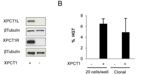
We recently reported the design of methylation-insensitive TALE nuclease using the unconventional TALE repeat N* [20]. In order to determine whether this approach could be used for the gene correction of XP4PA cells, we used the TALENTM(XPCT1) containing the N* residue previously described to induce up to 17% of TM frequency at the methylated XPCT1t in 293-H cells. As XPCT1 targets a genomic sequence (XPCT1t) overlapping the previous meganuclease target (Figure S1A), HGT experiments were performed with the same DNA repair matrix and therefore the same screening design as for gene correction experiment using meganuclease. XP4PA cells were transfected with the DNA matrix and a plasmid encoding the XPCT1. Three days post-transfection, a fraction of cells was recovered to verify the expression of TALENTMby western blot (Figure 3A), seeded the remaining cells at a density of 20 cells/well in a 96-well plate and characterized them three weeks later. Specific PCR screening for HGT events revealed that HGT occurred in 6.25% 60.95 (respectively 56/ 192, 69/192) of transfected cells, taking into account the number of cells per well and the efficiencies of transfection and cloning (Figure 3B).
A second series of three independent experiments was then performed in which transfected cells were seeded at low density in
Figure 1. Impact of demethylating treatment on targeted mutagenesis (TM) and homologous gene targeting (HGT) frequencies induced by engineered meganucleases in 293-H cells.(A) TM frequency was determined from cells grown with or without 0.2mM or 1mM
5-aza-dC and transfected with the engineered meganuclease (MN) XPCm, or RAG1 m and CAPNS1 m, two meganucleases targeting DNA sequences that lack methylated CpG. (B) Distribution of TM events in methylated (white) and unmethylated (black) sequences from cells transfected with XPCm with and without 5-aza-dC. (C) HGT frequency was determined from cells grown with 0.2mM (+) or without (2) 5-aza-dC and co-transfected with the DNA repair matrix (RM) and the XPCm engineered meganuclease (+) or empty vector (2).
Petri dishes allowing for clonal cell expansion. Independent clones were isolated and characterized. Under these conditions, 4.9% 62.5 (respectively, 33/493, 49/796, 16/819) of clones were positive for HGT events (Figure 3B). As expected, no positive clone was obtained when cells were co-transfected with a non related TALENTMand the DNA matrix. In order to determine the frequency of DTG correction, DNA sequencing of the genomicXPCgene in 45 randomly chosen HGT-positive clones
was performed. The presence of the wild type sequence (i.e. the corrected sequence) was observed in 53% (24/45) of the HGT positive clones.
Phenotypic Correction of Transfected XP4PA Cells In order to determine whether theXPCgene correction induced
by our engineered nucleases led to rescue of the NER pathway, we first examined the ability of the corrected clones to express the wild type XPC protein (Figure 4A and 4B). All clones showing genotypic correction displayed full-length XPC protein expression,
similar to that of the MRC5 control cell line, proficient forXPC
(Figure 4B). This UV-C survival rescue was confirmed in corrected clones obtained from 3 independent experiments (Figure S4). As expected, clones negative for HGT events and TG correction displayed high sensitivity to UV-C. Taken together, these results provided the first demonstration of a stable correction of XPC
mutation using sequence-specific engineered nucleases.
Discussion
In this study, we showed that both meganuclease- and TALENTM-assisted targeted approaches allowed efficient correc-tion of the XPC founder mutacorrec-tion in an XP4PA cell line derived from the fibroblasts of XP-C patients. The successful correction of
XPC, enabled stable re-expression of the full-length XPC protein
and allowed XP4PA cells to recover their fully functional NER pathway. Because of the methylation status of the sequences surrounding the XPC mutation, we opted for two independent strategies to overcome the nucleases’ sensitivity to methylation and enhance their activityin vivo.
In the case of the meganuclease XPCm, we used the epigenetic drug 5aza-dC to demethylate the XPC locus and rescue its
nuclease activity. As expected, such treatments led to a significant enhancement of HGT frequency in 293-H cells (12-fold, 12% HGT, figure 1D). Although to a much lower extent, an increase of HGT frequency was also observed in XP4PA patient cell line as HGT-positive clones were obtained only after 5-aza-dC treatment. In addition, the XP4PA cells treated with 5-aza-dC displayed an altered cellular proliferation compared to their untreated coun-terparts. These two results might be explained by the co-existence of two deleterious factors in our experiment: the NER deficiency of XP4PA cell line and the well known ability of 5-aza-dC to alter DNA structure, especially by being inserted into DNA as a nucleotide analogue and by promoting DSB, gene expression modification, cell cycle disruption [26]. Indeed, we hypothesized that due to their NER deficiency, XP4PA cells would be unable to proficiently process altered DNA structures generated by 5-aza-dC, leading to significant cell death. Because the vast majority of corrected cells came from cells with a high level of demethylation (Figure 1B), we made the assumption that a high proportion of corrected cells were dying. Thereby, due to its deleterious effect, the use of 5-aza-dC, at least at the dose used in this study, may represent a hurdle for its application in primary keratinocytes, the relevant primary cells for genetic correction.
An alternative strategy was to use an engineered TALENTM (XPCT1) insensitive to 5mC. This TALENTM, previously described to induce up to 17% of TM frequency at the methylated
XPClocus in 293-H cells [20], induced about 2.5% of genetically Figure 2. Efficacy of XPCm in XP4PA cells after 5-aza-dC
treatment. (A) Chromatogram showing the impact of 5-aza-dC treatment on methylating status of the XPCt. Cells were grown with 0.2mM (+) or without (2) 5-aza-dC and transfected with empty vector under the same conditions as in TM or HGT expriments. While the CpGs present in XPCt was fully methylated under non-treated conditions, the 5-aza-dC treatment induced partial demethylation as shown by the presence of a double peak. (B) TM frequency was determined from XP4PA cells grown with 0.2mM (+) or without (2) 5-aza-dC and
transfected with XPCm (+) or empty vector (2). (C) Design of the DNA correction matrix used for HGT experiments, which was composed of two arms of 1,579 bp and 1,830 bp, homologous to theXPCsequences and separated by the underlined meganuclease-recognizing site (part of the normal wild type sequence ofXPC). The DNA sequence that was recognized by the meganuclease was modified by producing silent mutations (in red letters) to avoid any cleavage of the matrix by XPCm. (D) Sequencing of HGT-PCR products from one corrected population (CP). These sequences were compared to the sequences obtained in the MRC5 cell proficient for XPC (+) and in the parental cell line XP4PA carring the TG deletion (2).
corrected XP4PA cells in the presence of a repair matrix lacking selection marker. Because XPC is an autosomal recessive disease, a monoallelic correction of only few keratinocytes may be sufficient for clinical application providing a safe selection method is available [27].
Overall, with these two independent strategies, the TALENTM and the meganuclease succeeded at correcting the XPC locus and
restoring the full NER pathway of XP4PA cells, as shown by an UV-C survival equivalent to that of MRC5 cells. Interestingly, among the HGT-positive clones identified by HGT-specific PCR, we found that 47% still exhibited the DTG mutation. Such a peculiar result could be explained by the fact that the length of the conversion tract during the homologous recombination mecha-nism is a function of the distance from the cleavage site [28]. For a
Figure 3. Homologous gene targeting (HGT) induced by the TALENTM(XPCT1) in XP4PA cells.(A) Western blot performed on protein extracts from cells transfected with XPCT1 (+) or empty vector (2). Each monomer, XPCT1R and XPCT1L was tagged with S-tag and HA-tag, respectively. (B) HGT frequency was determined from XP4PA cells transfected with XPCT1 (+) or non-related TALENTM(2) in the presence of the DNA
correction matrix described in Figure 2. The transfected cells were seeded at a density of 20 cells/well or lower, enabling the formation of individual clones.
doi:10.1371/journal.pone.0078678.g003
Figure 4. Phenotypic correction of XP4PA cells.(A) Western blot performed on protein extracts from clones derived from transfection with the meganuclease XPCm in the presence of demethylating treatment (left panel) or from transfection with the TALENTMXPCT1 (right panel). XPC
expression of corrected clones (Corr) was compared to negative controls, XP4PA (1), to uncorrectedDTG clones (3), and to a positive control MRC5, proficient for XPC (2). In the left panel, an additional band is revealed by the XPC antibody. This band is most probably due to the non-specific binding of the antibody. Furthermore, this could be heightened by the 5-aza-dC treatment, as the band seems to appear only in treated samples. (B) UV-C survival assay on clones derived from gene correction experiment using XPCm (left panel) or using XPCT1 (right panel). The percentage of cell survival after exposure to UV-C of XPC corrected clones (closed triangle and lozenge)) was compared to two negative controls, XP4PA and uncorrectedDTG clone (open triangle and lozenge, respectively) and one positive control MRC5 (closed square).
it is corrected in situ, the functional gene benefits from the natural chromosomal context and regulatory regions (endogenous pro-moter, terminator, and enhancer), known to play key roles in the fine tuning of gene expression. This is particularly true for the XPC promoter region, shown to contain regulatory elements located 1,700 bp upstream from the Transcription Start Site (TSS) [30]. XPC is induced following UV radiation, and the response seems to be substantial after repeated daily exposures. Likewise, our data showed that gene therapy using nucleases enabled the full-length XPC protein to be re-expressed at a level compatible with normal UV-dependent DNA damage repair. Physiological level of XPC expression is finely regulated and maintained at low background level. When XPC-GFP or HA-RAD4 were overex-pressed in murine fibroblasts or in yeast respectively, a rapid degradation of these proteins by the proteasome was observed [31,32]. Overexpression of XPC could be detrimental due to its versatile capacity to recognize physiological distortions in the DNA double helix and to bind to DNA mismatch with high affinity. Finally, we observed a steady expression of the protein at least up to 125 population doubling (Figure S4), which indicated that the expression of the corrected XPC was not down-regulated with time. This suggests that targeted nuclease approaches are unlikely to trigger epigenetic silencing of the corrected gene, as reported in complementation using retroviral approaches [33].
In summary, our work provides the first evidence that nuclease-assisted targeted approaches promote successful correction of the
XPC founder mutation and enable restoration of the NER
pathway in XP4PA cells. This study represents a strong framework for further research into xeroderma pigmentosum gene therapy.
Materials and Methods
Engineered Nucleases
The XPCm, RAG1m, and CAPNS1m meganucleases used in this study are derived from I-CreI and were engineered as described previously [15]. They are designed to recognize sequences within the genes XPC (NM_004628), RAG1 (NM_000448.2) and CAPNS1 (NM_001749.2), respectively. The XPCT1 TALENTM was derived from TALE AvrBs3 and obtained from Cellectis Bioresearch [20].
Cell Culture
Human 293-H cells (Life Technologies, Carlsbad, CA), human XP4PA and hamster CHO-KI cells (ATCC) were cultured at 37uC with 5% CO2 in complete medium DMEM for human cells and F12-K for hamster cells, supplemented with 2 mM L-glutamine, penicillin (100 IU/ml), streptomycin (100mg/ml), amphotericin B (Fongizone: 0.25mg/ml, Life Technologies,) and
and XP4PA cells were pre-treated 48 hours before transfection with 0.2mM or 1mM of 5-aza-dC (Sigma) and the treatment was maintained 48 hours post-transfection. The medium was changed every day. Two days post-transfection, genomic DNA was extracted. The monitoring of demethylation treatment was performed via bisulfite treatment, which converts cytosine (C) but not 5-methylcytosine into Uracil, using to the DNA methylation Gold Kit (Zymo Research). DNA was then amplified via PCR using specific primers (Table S1). PCR amplicons were analyzed via regular or deep sequencing using specific primers (Table S1).
Monitoring of Nuclease Activity at Endogenous Loci In order to evaluate the ability of nucleases to induce TM, 293-H or XP4PA cells were transfected with 3mg of meganuclease expression vector or with 5mg of each monomer of TALENTM expression vector. As a control, cells were transfected with empty vector or non related TALENTM (targeting a different genomic locus). Three days post-transfection, genomic DNA was extracted and the study targets were amplified using specific primers (Table S1) flanked by specific adapters needed for HTS sequencing, as described in Daboussiet al[15]. An average of 10,000 sequences
per sample were analyzed.To evaluate the ability of nucleases to induce HGT at the XPC endogenous locus, cells were
co-transfected with 3mg of meganuclease expression vector and 2mg of DNA circular matrix, or with 5mg of each monomer of TALENTMexpression vector and 5mg of DNA circular matrix. In 293-H cells, the matrix was composed of two homologous arms (980 bp and 1,000 bp) separated by 29 bp of an exogenous sequence. In XP4PA cells, the matrix was composed of two arms of 1.8 and 1.5 Kb homologous to theXPCsequences, separated by
Phenotypic Characterizaton ofXPCCorrected Clones XPC expression was revealed by western blot using an XPC specific antibody 1:1000 (Abcam Ab6264). Actin antibody 1:10000 (Sigma A1978) was used as a loading control. For the survival assay, cells were seeded at a density of 1.105cells per well in 6-well plates and exposed the following day to different doses of UV-C (254nm) at a fluency of 0.3 J/m2/sec. Three days post-irradiation, cells was counted and the survival frequency was determined by the ratio between irradiated and non-irradiated cells.
Statistical Analysis
Data depicted in the Figure 1B were analysed using Chi2 test. All the other statistic analysis were performed using Student’s t-test.
Supporting Information
Figure S1 Engineering of nucleases with recognition of theXPCsequence.(A) Description of the sequences targeted by
the XPCm meganuclease and the XPCT1 TALENTM. The two CpG sequences are underlined. (B)In vivocleavage activity of the
XPCm, I-SceIm and RAG1 m engineered meganucleases mon-itored in an extrachromosomal SSA assay. TM (C) and HGT (D) frequencies were determined from 293-H cells transfected with XPCm or RAG1 m meganucleases.
(TIF)
Figure S2 Impact of demethylating treatment on XPCt methylation status and biological consequences, in 293-H cells. (A) Chromatogram showing the impact of 5-aza-dC treatment on methylating status of XPCt. Cells were grown with 0.2mM (+) or without (2) 5-aza-dC and transfected with empty vector under the same conditions as in TM or HGT expriments. While the CpGs present in XPCt were fully methylated under non-treated conditions, the 5-aza-dC treatment induced partial demethylation as shown by the presence of a double peak. This demethylation frequency was quantified after bisulfite treament by deep sequencing (B). (C) Monitoring of non-processed DNA ends by LM-PCR in cells grown with 0.2mM (+) or without (2) 5-aza-dC, and transfected with XPCm or RAG1 m.
(TIF)
Figure S3 Long-term expression of the XPC protein in XP4PA corrected cells. Two corrected clones (Corr1 and Corr2) from transfection with XPCT1 and one clone from transfection with non-related TALENTM(controlDTG) were kept in culture for 3 months. Protein extracts were prepared at PD35, PD65, PD95 and PD125 following transfection and XPC protein expression was monitored by western blot. XP4PA and MRC5 were used as negative and positive controls, respectively. Beta-actin was used as a loading control.
(TIF)
Figure S4 UV-C survival assay on clones derived from gene correction experiments using XPCT1. Clones cor-rected for TG mutation from experiments 1, 2 and 3 as well as uncorrected clones from experiments 1 and 2, parental cells XP4PA (negative control) or MRC5 cells, proficient for NER, were irradiated with UV-C. Three days post-irradiation, cells were counted. Cell survival was calculated as a ratio of number of cells counted after UV exposure to the number of cells counted in absence of exposure. This percentage was related to the percentage of survival of MRC5 cells.
(TIF)
Table S1 Names and sequences of oligonucleotides used to perform bisulfite sequencing analysis of XPC locus, LM-PCR and Q-PCR of XPC and RAG1 loci and to monitor TM (Targeted Mutagenesis) and HGT (Homologous Gene Targeting) events at different endogenous loci in 293-H and XP4PA cells.
( )
Materials and Methods S1 Methodologies used to per-form ligation-mediated PCR (LM-PCR) and to assess meganuclease and TALENTM activities using an extra-chromosomal assay.
(DOC)
Author Contributions
Conceived and designed the experiments: AD JV RG AG A. Stary FP PD A. Sarasin FD. Performed the experiments: AD SL JA RG CL A. Stary FD. Analyzed the data: AD JV SL JA RG AG CL A. Stary FP PD FD. Contributed reagents/materials/analysis tools: AD SL JA RG CL A. Stary FD. Wrote the paper: JV RG PD A. Sarasin FD.
References
1. Stary A, Sarasin A (2002) The genetics of the hereditary xeroderma pigmentosum syndrome. Biochimie 84: 49–60.
2. Bradford PT, Goldstein AM, Tamura D, Khan SG, Ueda T, et al. (2013) Cancer and neurologic degeneration in xeroderma pigmentosum: long term follow-up characterises the role of DNA repair. J Med Genet 48: 168–176. 3. Hoeijmakers JH (2001) From xeroderma pigmentosum to the biological clock
contributions of Dirk Bootsma to human genetics. Mutat Res 485: 43–59. 4. Soufir N, Ged C, Bourillon A, Austerlitz F, Chemin C, et al. (2010) A prevalent
mutation with founder effect in xeroderma pigmentosum group C from north Africa. J Invest Dermatol 130: 1537–1542.
5. Ergun SS, Cek DI, Demirkesen C (2002) Is facial resurfacing with monobloc full-thickness skin graft a remedy in xeroderma pigmentosum? Plast Reconstr Surg 110: 1290–1293.
6. Warrick E, Garcia M, Chagnoleau C, Chevallier O, Bergoglio V, et al. (2012) Preclinical corrective gene transfer in xeroderma pigmentosum human skin stem cells. Mol Ther 20: 798–807.
7. Hacein-Bey-Abina S, Von Kalle C, Schmidt M, McCormack MP, Wulffraat N, et al. (2003) LMO2-associated clonal T cell proliferation in two patients after gene therapy for SCID-X1. Science 302: 415–419.
8. Fischer A, Cavazzana-Calvo M (2005) Integration of retroviruses: a fine balance between efficiency and danger. PLoS Med 2: e10.
9. Adimoolam S, Ford JM (2002) p53 and DNA damage-inducible expression of the xeroderma pigmentosum group C gene. Proc Natl Acad Sci U S A 99: 12985–12990.
10. Rezvani HR, Dedieu S, North S, Belloc F, Rossignol R, et al. (2007) Hypoxia-inducible factor-1alpha, a key factor in the keratinocyte response to UVB exposure. J Biol Chem 282: 16413–16422.
11. Urnov FD, Miller JC, Lee YL, Beausejour CM, Rock JM, et al. (2005) Highly efficient endogenous human gene correction using designed zinc-finger nucleases. Nature 435: 646–651.
12. Yusa K, Rashid ST, Strick-Marchand H, Varela I, Liu PQ, et al. Targeted gene correction of alpha1-antitrypsin deficiency in induced pluripotent stem cells. Nature 478: 391–394.
13. Sebastiano V, Maeder ML, Angstman JF, Haddad B, Khayter C, et al. In situ genetic correction of the sickle cell anemia mutation in human induced pluripotent stem cells using engineered zinc finger nucleases. Stem Cells 29: 1717–1726.
14. Connelly JP, Barker JC, Pruett-Miller S, Porteus MH (2010) Gene correction by homologous recombination with zinc finger nucleases in primary cells from a mouse model of a generic recessive genetic disease. Mol Ther 18: 1103–1110. 15. Daboussi F, Zaslavskiy M, Poirot L, Loperfido M, Gouble A, et al. (2012)
Chromosomal context and epigenetic mechanisms control the efficacy of genome editing by rare-cutting designer endonucleases. Nucleic Acids Res 40: 6367–6379.
16. Miller JC, Tan S, Qiao G, Barlow KA, Wang J, et al. (2011) A TALE nuclease architecture for efficient genome editing. Nat Biotechnol 29: 143–148. 17. Urnov FD, Rebar EJ, Holmes MC, Zhang HS, Gregory PD. (2010) Genome
editing with engineered zinc finger nucleases. Nat Rev Genet 11: 636–646. 18. Brenneman M, Gimble FS, Wilson JH (1996) Stimulation of intrachromosomal
homologous recombination in human cells by electroporation with site-specific endonucleases. Proc Natl Acad Sci U S A 93: 3608–3612.
wide DNA damage that is distinctly influenced by DNA methyltransferases 1 and 3B. Mol Cell Biol 28: 752–771.