The objectives of this study were to evaluate physical properties and antibacterial activity of a light-activated composite modified with silver nanoparticles. Discs were produced with unmodified resin (control group - CG) and modified resin with silver nanoparticles at two concentrations, 0.3% wt (MR03) and 0.6% wt (MR06). Streptococcus mutans
and Lactobacillus acidophilus biofilms were induced in vitro by incubation of discs in a 20% sucrose medium, followed by sonication and counting of viable cells after 1, 4 and 7 days (n=9). The arithmetic roughness of all three groups was evaluated by atomic force microscopy (n=9). Compression assay was conducted in all groups to measure the compressive strength at failure and elasticity modulus (n=5). Data were subjected to ANOVA and Tukey’s tests (α=0.05%). At all three time points the number of viable cells was statistically lower for MR03 and MR06 compared with CG, for both specimens. MR03 and MR06 showed no significant differences. Microscopic analysis demonstrated no significant differences for roughness among the three groups (p>0.05). The MR03 was stronger to compression than CG, and MR06 was statistically lower than CG and MR03. It was concluded that the MR03 were less conducive to biofilm growth, without compromising the strength in compression and surface roughness.
Addition of Silver Nanoparticles to
Composite Resin: Effect on Physical
and Bactericidal Properties
In Vitro
Patrícia Bolzan Agnelli das Neves1, José Augusto Marcondes Agnelli2, Cristina Kurachi3, Clovis Wesley Oliveira de Souza1
1Department of Morphology and
Pathology, UFSCar - Federal University of São Carlos, São Carlos, SP, Brazil
2Department of Materials Engineering,
UFSCar - Federal University of São Carlos, São Carlos, SP, Brazil
3Department of Physics and
Materials Science, Physics Institute of São Carlos, USP - University of São Paulo, São Carlos, SP, Brazil
Correspondence: Dra. Patrícia Bolzan Agnelli das Neves, Rodovia Washington Luis, 235, 13565-905, CP: 676, São Carlos, SP, Brasil. Tel: +55-16-3351-8764. E-mail: [email protected]
Key Words: silver, nanoparticles, composite, bactericidal.
Introduction
The accumulation of microorganisms on the teeth and soft tissues causes various diseases. Whereas microorganisms can also colonize restorative materials, the prevention of biofilm growth on these materials is also very important (1,2). The cariogenic biofilm attached to the restorative materials or at the tooth-restoration interface can cause caries, reducing the durability of the restoration (3-5). Thus, several studies have attempted to reduce microbial adhesion to restorative materials, and the results are promising. Theses studies showed the development of resin-based restorative materials with antibacterial effect reached by addition of some reagents as antibacterial monomer, chlorhexidine or titanium dioxide nanoparticles (3-5).
Silver is a metal known for its broad spectrum of activity against bacteria, fungi and certain viruses and in the form of nanoparticles its properties are considerably enhanced (6-8). Silver nanoparticles have affinity with molecular groups that contain sulfur and phosphorus, which are found in bacterial membranes and within bacterial cells. These nanoparticles release silver ions, damaging transmembrane electron transportation and preventing DNA replication (8-10). On the other hand, several studies have demonstrated that silver nanoparticles are non-toxic to both animals and humans cells (11-13). Indeed, nowadays silver nanoparticles are being applied as antimicrobials incorporated in biomaterials and food packaging, and in
coatings for water filters, washing machines, refrigerator and air purifiers, in order to reduce surface microbial biofilm formation (8-10).
However, few studies have investigated dental resins modified with silver nanoparticles and their bactericidal action. One study demonstrated bactericidal activity against
Streptococcus mutans of a light-activated composite resin modified with silver nanoparticles (14). Others studies reported the antibacterial action of silver-modified adhesives without damaging bond strength (15,16).
The aim of this research was to evaluate the reduction in
S. mutans and Lactobacillus acidophilus biofilm, the surface roughness and the compressive strength on light-activated composite resin when modified with bactericidal silver nanoparticles. The hypothesis was that the antibacterial activity of the silver nanoparticles would be maintained on the surface of the composite after adding them as a new component.
Material and Methods
Sample Preparation142
P
.B
.A. das Neves et al
(2 mm high and 4 mm diameter) were produced with unmodified resin (control group - CG) and modified resin with silver nanoparticles at two different concentrations, 0.3% wt (MR03) and 0.6% wt (MR06). Metal matrix (base 2 cm diameter, central hole 4 mm diameter, height 2 mm) was used to accommodate the material and standardize the size of all specimens used in the biofilm experiment and surface roughness analysis (171 resin discs in total).
During preparation of discs with unmodified resin (CG), the resin was placed in increments within the matrix. After filling, two glass plates were placed below and above the matrix to result in flat circular faces. Then, the resin was cured by light exposure for 40 s with Optilight LD III (15 VA power, 50 Hz frequency; Gnatus, Ribeirão Preto, SP, Brazil) and removed for a new matrix filling, repeatedly until achieving the necessary number of discs.
To incorporate nanoparticles within the composite resin, the required amount of nanoparticles was weighed for every 1 g of resin in order to provide the desired concentration of modified resin. A previous merger between the two materials was made with a metal spatula on a glass plate. After, a manual mixing was performed for 1 min. The modified resin was protected from light inside a small darkened box to prevent premature polymerization until placement within of matrix. The resin was taken in increments from the darkened box and placed within the matrix. Glass plates were placed below and above the matrix. Then the resin was cured and removed for a new matrix filling. The procedures were repeated until obtaining the desired number of discs. Finally, all resin discs were polished with Enhance silicone tips (Dentsply, Adlestone, Surrey, UK).
Biofilm Formation
S. mutans strain ATCC 25175 was cultivated in 10 mL of the culture medium MRS broth (Oxoid, Basingstoke, HAM, UK), modified with 20% sucrose. The culture was incubated at 37 °C for 24 h. After incubation a bacterial suspension was prepared, with standardized absorbance. The culture was centrifuged at 3000 rpm for 20 min (Excelsa II centrifuge, 206-BL model; FANEM, São Paulo, SP, Brazil). Next the supernatant was discarded and the precipitated biomass was resuspended in phosphate buffered saline (PBS) until it reached an absorbance corresponding to McFarland scale 0.5 (number of cells in the order of 108 colony-forming units -CFU/mL).
For L. acidophilus strain ATCC 4356 the same procedures were performed, but
the culture was incubated for 48 h within the anaerobic jar containing a microareophilic atmosphere generator (Microaerobac, Probac, São Paulo, SP. Brazil) at 37 ºC, and after a bacterial suspension was likewise prepared. Microaerobac generates an atmosphere with 10% CO2 and 5% O2. The ampoules with strais were acquired from Oswaldo Cruz Foundation (FIOCRUZ, Rio de Janeiro, RJ, Brasil).
The resin discs were placed in a 24-well polystyrene microtiter plate (NunclonTM, Nunc, Rochester, NY, USA). The wells were filled with 1 mL of 20% sucrose MRS and 200 µl of standardized baterial suspensionin PBS. Biofilms were grown on nine discs for each time point from all three groups, for both species. To induce biofilm formation the plates were incubated at 37°C, and the time points chosen for sonication of biofilms were 1, 4 and 7 d. Only microtiter plates with L. acidophilus were incubated within jar with 10% CO2. In order to avoid nutrient depletion, the medium in all wells was changed after the first 48 h to sterilized culture medium, and then changed successively every 48 h.
Biofilm Quantification
After each incubation period, the discs of all groups were washed three times with sterilized PBS to remove loosely adherent cells, then placed in tubes with 10 mL of PBS. These tubes were shaken for 1 min and placed in an ultrasonic tank for 8 min (Digital Ultrasonic Cleaner, with cleaning power of 160 W; Kondortech, São Carlos, SP, Brazil). This procedure causes the biofilm disaggregation and releases the bacterial cells adherent to the discs. The viable cells in the resulting suspension were quantified
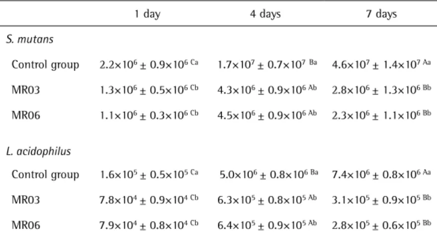
Table 1. Viable Streptococcus mutans and Lactobacillus acidophilus cells recovered from the biofilm adhered to the surfaces of specimens after 3 incubation periods, expressed as colony forming units per mL of culture medium (CFU/mL)
1 day 4 days 7 days
S. mutans
Control group 2.2×10⁶ ± 0.9×10⁶ Ca 1.7×10⁷ ± 0.7×10⁷Ba 4.6×10⁷ ± 1.4×10⁷ Aa
MR03 1.3×10⁶ ± 0.5×10⁶ Cb 4.3×10⁶ ± 0.9×10⁶ Ab 2.8×10⁶ ± 1.3×10⁶ Bb
MR06 1.1×10⁶ ± 0.3×10⁶ Cb 4.5×10⁶ ± 0.9×10⁶ Ab 2.3×10⁶ ± 1.1×10⁶ Bb
L. acidophilus
Control group 1.6×10⁵ ± 0.5×10⁵ Ca 5.0×10⁶ ± 0.8×10⁶ Ba 7.4×10⁶ ± 0.8×10⁶ Aa
MR03 7.8×10⁴ ± 0.9×10⁴ Cb 6.3×10⁵ ± 0.8×10⁵ Ab 3.1×10⁵ ± 0.9×10⁵ Bb
MR06 7.9×10⁴ ± 0.8×10⁴ Cb 6.4×10⁵ ± 0.9×10⁵ Ab 2.8×10⁵ ± 0.6×10⁵ Bb
143
Antibacterial restorative material
after disaggregation, by counting of colony-forming units (CFU). Five dilutions were made from this initial suspension. In the first, 100 µL from the initial suspension was inoculated into 900 µL of PBS (10-1 dilution), with the final dilution being 10-5. From each dilution, 100 µL was pipetted on to MRS agar on a Petridish (90 x 15 mL), and this inoculum was spread over the surface with a Drigalsky handle. The Petri dishes with S. mutans were incubated directly in the oven at 37°C for 48 h and dishes with L. acidophilus were incubated under microaerophilic atmosphere (10% CO2) at 37°C for 48 h. After incubation, dishes with 30-300 colonies were selected for counting. This number was multiplied by the dilution factor and then by 10 in order to obtain the number of CFU present in 1 mL of the initial suspension. Tables were structured with the CFU number per mL. Statistical analysis was performed by two-way analysis of variance and Tukey’s test pairwise comparison (α=0.05%).
Surface Roughness Analysis
Atomic force microscopy analysis (AFM) was performed to measure surface roughness (Ra, arithmetic roughness) at three sites (10 µm2) of three discs from each group and the values were statistically analyzed by ANOVA one-way and Tukey’s test (α=0.05%). Surfaces were analyzed using a Nanoscope V Multimode microscope (Bruker, Billerica, MA, USA).The resin discs previously formed did not need to be cut to fit this microscope. To operate the microscope, the method chosen was tapping mode, in air. NanoScope analysis© (Version 1.40, Bruker Corporation, Billerica, MA, USA) is a specific software for the analysis of data provided by Bruker for scanning probe microscopes.
Compression Assay
Compression assay was carried out in order to check whether there was damage to the mechanical strength of the modified resin. Five cylindrical specimens with circular base 1 cm diameter and height 1 cm were produced for each experimental group. The specimens had to be larger for a
good fit in the testing machine. One polytetrafluoroethylene matrix was machined with cylindrical perforations of desired dimensions. The composite resin was condensed in small increments in the spaces of the matrix and then polymerized. The compression tests were performed using an Instron 5569 universal testing machine (Instron Corp., Norwood, MA, USA). The procedure adopted was based on ASTM D 695-10 guideline (17), at compression speed 1.3 mm/min and load cell 10 KN. Two parameters were measured for all samples: compressive strength at failure and elasticity modulus in compression. Statistical analysis was performed by one-way ANOVA and Tukey’s test (α=0.05%).
Results
Viable Cell CountTable 1 shows the viable cells of S. mutans and L. acidophilus recovered from the biofilms adherent to the surfaces of specimens after the three incubation periods, expressed as CFU/mL. The results showed that for the three incubation periods, CFU numbers from biofilms were significantly lower in MR03 and MR06 than in CG, for both studied species (p<0.05). MR03 and MR06 showed no statistically significant differences (p>0.05). Within each material group, CFU numbers were significantly different for the three incubation periods, for both species (p<0.05).
Surface Roughness Analysis
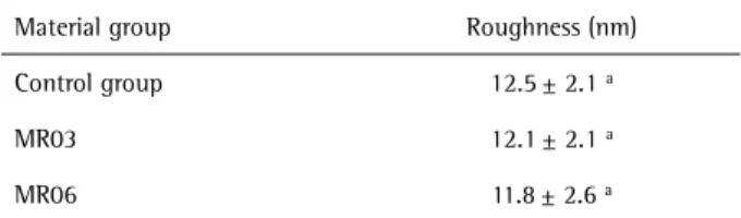
Table 2 shows the values for surface roughness obtained by NanoScope software expressed as arithmetic roughness (Ra). The result demonstrates that there was no difference among the three material groups according to statistical analysis (p>0.05).
Compression Assay
The values of compressive strain at failure and elasticity modulus in compression are shown in Table 3 for the three groups. The table demonstrates that compressive strength
Table 2. Values of surface roughness (Ra)
Material group Roughness (nm)
Control group 12.5± 2.1 a
MR03 12.1± 2.1 a
MR06 11.8± 2.6 a
The values are presented as means and standard deviations (n=9). Same letters indicate no statistically significant difference at 5% (ANOVA and Tukey’s test). Control Group, MR03 and MR06 represent unmodified resin, and modified resins with 0.3% and 0.6% of silver nanoparticles by weight, respectively.
Table 3. Values of compression assay
Material group Strength at failure (MPa)
Elasticity modulus (MPa)
Control group 85.4± 5.3 b 1466.0 ± 72.1 b
MR03 96.0± 4.9 a 1725.0 ± 54.0 a
MR06 72.8± 4.2 c 1205.0 ± 84.9 c
144
P
.B
.A. das Neves et al
at failure and elasticity modulus in compression were statistically different among the three groups (p<0.05).
Discussion
The biofilm reduction in vitro verified on the modified resins is an important result because a composite resin with these properties will enable lower bacterial accumulation on the restored areas of the teeth, helping to prevent recurrent caries.After the 3 incubation periods, CFU counts from biofilms were similar in MR03 and MR06, and lower in these groups than in CG, for both species studied. This result supports the hypothesis that the antibacterial action of silver nanoparticles, previously demonstrated in the literature (8-10), remains occurring after mixing with the composite resin and affects the microorganisms that are in contact with the modified material, similarly to others
in vitro investigations (14-16).
In addition, the roughness results obtained by AFM analysis demonstrate that there was no significant difference among the three material groups. Therefore, the surface roughness of composite resin was not increased by the presence of silver nanoparticles, in both concentrations of modified resin tested. These findings are important because a change in the roughness of modified resin could increase the biofilm accumulation. In accordance with this, several studies have shown that a rougher surface favors the biofilm growth on restorative materials (18-20). In contrast, an experimental composite adhesive containing silver nanoparticles that also presented bactericidal property showed rougher surface than conventional adhesive (15).
The compression assay showed that MR03 achieved the best mechanical performance for two measured parameters, and MR06 the worst, with a statistical difference. MR03’s better performance in the compression assay compared with CG suggests that the 0.3% silver nanoparticle concentration should be within the ideal range of modification for optimization of the composite mechanical properties on compression. About the change of filler volume-fractions in composite resins, Masouras et al. (21) found a correlation of inorganic filler content and elastic properties of fifteen trademarks of composite resins, considering three parameters. Elasticity modulus varied with statistical significance and a positive correlation occurred between modulus and inorganic filler fraction in the materials evaluated.
On the other hand, the results of the compressive assay for MR06 indicate that the concentration of nanoparticles was higher than that appropriate for maintaining the efficiency of resin reinforcement. The main limitation of the compression assay is that most assays about composite resin resistance have not used the same norm. This fact
impairs the comparison of values obtained here with the values already reported about the minimum acceptable resistance values. Nevertheless, the comparison of the experimental groups with the control group was carried out, and no reduction in resistance to compression is desired.
In terms of bacterial effect, the modified resins used in MR03and MR06 would be advantageous. Thus, MR03 is more appropriate for clinical application because it requires fewer nanoparticles and has lower cost. Furthermore, the results of the compression assay suggest that MR06 is more prone to suffer impairment of physical properties than MR03. This research shows promising outcomes, but further investigation are necessary in order to determine whether a new dental restorative material can be used in the oral cavity. Future studies will be performed to quantify the release of silver to saliva. They will be references for comparison with studies that described a non-toxic amount of silver nanoparticles to human cells (11-13). Additional studies will also be conducted to assess the cytotoxicity of modified resin through contact with living tissue in comparison with unmodified resin, and it will be essential to investigate other physical properties and color change.
In conclusion, the experimental composite resins with 0.3% and 0.6% wt of silver nanoparticles were less conducive to biofilm growth on their surfaces than unmodified resin and suffered no increase of surface roughness. Moreover, the modified resins with 0.3% wt of silver nanoparticles achieved improvement of compression resistance. Continuation of this study is very important because a restorative composite with these properties may benefit patients by helping maintaining good oral hygiene along with regular toothbrushing and by assisting in the prevention of caries recurrency.
Resumo
145
Antibacterial restorative material
crescimento dos biofilmes, sem comprometimento da resistência à compressão e da rugosidade superficial.
Acknowledgements
This research was financially supported by the Brazilian Government research funding agency CAPES.
References
1. Glasspoole EA, Erickson RL, Davidson CL. A fluoride-releasing composite for dental applications. Dent Mater 2001;17:127-133.
2. Creanor SL. Fluoride uptake and release characteristics of glass ionomer cements. Caries Res 1994;28:322-328.
3. Imazato S. Bio-active restorative materials with antibacterial effects: new dimension of innovation in restorative dentistry. Dent Mater 2009;28:11-19.
4. Sanders BJ, Gregory RL, Moore K, Avery DR. Antibacterial and physical properties of resin modified glass-ionomers combined with chlohexidine. J Oral Rehab 2002;29:553-558.
5. Elsaka SE, Hamouda IM, Swain MV. Titanium dioxide nanoparticles addition to a conventional glass-ionomer restorative: influence on physical and antibacterial properties. J Dent 2011;39:589-598. 6. Morones J, Elechiguerra J, Camacho A, Holt K, Kouri J, Ramirez J. The
bactericidal effect of silver nanoparticles. Nanotech J 2005;16:2346-2353.
7. Kalishwaralal K, BarathManiKanth S, Pandian SRK, Deepak V, Gurunathan S. Silver nanoparticles prevents the biofilm formation by Pseudomonas aeruginosa and Staphylococcus epidermidis. Coll Surf B: Biointer 2010;79:340–344.
8. Li Q, Mahendra S, Lyon DY, Brunet L, Liga MV, Li D et al. Antimicrobial nanomaterials for water disinfection and microbial control: Potential applications and implications. Water Res 2008;42:4591-4602. 9. Allaker RP. The use of nanoparticles to control oral biofilm formation.
J Dental Res 2010;89:1175-1186.
10. Blecher K, Nasir A, Friedman A. The growing role of nanotechnology in
combating infectious disease. Virulence 2011;2:1-7.
11. Sayes CM, Fortner JD, Guo W, Lyon D, Boyd AM, Ausman KD et al. The differential cytotoxicity of water-soluble fullerenes. Nano Lett 2004;4:1881–1887.
12. Sondi I, Salopek-Sondi B. Silver nanoparticles as an antimicrobial agent: a case study on E. coli as a model for Gram-negative bacteria. J Coll Inter Sci 2004;275:177-182.
13. Yudovin-Farber I, Beyth N, Nyska A, Weiss EI, Golenser J, Domb AJ. Surface characterization and biocompatibility of restorative resin containing nanoparticles. Biomacromolecules 2008;9:3044-3050. 14. Burguers R, Eidt A, Frankenberg R, Rosentritt M, Schweiki H, Handel
G, et al.. The ant-adherence activity and bactericidal effect of microparticulate silver additives in composite resin materials. Arch Oral Biol 2009;54:595-601.
15. Ahn SJ, Lee SJ, Kook JK, Lim BS. Experimental antimicrobial orthodontic adhesives using nanofillers and silver nanoparticles. Dent Mater 2009;25:206-213.
16. Melo MA, Cheng L, Zhang K, Weir MD, Rodrigues LK, Xu HH. Novel dental adhesives containing nanoparticles of silver and amorphous calcium phosphate. Dent Mater 2013;29:199-210.
17. ASTM D 695: Standard Test Method for Compressive Properties of Rigid Plastics. Annual Book of ASTM Standards Philadelphia, 2010. 18. Ikeda M, Matin K, Nikaido T, Foxton RM, Tagami J. Effect of surface
characteristics on adherence of S. mutans biofilms to indirect resin composites. Dent Mater 2007;26:915-923.
19. Kantorski KZ, Scotti R, Valandro LF, Bottino MA, Koga-Ito CY, Jorge AO. Adherence of Streptococcus mutans to uncoated and saliva-coated glass-ceramics and composites. Gen Dent 2008;56:740-747. 20. Aykent F, Yodem I, Ozyesil AG, Gunal SK, Avunduk MC, Ozkan S. Effect
of different finishing techniques for restorative materials on surface roughness and bacterial adhesion. Prosthet Dent 2010;103:221-227. 21. Masouras K, Silikas N, Watts DC. Correlation of filler content and elastic
properties of resin-composites. Dent Mater 2008;24:932-939.