271 271 271 271 271 Mem Inst Oswaldo Cruz, Rio de Janeiro, Vol. 95(2): 271-277, Mar./Apr. 2000
Nuclear Phenotype Changes after Heat Shock in
Panstrongylus megistus
(Burmeister)
Simone L Garcia, Maria Luiza S Mello/
+,
Vera Lúcia CC Rodrigues*,
Nancy L Garcia**
Departamento de Biologia Celular e Parasitologia, Instituto de Biologia ** Departamento de Estatística, Instituto de Matemática, Estatística e Computação Científica, Unicamp, 13083-970 Campinas, SP, Brasil
*Sucen, Mogi-Guaçu, SP, Brasil
The nuclear phenotypes of Malpighian tubule epithelial cells of male nymphs of the blood-sucking insect, Panstrongylus megistus, subjected to short- and long-duration heat shocks at 40oC were ana-lyzed immediately after the shock and 10 and 30 days later. Normal nuclei with a usual heterochromatic body as well as phenotypes indicative of survival (unravelled heterochromatin, giants) and death (apoptosis, necrosis) responses were observed in control and treated specimens. However, all nuclear phenotypes, except the normal ones, were more frequent in shocked specimens. Similarly altered pheno-types have also been reported in Triatoma infestans following heat shock, although at different frequen-cies. The frequency of the various nuclear phenotypes observed in this study suggests that the forms of cell survival observed were not sufficient or efficient enough to protect all of the Malpighian tubule cells from the deleterious effects of stress. In agreement with studies on P. megistus survival following heat shock, only long-duration shock produced strongly deleterious effects.
Key words: Pantrongylus megistus - heat shock - nuclear phenotypes - cell survival - apoptosis - necrosis
The effect of heat shock on survival and molt-ing incidence in Panstrongylus megistus varies with
the duration of the shock, the developmental stage and sex of the specimens, and in certain cases, the insect’s habits and nutritional states (Garcia et al. 1999).
P. megistus is less resistant to heat shock than Triatoma infestans, indicating that no
generaliza-tion can be made about the responses of different reduviid species to temperature shocks (Rodrigues et al. 1991, Garcia et al. 1999).
As with other stress factors, heat shocks induce
cytological changes in T. infestans, including
nuclear fusion, heterochromatin unravelling, and cell necrosis and apoptosis, as part of the mecha-nisms of cell survival and cell death, respectively (Mello 1989, Dantas & Mello 1992, Mello et al.
1995, Tavares et al. 1997).
The Malpighian tubule cells in late nymphs of
P. megistus are highly polyploid (Mello 1975). In
males, the most usual nuclear phenotype of this organ has a homogeneous distribution of granulous chromatin and a small but conspicuous heteromatic body formed by several copies of the Y chro-mosome (Mello et al. 1986).
Since P. megistus and T. infestans differ in their
normal nuclear phenotypic characteristics (Mello 1971, 1975, Mello et al. 1986), these nuclear phe-notypes may be affected differently by heat shock treatment.
In the present study, the nuclear phenotypes of
P. megistus nymphs were determined after heat
shock and the changes compared with those for T.
infestans under similar temperature conditions
(Dantas & Mello 1992).
MATERIALS AND METHODS
Fifth instar male nymphs of a domestic popu-lation of P. megistus (Hemiptera, Reduviidae),
de-scended from insects obtained in Fazendas Pedra Balão and Pedra Branca in São João da Boa Vista (State of São Paulo) and reared in the laboratory at Sucen (Mogi Guaçu, SP), were used. The control
nymphs were maintained at 28oC and 80%
rela-tive humidity, conditions which have traditionally been used for rearing this species in the labora-tory at Sucen since 1980.
This work was supported by the State of São Paulo Re-search Foundation (Fapesp, grants 95/1954-8, 95/6629-8 and 99/02547-95/6629-8) and the Brazilian National Research and Development Council (CNPq).
This study was part of a thesis presented by SLG to the Instituto de Biologia, Unicamp, in partial fulfillment of the requirements for the Masters degree.
+Corresponding author. Fax: + 55-19-788.7821. E-mail:
272 272 272 272
272 Changes with Heat Shock in P. megistus Simone L Garcia et al.
The treated specimens underwent heat shock at 40oC for 1 h and 12 h. The choice of this
tempera-ture was based on a previous study of survival and molting incidence in P. megistus after heat shock
(Garcia et al. 1999). The specimens were fasted for 15 days before the shock and after treatment were returned to a normal diet of hen blood once a week. Malpighian tubule preparations were obtained immediately after heat shock and 10 and 30 days later. The organs from at least three specimens were used for each experimental condition and the corresponding control.
Whole Malpighian tubules were mounted on glass slides, immediately fixed in acetic ethanol for 1 min, rinsed in 70% ethanol for 5 min and air dried at room temperature. The material was then subjected to the Feulgen reaction, with hydrolysis
in 4 M HCl at 25oC for 1 h and 5 min. The
Feul-gen-stained material was rinsed in sulfurous and
distilled water, air dried, cleared in xylene and mounted in Canada balsam.
A Zeiss light microscope was used to count the total number of Malpighian tubule epithelial cell nuclei per specimen, to identify the different nuclear phenotypes and to evaluate their frequen-cies in each specimen. Photomicrographs were obtained with a Zeiss Axiophot II microscope.
A linear correlation was used to evaluate the relationship between the stress conditions and the various nuclear phenotypes.
RESULTS
The most frequent phenotype in the control and heat shocked specimens consisted of nuclei with a small heterochromatic body containing copies of the Y chromosome (Mello et al. 1986) in the middle of evenly distribution of lightly stained chromatin (Fig. 1, Table I).
TABLE I
Absolute frequencies of nuclear phenotypes in Malpighian tubule epithelial cells of Panstrongylus megistus 5th instar nymphs after heat shock at 40oC
Experimental Nuclear phenotypes
conditions A As NE G GNE GHD GS HD N Total
Control, t0 10 1324 1255 25 4 3 0 147 5302 8070
0 1345 192 1 1 0 0 54 8786 10378
6 2347 1037 0 0 0 0 35 10499 13951
1 h shock: t0 54 2405 2292 12 1 2 15 143 9934 14858
77 1295 655 11 2 0 0 284 7914 10238
18 1447 984 3 0 0 0 340 13714 16506
12 h shock: t0 16 2661 1402 1 0 0 4 119 13354 17557
3 2244 676 13 7 2 4 224 15389 18562
5 2954 1655 12 4 3 51 1048 11678 17410
Control, t10 days 7 1729 325 6 0 0 0 98 9039 11204
9 2482 116 0 0 0 0 76 6479 9162
16 3252 728 0 0 0 0 268 11428 15692
1 h shock: t10 days 37 2783 1477 14 0 3 17 667 9904 15236
25 1385 1272 68 20 17 22 275 3978 7062
35 2703 1696 229 87 33 20 525 10834 16162
Control, t30 days 25 2587 3 0 0 0 0 173 10242 14506
11 1351 1882 10 0 0 8 295 8169 11726
23 2704 1962 5 0 0 0 424 10673 15791
8 1408 1522 5 0 1 0 164 7271 10379
1 h shock: t30 days 11 3960 1164 1 0 0 0 864 11206 17224
23 3973 1576 0 0 0 0 335 10386 16283
8 2754 866 3 0 0 0 130 7122 10239
19 2218 745 5 0 0 0 130 7122 10239
12 h shock: t30 days 93 2329 2995 43 75 13 165 148 11894 17755
96 4787 840 19 15 0 12 124 12446 18339
47 3046 2445 14 3 3 17 204 7766 13555
273 273273 273 273 Mem Inst Oswaldo Cruz, Rio de Janeiro, Vol. 95(2), Mar./Apr. 2000
Other nuclear types observed in all the experi-mental conditions were generally more frequent after heat shock (Table I). These included nuclei with heterochromatin unravelling (Fig. 2), apoptotic nuclei (Fig. 3), nuclei suspected of apoptosis (Fig. 4), necrotic nuclei (Fig. 5) and gi-ant nuclei (Fig. 6). In some cases gigi-ant nuclei ex-hibited signs of necrosis or were suspected of apoptosis (Fig. 6). Some giant nuclei were also observed in which heterochromatin decondensation
was suspected. Apoptosis and necrosis were de-fined here in terms of their classic morphological characteristics (Kerr 1971, Kerr et al. 1972). There was a significant decrease in the relative frequency of normal nuclei with heat shock and its duration (Fig. 7a, Table II). The absolute frequency of nuclei with heterochromatin unravelling in-creased just after the short and long shocks, and 10 and 30 days after the short shock (Table I). However, in terms of relative frequencies, the
in-Figs 1-6: nuclear phenotypes in Feulgen-stained Malpighian tubules of Panstrongylus megistus specimens subjected to heat shock.
274 274 274 274
274 Changes with Heat Shock in P. megistus Simone L Garcia et al.
crease in nuclei with heterochromatin unravelling was significant only immediately after short and long shocks (Fig. 7b, Table II); the frequency of this phenotype decreased after long shocks com-pared to short shocks (Fig. 7b). There was no sig-nificant correlation with the time after the shock (Table II). The appearance of heterochromatin decondensation was moderately correlated with that of apoptotic nuclei (Table II).
A few giant nuclei were detected (Table I). The relative frequency of this nuclear phenotype de-creased immediately after long shocks when com-pared to short shocks (Fig. 7c), i.e. there was a slight negative correlation between shock duration and phenotype frequency (Table II). There was no cor-relation between the relative frequency of morpho-logically normal giant nuclei and the time after the shock or the shock temperature, but a moderate correlation was observed between giant nuclei with heterochromatin decondensation and the time af-ter the shock (negative), between the occurrence of necrotic giants and shock temperature (positive) and between giants suspected of apoptosis and the shock temperature and duration (positive) (Table II).
There was a high correlation between giant nuclei with normal morphological characteristics and necrotic giant nuclei as well as between ne-crotic giants and giants suspected of apoptosis (Table II).
The absolute and relative frequencies of apoptotic nuclei increased immediately after heat shock (Fig. 8a, Tables I, II), but there was no sig-nificant correlation between these frequencies and the time after shock or the duration of the shock (Table II).
The relative frequency of nuclei suspected of apoptosis did not increase with the heat shock tem-perature or duration, but there was a moderately significant correlation between the increase in this frequency and the time after shock (Fig. 8b, Table II). The absolute and relative frequencies of ne-crotic nuclei increased significantly 10 and 30 days after the heat shock (Tables I, II, Fig. 8c).
DISCUSSION
The number of nuclei in the Malpighian tubules of fully-nourished laboratory-reared P. megistus is
about 18,000 (Mello et al. 1986). In the present study, control specimens had a much smaller nuclear frequency, indicating that nuclear fusion and cell death induced by other stressors may have occurred under laboratory conditions. Indeed, the specimens used in this investigation had been mod-erately fasted prior to the heat shock. Fasting is a stress agent in blood-sucking insects (Andrade & Mello 1987, Mello 1989). The choice of a slight fasting condition for the present study was based on the finding that only after it some 5th instar specimens are capable of surviving a long heat
TABLE II
Linear correlation between stress factors and nuclear phenotypes
D oC H A A
S NE G GD GHD GS HD oC .05
A -.03 .29 -.18
AS .38 .08 -.02 .06
NE .30 .13 .06 .12 -.13
G -.18 .12 -.22 .06 -.01 .26
GD -.02 .23 .03 .07 -.07 .30 .86
GHD -20 .07 -.18 .08 .01 .27 .93 .79
GS .13 .31 .37 .18 -.14 .47 .24 .63 .33
HD .09 .38 -.34 .25 .05 .15 .20 .06 .18 .08
N -.47 -.27 .07 -25 -.56 -70 -.31 -.27 -.33 -.33 -.39
D: days after shock; oC: temperature; H: shock duration; A: apoptosis; A
s: suspected apoptosis; NE: necrosis;
G: giant nuclei; GD: necrotic giant nuclei; GHD: giant nuclei with heterochromatin unravelling; Gs: giant nuclei with suspected apoptosis; HD: heterochromatin unravelling; N: normal with usual heterochromatin body.
275 275275 275 275 Mem Inst Oswaldo Cruz, Rio de Janeiro, Vol. 95(2), Mar./Apr. 2000
(%)
Duration Temp
Days
12 1
0 40 28
30 10 0
75.8
73.6
71.4
69.2
67.0
Duration Temp
Days
12 1
0 40 28
30 10 0
3.1
2.7
2.3
1.9
1.5
(%
)
Duration Temp
Days
12 1
0 40 28
30 10 0
0.42
0.34
0.26
0.18
0.10
(%
)
A
B
C
Fig. 7: influence of time after shock, shock temperature and shock duration (h) on the relative frequency of normal nuclei (A), normally-sized nuclei showing heterochromatin unravelling (B) and giant nuclei (C) of Panstrongylus megistus.
shock (Garcia et al. 1999). The observation that some altered nuclear phenotypes also occurred in the control specimens supports the idea that some stressing effect other than heat shock was involved. Maybe an occasional and unexplained refusal of some specimens to feed a blood meal added some fasting period to that intentionally provoked.
Nuclear phenotypes differing from the normal phenotype seen in P. megistus have also been
re-ported for T. infestans subjected to heat shock or
other stressing agents (Álvares-Garcia 1988, Mello 1989, Dantas & Mello 1992, Mello et al. 1995). Such phenotypes may reflect mechanisms of cell survival (heterochromatin unravelling, nuclear fu-sion) or cell death (apoptosis, necrosis) in response
to stress (Dantas & Mello 1992, Tavares et al. 1997).
Heterochromatin unravelling occurred even long after heat shock in P. megistus. This situation
differs from that for T. infestans in which such
unravelling is most frequent 10-120 min after the shock (Dantas & Mello 1992). If heterochromatin unravelling leads to the activation of silent genes during stress (Simões et al. 1975), its effects may be longer-lasting in P. megistus than in T. infestans.
It is not known whether heterochromatic zones contain genes for heat shock proteins (hsp).
276 276 276 276
276 Changes with Heat Shock in P. megistus Simone L Garcia et al.
Mello 1989, Dantas & Mello 1992). These nuclei have been suggested to be involved in cell or organ survival mechanisms under unfavorable conditions. In this regard, the high mortality rates seen in P. megistus subjected to long heat shock (Garcia et al.
1999) suggest that giant nuclei were not present in sufficient numbers and/or were not efficient enough to protect the insects from heat shock-induced dam-age. Indeed, the relative frequency of giant nuclei decreased significantly with the shock duration whereas the number of giants suspected of apoptosis was weakly correlated with this factor.
P. megistus survivors of heat shock showed a
significant decrease in the frequency of giant
nu-clei 10 to 30 days after the shock. In T. infestans,
the highest frequency of giants occurs 30 days af-ter the shock (Dantas & Mello 1992). In addition to a decrease in new fusions, the fact that some giant nuclei in P. megistus exhibited necrosis or
were suspected of apoptosis probably also contrib-uted to the reduction in their frequency. It is pos-sible that in P. megistus cells and nuclei resulting
from fusion may be more susceptible to metabolic failure than in T. infestans.
When the stress is enhanced beyond a certain level, the presence of hsp is incapable of protect-ing the cells and apoptosis program is initiated (Lindquist & Craig 1988, Maihos et al. 1993,
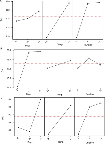
Fig. 8: influence of time after shock, shock temperature and shock duration (h) on the relative frequency of apoptotic (A), sus-pected apoptotic (B) and necrotic (C) nuclei of Panstrongylus megistus.
Duration Temp
Days
12 1
0 40 28
30 10 0
0.26
0.22
0.18
0.14
0.10
(%)
Duration Temp
Days
12 1
0 40 28
30 10 0
20.0
18.5
17.0
15.5
14.0
(%
)
Duration Temp
Days
12 1
0 40 28
30 10 0
10.4
9.8
9.2
8.6
8.0
(%
)
277 277277 277 277 Mem Inst Oswaldo Cruz, Rio de Janeiro, Vol. 95(2), Mar./Apr. 2000
Samali & Cotter 1996). If the stress exceeds this level, cell death by necrosis predominates (Samali & Cotter 1996). Thus, murine mastocytoma cells
subjected to a temperature of 43-44oC show an
increase in apoptotic index. At a temperature of 45oC, both apoptosis and necrosis are observed in
these cells while at 46-47oC only necrosis is found (Harmon et al. 1990). Similar responses have been described in other cell types (Sakaguchi et al. 1995).
In the Malpighian tubules of P. megistus,
apoptosis and necrosis occurred simultaneously, especially after hyperthermia. Only the apoptosis program intensified immediately after the heat shock. The frequency of the apoptotic nuclei then remained unchanged whereas necrosis, which was not significantly affected immediately after heat shock, intensified with the shock duration and time after shock.
Hsp expression, apoptosis, heterochromatin unravelling and nuclear fusion were thus not ap-parently sufficient to protect all P. megistus
Mal-pighian tubule cells from the deleterious effects of heat shock. In terms of insect survival, only long shocks proved to be strongly deleterious (Garcia et al. 1999). In the few survivors of long shocks, there was probably no additional degeneration since no significant difference in the frequency of necrotic nuclei was observed compared to insects subjected to short shocks.
The individual variations in response to hyper-thermia were similar to those seen in T. infestans
after other stressing agents and suggest that speci-mens of P. megistus may also vary in their
resis-tance to different stressors, including heat shock. ACKNOWLEDGMENTS
To Mr Pedro Ribeiro da Silva for his technical sup-port.
REFERENCES
Álvares-Garcia RS 1988. Efeitos da Radiação Gama sobre os Fenótipos Nucleares de Alguns Tipos Celulares deTriatoma infestans Klug (Hemiptera, Reduviidae),MSc Thesis, Unicamp, Campinas, 104
pp.
Andrade CGTJ, Mello MLS 1987. Phenotypes and num-ber of Malpighian tubule nuclei in Triatoma infestans
Klug along development and starvation. Rev Bras Genet10: 449-457.
Dantas MM, Mello MLS 1992. Changes in the nuclear phenotypes of Triatoma infestans Klug, induced by
thermal shocks. Rev Bras Genet15: 509-519. Garcia SL, Rodrigues VLCC, Garcia NL, Ferraz-Fillho
AN, Mello MLS 1999. Survival and molting inci-dence after heat and cold shocks in Panstrongylus megistus Burmeister. Mem Inst Oswaldo Cruz 94:
131-137.
Harmon BV, Corder AM, Collins RJ, Gobé GC, Allan
DJ 1990. Cell death induced in a murine mastocy-toma by 42-47oC heating in vitro: evidence that the
form of death changes from apoptosis to necrosis above a critical heat load. Int J Radiat Biol58:
854-858.
Kerr JFR 1971. Shrinkage necrosis: a distinct mode of cellular death. J Pathol105: 13-20.
Kerr JFR, Wyllie AH, Currie AR 1972. Apoptosis: a basic biological phenomenon with wide-ranging implications in tissue kinetics. Br J Cancer26:
239-257.
Lindquist S, Craig EA 1988. The heat shock proteins.
Ann Rev Genet22: 631-677.
Mailhos C, Howard MK, Latchman DS 1993. Heat shock protects neuronal cells from programmed cell death by apoptosis. Neuroscience55: 621-627.
Mello MLS 1971. Nuclear behaviour in the Malpighian tubes of Triatoma infestans (Reduv., Hemiptera). Cytologia 36: 42-49.
Mello MLS 1975. Feulgen-DNA values and ploidy de-grees in the Malpighian tubes of some triatomids.
Rev Bras Pesq Med Biol8: 101-107.
Mello MLS 1989. Nuclear fusion and change in chro-matin packing state in response to starvation in Tri-atoma infestans. Rev Bras Genet12: 485-498.
Mello MLS, Raymundo HH 1980. Nuclear fusion in the Malpighian tubes of a blood-sucking hemipteran.
Cytologia 45: 203-209.
Mello MLS, Randi MA, Giorgio S, Ferraz-Filho AN, Rodrigues VLCC, Rocha-e-Silva EO, Cordeiro JA 1986. Number of chromosomes, Feulgen-DNA con-tent and nuclear phenotypes in domestic and wild specimens of Panstrongylus megistus. Ann Trop Med Parasitol 80: 641-648.
Mello MLS, Kubrusly FS, Randi MA, Rodrigues VLCC, Ferraz Filho AN 1995. Effects of heavy metals on chromatin supraorganization, nuclear phenotypes, and survival of Triatoma infestans. Entom Exp Appl 74: 209-21.
Rodrigues VLCC, Mello MLS, Ferraz Filho AN, Dantas MM 1991. Sobrevivência e ocorrência de muda em
Triatoma infestans Klug (Hemiptera, Reduviidae)
após choque de temperatura. Rev Saúde Pública (São Paulo)25: 461-467.
Sakaguchi Y, Stephens LC, Makino M, Kaneko T, Strebel FR, Danhauser LL, Jenkins GN, Bull JMC 1995. Apoptosis in tumors and normal tissues induced by whole body hyperthermia. Cancer Res55:
5459-5464.
Samali A, Cotter TG 1996. Heat shock proteins increase resistance to apoptosis. Exp Cell Res 223: 163-170.
Simões LCG, Amabis JM, Cestari NA 1975. Puffs in the heterochromatin in chromosomes of
Rhynchosciara. Ciênc Cult São Paulo27: 159-161.
Tavares MCH, Dantas MM, Rodrigues VLCC, Mello MLS 1997. Alterações em fenótipos nucleares, com ênfase na apoptose, em túbulos de Malpighi de Tri-atoma infestans Klug após choque hipertérmico, 43o
Congresso Nacional de Genética, Goiânia, p. 65. Wigglesworth VB 1967. Polyploidy and nuclear fusion