online | memorias.ioc.fiocruz.br
Chagas disease is a neglected parasitic disease that was discovered 100 years ago (Lannes-Vieira et al. 2009). It is caused by the intracellular parasite Trypanosoma cru-zi and is most frequently associated with cardiac-related manifestations. T. cruzi initially invades myocardial cells, resulting in a diffuse and severe neutrophilic and mono-cytic inflammatory infiltrate in addition to myofibrillar lesions (Bilate & Cunha-Neto 2008). At the acute phase, patients can present with pericardial effusion, dyskinesis or ventricular dilatation detected by echocardiography. Electrocardiographic (ECG) alterations are also observed in patients; these include diffuse ST-T repolarization changes as well as low voltage, conduction problems, left anterior hemiblock and combined blocks (Blum et al. 2008). At the chronic phase, roughly 30% of patients pres-ent cardiac dysrhythmia, heart failure and thromboembo-lism (both systemic and pulmonary) (Rassi et al. 2009). A depressed left-ventricular ejection fraction and increased left-ventricular internal dimensions have been shown to be associated with increased morbidity and mortality (Rassi et al. 2006, Acquatella 2007).
Evidence has shown that oxidative stress resulting from augmentation of free radical production plays an important role in the pathogenesis of some heart diseases (Gupta et al. 2009). In an experimental model of T. cruzi infection, it was reported that increased oxidative stress and antioxidant insufficiency are associated with myocar-dial oxidative damage and mitochondrial functional de-cline, findings that might be of pathological significance in Chagas disease (Wen et al. 2004, Gupta et al. 2009).
We have previously investigated the role of selenium (Se), an essential trace element with antioxidant prop-erties, in both human and experimental infection of T. cruzi. We demonstrated that a significant percentage of patients with advanced chronic chagasic cardiomyopa-thy exhibited low Se levels that positively correlated with cardiac insufficiency (Rivera et al. 2002). Moreover, we demonstrated that a Se-deficient diet contributes to an increased susceptibility to this infection in experimental models (de Souza et al. 2002), whereas oral Se supple-mentation at low doses alleviates heart damage (de Sou-za et al. 2003). Gomez et al. (2002) have also reported an increased severity of myopathy in Se-depleted chroni-cally infected mice; furthermore, Davis et al. (1998) showed that Se supplementation reduced parasitaemia and mortality in infected mice. Based on these findings and taking into account the association of Se intake with the prevention of cardiac diseases (Korpela et al. 1989, Reeves et al. 1989, Foster & Sumar 1997, Saito et al. 1998), we investigated the effect of Se treatment on the development of chronic cardiopathy in T. cruzi-infected mice in both prevention and reversion schemes.
Financial support: FIOCRUZ-PDTSP, IOC-FIOCRUZ, FAPERJ, CNPq, MS-DECIT, CAPES, NIH (AI-052739, AI-12770, AI-062730) + Corresponding author: [email protected]
Received 8 February 2010 Accepted 20 July 2010
The benefits of using selenium in the treatment of Chagas disease:
prevention of right ventricle chamber dilatation and reversion of
Trypanosoma cruzi
-induced acute and chronic cardiomyopathy in mice
Andréa P de Souza1, Linda A Jelicks2, Herbert B Tanowitz3,Bianca P Olivieri1, Monica M Medeiros1, Gabriel M Oliveira4, Andrea Rodrigues Cordovil Pires5,
Alessandro M dos Santos1, Tania C Araújo-Jorge1/+
1Laboratório de Inovações em Terapias, Ensino e Bioprodutos 4Laboratório de Biologia Celular, Instituto Oswaldo Cruz-Fiocruz,
Av. Brasil 4365, 21040-360 Rio de Janeiro, RJ, Brasil 2Department of Physiology and Biophysics 3Department of Pathology and Medicine,
Albert Einstein College of Medicine, New York, NY, USA 5Fonte Medicina Diagnóstica, Niterói, RJ, Brasil
Cardiac damage is a frequent manifestation of Chagas disease, which is caused by the parasite Trypanosoma cruzi. Selenium (Se) is an essential micronutrient, the deficiency of which has been implicated in the development of cardio-myopathy. Our group has previously demonstrated that Se supplementation prevents myocardial damage during acute T. cruzi infection in mice. In this study, we analyzed the effect of Se treatment in cases of T. cruzi infection using prevention and reversion schemes. In the Se prevention scheme, mice were given Se supplements (2 ppm) starting two weeks prior to inoculation with T. cruzi (Brazil strain) and continuing until 120 days post-infection (dpi). In the Se reversion scheme, mice were treated with Se (4 ppm) for 100 days, starting at 160 dpi. Dilatation of the right ventricle was observed in the infected control group at both phases of T. cruzi infection, but it was not observed in the infected group that received Se treatment. Surviving infected mice that were submitted to the Se reversion scheme presented normal P wave values and reduced inflammation of the pericardium. These data indicate that Se treatment prevents right ventricular chamber increase and thus can be proposed as an adjuvant therapy for cardiac alterations already established by T. cruzi infection.
SUBJECTS, MATERiALS And METHOdS
Infection and Se supplementation - Male CD1 (WT) (Jackson Laboratories) and Swiss (CECAL-Fiocruz) mice were separated into two cohorts that either re-ceived treatment with sodium selenate (Sigma Chemical Co, MO, USA), an inorganic form of Se or no treatment. The first cohort was placed in the Se prevention scheme, aiming to prevent cardiac alteration resulting from T. cruzi infection. CD1 mice were treated with sodium sel-enate added at a concentration of 2 ppm to the drink-ing water (ad libitum) two weeks prior to infection, as previously described (de Souza et al. 2003). Treatment with Se was carried out for 100 days. These groups were named Inf-Se0(infected untreated, n = 11)andInf-Se2 (infected and treated with 2 ppm Se, n = 10). Uninfect-ed animals (5 mice/group) supplementUninfect-ed with the same doses (0 and 2 ppm) were used as controls (Cont-Se0 and Cont-Se2). The consequences of infection in terms of parasitaemia and mortality were monitored until 100 days post infection (dpi). The second cohort was placed in the Se reversion scheme to evaluate the effects of Se treatment on cardiac damage already established by the infection. Swiss mice at the chronic phase were treated with sodium selenate for 100 days, starting at 160 dpi. These mice were divided into three groups: infected (Inf), infected and treated with 4 ppm Se (InfSe4) and uninfected (Cont). Two weeks after beginning the Se treatment, the CD1 mice in the Se prevention scheme were infected by intra-peritoneal (ip) injection with 5 x 10⁴ bloodstream forms of the Brazil strain of T. cruzi, as described byHuang et al. (1999), the same inoculum and T. cruzi strain used in the reversion scheme. These mice were fed standard rodent chow. Parasitaemia and mor-tality were determined and cumulative mormor-tality (CM) and survival rate were calculated.
Magnetic resonance imaging(MRI) - Mice from the Se prevention scheme group was anaesthetized with aver-tin and a set of Gould ECG leads with custom electrodes were attached to their limbs for cardiac gating. The ECG signal was transmitted to a MRI spectrometer through a cardiac gating box as well as a PC running Ponemah Physiology Suite software. Mice were positioned in a 35 mm home-built MRI coil in a 9.4T GE Omega verti-cal-bore imaging system. Heart rate and ECG were moni-tored continuously. Gating trigger delays were controlled by the pulse sequence and were set to acquire images in either systole or diastole. After the imaging experiments, the mice were allowed to recover from the anaesthesia and were returned to their cages in the Animal Institute. All MRI experiments were performed at the Albert Ein-stein College of Medicine using a GE Omega 9.4T verti-cal wide-bore nuclear magnetic resonance spectrometer equipped with a microimaging accessory.
Cardiac gated MRI - Scout images of the thorax were acquired. Once the specific region of interest in the heart was identified, images were acquired in diastole. Images were analyzed using MATLAB-based software. Cardiac dimensions were recorded. The left ventricular inner dimension (LVID) corresponds to the average of
the septum-lateral and anterior-posterior LVDI. The LV wall is the average of the anterior, posterior, lateral and septum wall dimensions. The right ventricular inner mension (RVID) is the measurement of the chamber di-mension at the widest point of the RV. Cardiac volumes were not calculated from the multi-slice images because there is a slight time shift during each slice acquisition, such that all slices from a single imaging data set are not collected at the same position in the cardiac cycle. Instead, we evaluated all images acquired with different delays, selecting the centre slice of the heart exhibiting the largest LVID to represent the end-diastolic image.
ECG study - Mice from the Se reversion scheme were tranquilized with diazepam (20 mg/Kg ip) and transduc-ers were carefully placed on the skin in accordance with the chosen preferential derivation (DII). Traces were recorded using a digital system (Power Lab 2/20) con-nected to a bio-amplifier (Pan Lab Instruments). Trac-es were analyzed using the Scope software for Windows. We measured the duration of the PR, QRS and QT in-tervals (QTc) and the P wave in milliseconds (ms) at the end of the experiment (237-259 dpi). The relationship be-tween the QT and RR intervals (RR0) was individually assessed. To obtain physiologically relevant values for the heart rate-corrected QTc in units of time rather than time to a power not equal to 1, the observed RR0 was first expressed as a unit-less multiple of 100 ms, giving a normalized RR0 (RR100 = RR0/100 ms). Next, the value of the exponent (y) in the formula QT0 = QTc x RRy
100
was assessed, where QT0 is the observed QT and both QT and QTc are expressed in milliseconds. Taking the natural logarithm of each side of the formula (QT0) = In (QTc) + yln(RR100), the slope of the linear relationship between the log-transformed QT and RR100 thus defined the y to which the RR0 ratio should be raised to correct QT for heart rate (Mitchell et al. 1998).
To avoid measurement bias in MRI and ECG stud-ies, treated and untreated mice received a code number and experts from each area performed measurements and analysis.
Histological examination - Before starting the Se re-version scheme, six infected mice and four uninfected mice were sacrificed at 150 dpi; at the end of the ex-periment (273 dpi), the surviving mice were sacrificed to collect the heart. The heart tissue was processed in paraffin-embedded sections stained with hematoxilin-eosin. The presence of necrosis was determined when cell debris and a faint coloration were detected in the sections; foci of inflammatory infiltrate (containing at least 10 mononuclear cells) were also investigated.
Statistical analysis - Statistical significance (p < 0.05) was evaluated using the Mann Whitney test for the analysis of parasitaemia, cardiac dimension and the log Rank (Gehan-Breslow-Wilcoxon) test for survival of the animals. A t test was used for the ECG analysis. The sta-tistical significance of variations in RV dimensions be-tween groups was evaluated using analysis of variance.
and approved by The Institutional Animal Care and Use Committee of the Albert Einstein College of Medicine and by the Fiocruz Committee of Ethics in Animal Use, license 0099/01 for the research, resolution 6.899/09.
RESULTS
Parasitaemia and mortality - In the Se prevention scheme, all infected mice presented a peak of parasitae-mia between 20-24 dpi; this was observed in both sup-plemented and unsusup-plemented groups, with no statisti-cally significant differences observed between the two. The median parasitaemia (75th percentile) was similar between Inf-Se0 and Inf-Se2 groups, with values of 11.8 x 10⁵ ± 4.3/mL and 7.1 x 10⁵ ± 3.6/mL, respectively.
Both groups presented a low mortality rate at the acute phase. The CM of the Inf-Se0 group was 18.2% at 40 dpi, whereas all mice supplemented with 2 ppm Se (Inf-Se2) survived beyond that day. At the end of the experi-ment (120 dpi), the Inf-Se0and Inf-Se2 groups exhibited a CM of 18.2% and 22%, respectively.
In cohort 2, infected mice presented a peak of para-sitaemia at 27 dpi (54.8 x 10⁴ mL) and a CM of 67.9%
at 150 dpi. At 160 dpi, infected mice were divided into groups to begin the Se treatment and CM measure-ments were restarted. Fifty days after Se treatment (240 dpi), the InfSe4 group presented lower CM rates (11%), though this was not significantly different from the Inf group (28.6% CM).
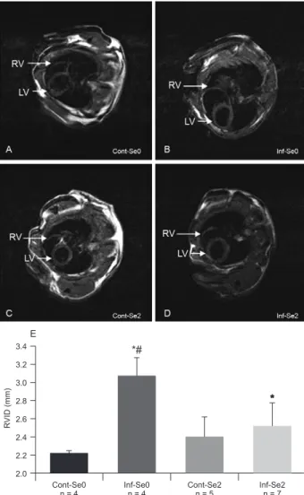
MRI - Cardiac-gated MRI of mice in the Se preven-tion scheme was performed during the acute and chronic phases (Fig. 1, Table). Compared with Cont-Se0, the RVID of Inf-Se0 mice was 1.3 times higher (3.1 ± 0.2 mm vs. 2.3 ± 0.2 mm; p = 0.04) at the acute phase. At the acute phase, the hearts of Inf-Se2 mice were not signifi-cantly different from those of Inf-Se0 mice,exhibiting the same infected profile of a significant increase of the RVID compared to Cont-Se2mice (2.9 ± 0.3 mm vs. 2.0 ± 0.1 mm; p = 0.01). During this period of the infection, the variance within groups was 0.1081, 0.0129, 0.3775 and 0.5089 for Cont-SeO, Cont-Se2, Se0 and Inf-Se2, respectively.
Treatment with Se over a long period of time did not induce any cardiac alterations in Cont-Se2mice. In rela-tion to the chronic phase (100 dpi), Inf-Se0 mice exhib-ited a significant enlargement of RVID (Fig. 1B) when compared to Cont-Se0 (Fig. 1A, Table). Critically, the RVID of infected mice treated with Se did not increase any further (Fig. 1D) and remained similar in size to that of the uninfected control mice in the chronic phase (Fig. 1C). The graphical comparison of mean RVID measures (Fig. 1E) clearly shows the effect of Se treatment on the prevention of right ventricular dilatation during experi-mental Chagas cardiomyopathy. The variance within groups was 0.0034 for Cont-Se0, 0.0757 for Cont-Se2, 0.1933 for Inf-Se0 and 0.0975 for Inf-Se2.
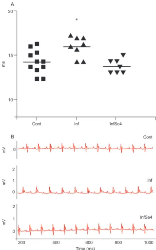
ECG findings-During the period between 237-259 dpi that corresponded to 76-98 days post Se treatment, mice in cohort 2 were subjected to ECG evaluations to study reversion of heart lesions. Se treatment resulted in a significant reversion of the increase of the P wave
duration (Fig. 2) in the InfSe4group (13.8 ± 0.7 ms, n = 7) in comparison to the Inf group (16.0 ± 1.1 ms, n = 8). No difference was observed between the Cont (14.2 ± 1.3 ms, n = 11) and InfSe4groups.
Histopathological analysis-At 150 dpi, uninfected mice did not present any histological alterations (Fig. 3A), whereas 5-6 Inf mice exhibited pericarditis and 1 exhibited myocarditis in the atrium (Fig. 3B). At the end of the experiment (dpi 273), no important histological findings were made in the hearts of Cont group mice (not shown), whereas two surviving untreated Inf mice exhibited moderate pericarditis (Fig. 3C). In contrast,
R
VID (mm)
Cont-Se0
n = 4 n = 4 n = 5 n = 7
Cont-Se2
Inf-Se0 Inf-Se2
3.4
3.2
3.0
2.8
2.6
2.4
2.2
2.0
*# E
Fig. 1: effect of selenium (Se) supplementation on hearts of mice in-fected with Trypanosoma cruzi at the chronic phase, 100 days after infection. A, C: magnetic resonance imaging (MRI) of non-infected mice supplemented (Cont-Se2) (C) or not (Cont-Se0) (A) with 2 ppm sodium selenate in drinking water; B, D, E: cardiac gated MRI of
ture and function of the hearts of infected mice (Hoit 2001, Jelicks et al. 2002). We recently proposed that en-largement of the RV may represent a marker for chagasic cardiomyopathy in mice (Huang et al. 1999, de Souza et al. 2005)andsome studies have addressed the possible reduction of the severity of this cardiomyopathy using various strategies(Jelicks et al. 2002, Goldenberg et al. 2008). In the present study, the Se prevention scheme was able to significantly reduce the infection-induced in-crease of the RVID at the acute and chronic phases of T. cruzi infection. Although the analysis of variance within the Cont-Se0 group was 0.0034 at the chronic phase, this group presented an average RVID of 2,226 mm with a standard deviation of 0.050 mm. The mechanisms underlying the effect of Se on the prevention of heart enlargement are currently under investigation. To our knowledge, this is the first MRI-based description of the preventive effects of Se treatment on ventricular dilata-tion. The disease progression observed in the experi-mental model used in this study may be analogous to the slow changes that occur during the early chronic phase of infection in humans; therefore, Se treatment could be used to halt disease progression in patients at this stage of infection.
Our findings using the Se prevention scheme en-couraged us to study the effectiveness of Se treatment in reducing cardiac alterations already established in the chronic phase of infection as a model for chronic human Chagas heart disease. In these experiments, we initiated Se treatment starting at 160 dpi, 10 days after heart alterations can be observed histologically TABLE
Cardiac dimensions in mice in the acute and chronic phasesof Trypanosoma cruzi infection with or without selenium (Se) supplementation
Cont-Se0 Cont-Se2 Inf-Se0 Inf-Se2
Acute phase
RVID (mm) IV
2.3 ± 0.2a
1.00
2.0 ± 0.1 0.86
3.1 ± 0.2c
1.34
2.9 ± 0.3b
1.26 LVID (mm)
IVa
4.3 ± 0.3 1.00
4.0 ± 0.1 0.94
3.9 ± 0.2 0.91
3.9 ± 0.2 0.91
n 4 5 7 6
Chronic phase
RVID (mm) IV
2.2 ± 0.0a
1.00
2.4 ± 0.1 1.09
3.2 ± 0.2c,d
1.45
2.5 ± 0.1 1.13 LVID (mm)
IV
4.0 ± 0.2 1.00
4.2 ± 0.2 1.05
4.3 ± 0.1 1.07
4.2 ± 0.20 1.05
n 4 4 7 5
a: results in average ± standard error of the mean; b: p < 0.05 as compared to uninfected animals supplemented with 2 ppm Se (Cont-Se2); c:p < 0.05 as compared to uninfected animals (Cont-Se0); d: p < 0.05 as compared to infected animals treated with 2 ppm Se (Inf-Se2); Inf-Se0: infected untreated animals; IV: index of variation; LVID: left ventricular inner dimension; n: number of survivors mice in each group during the acute and chronic phases; RVID: right ventricular inner dimension.
two out of three InfSe4 mice did not present any his-tological alterations in the heart (Fig. 3D) and only one presented minimal pericarditis.
diSCUSSiOn
In the present study, we observed the course of infec-tion of two outbred mice lineages (CD1 and Swiss) by the Brazil strain of T. cruzi to study the potential beneficial effect of Se treatment in experimental Chagas disease; the experiments described revealed a striking effect of Se treatment on both the prevention and reversion of heart functional and histopathological damage. Based on our previous report (de Souza et al. 2003), we conducted the treatment study using the inorganic form of Se, sodium selenate. To investigate the preventative effects of Se, we administered a dose of 2 ppm (the lowest concentration assayed in preliminary experiments, data not shown) to CD1 mice infected with Brazil strain T. cruzi for long-term follow-up studies. For the Se reversion scheme, we used 4 ppm Se, a dose that was sufficient to prevent car-diac necrotic lesions in acute Swiss mice (de Souza et al. 2003). The course of the Brazil strain infection was similar in both cohorts, which presented low mortality rates and peaks of parasitaemia from 20-27 dpi, allowing monitoring of the mice until the late stage of the chronic phase. Se did not influence the survival of infected mice, as we reported before (de Souza et al. 2003); however, it exhibited positive effects on heart function.
struc-ginning of the stimulation in ventricles, subsequently af-fecting the contraction of ventricles. Unfortunately, the histology could only be evaluated in the small number of surviving infected mice; however, it was possible to ob-serve that Se treatment reversed the pericarditis in two out of three infected mice, whereas all of the non-treated infected mice exhibited this alteration.
The beneficial effects of treatment with the inor-ganic form of Se were demonstrated in experimental models with several cardiac injuries. Dietary supple-mentation of 100 µg Se (sodium selenite) in patients submitted to total parenteral nutrition was shown to revert arrhythmias and cardiomegalies and lead to an increase in left ventricle ejection fraction (Saito et al. 1998). In addition, the incidence of Keshan disease, an endemic dilated congestive myocardiopathy in areas of Se deficiency in China and Russia, has been shown to be prevented by oral Se supplementation at a dosage of 150-300 µg/week (Reeves et al. 1989). It should be not-ed that Se supplementation has also been suggestnot-ed as a strategy for prevention of myocardial diseases in other studies of human cardiopathy (Korpela et al. 1989). This strategy may be classified as either supplementation or treatment depending on the concentration of Se used and on the country-specific legal regulations on drugs that are different, for example, between the USA (Food and Drug Administration) and Brazil (National Health Surveillance Agency). Thus, we opted to describe the presented effects as resulting from Se treatment. Fig. 2:P wave of mice submitted to selenium (Se) reversion scheme.
A: P wave duration expressed in individual values with means; B: electrocardiographic recordings of three representative mice; Cont: uninfected; Inf: infected; InfSe4: infected and treated with 4 ppm Se. Asterisk means p < 0.05 as compared to Cont and InfSe4.
15 20
10
Cont Inf InfSe4
m
s
*
A
Cont
Inf
200 400
0
2
1
mV
mV
mV
0
2
1
0
600 Time (ms)
800 1000 InfSe4
B
(Chandra et al. 2002, Huang et al. 2003). Treatment with Se for 76-98 days reversed atrial alterations, ob-served in all infected non-treated mice; this effect was manifested as a normalization of the P wave duration that is typically prolonged in infected-untreated mice. As we were particularly interested in measuring the RVID using MRI, we focused on acquiring images at end diastole that showed the fullest expansion of the ventricular chambers. We did not acquire images at end systole and therefore could not accurately calculate the ejection fraction from the MRI data.
It has been reported that mice infected with the Brazil strain of T. cruzi presented inflammation and fibrosis of the myocardium at 150 dpi (Chandra et al. 2002, Huang et al. 2003). In the present work, infected mice at the late stage of the chronic phase presented pericarditis with a discrete infiltrated focus around the coronary (more precisely, close to the sinoatrial node). The pericarditis could affect the normal electric stimulation. We suggest that, even if discrete, the inflammation could impair the initiation of electric stimulation of the atrium and the
We conclude that Se treatment prevents right ventric-ular enlargement in experimental Chagas disease models and even after the establishment of cardiac damage, Se treatment reverses the inflammation in the pericardium close to the atrioventricular node. These data indicate that Se can be considered at least as an adjuvant for the treatment of cardiac alterations caused by T. cruzi infec-tion. A clinical trial investigating the effectiveness of Se supplementation in preventing heart dilation at adequate doses should be encouraged; particularly in endemic areas with high risks of T. cruzi infection and Chagas disease progression in chronic cases. We have recently shown that either an inorganic (sodium selenate) or an or-ganic form (Se-methylselenocysteine) of Se were able to reduce intestine lumen diameter and increase intestinal motility at the chronic phase of infection (de Souza et al. 2010). Based in our experimental findings reported both here and in previous works, a clinical trial is in prepara-tion to evaluate if Se treatment is capable of (i) impair-ing the progression of ventricular dysfunction in patients with mild heart dysfunction and (ii) improving cardiac function in patients with moderate heart dysfunction.
ACknOwLEdGEMEnTS
To Dr Baiyu Tang, Dazhi Zhao and Msc Marcelo Meuser, for expert technical assistances.
REfEREnCES
Acquatella H 2007. Echocardiography in Chagas heart disease. Cir-culation115:1124-1131.
Bilate AM, Cunha-Neto E 2008. Chagas disease cardiomyopathy: current concepts of an old disease. Rev Inst Med Trop Sao Paulo 50: 67-74.
Blum JA, Zellweger MJ, Burri C, Hatz C 2008. Cardiac involvement in African and American trypanosomiasis. Lancet Infect Dis8: 631-641.
Chandra M, Shirani J, Shtutin V, Weiss LM, Factor SM, Petkova SB, Rojkind M, Dominguez-Rosales JA, Jelicks LA, Morris SA, Wittner M, Tanowitz HB 2002. Cardioprotective effects of verapamil on myocardial structure and function in a murine model of chronic Trypanosoma cruzi infection (Brazil strain): an echocardiographic study. Int J Parasitol32: 207-215.
Davis CD, Brooks L, Calisi C, Bennett BJ, McElroy DM 1998. Ben- Ben-eficial effect of selenium supplementation during murine infec-tion with Trypanosoma cruzi. J Parasitol 84: 1274-1277.
de Souza AP, de Oliveira GM, Vanderpas J, de Castro SL, Rivera MT, Araújo-Jorge TC 2003. Selenium supplementation at low doses contributes to the decrease of heart damage in experimen-tal Trypanosoma cruzi infection. Parasitol Res 91: 51-54.
de Souza AP, Melo de Oliveira G, Nève J, Vanderpas J, Pirmez C, de Castro SL, Araújo-Jorge TC, Rivera MT 2002. Trypanosoma cruzi: host selenium deficiency leads to higher mortality but similar parasitemia in mice. Exp Parasitol 101: 193-199.
de Souza AP, Sieberg R, Li H, Cahill HR, Zhao D, Araújo-Jorge TC, Tanowitz HB, Jelicks LA 2010. The role of selenium in intesti-nal motility and morphology in a murine model of Trypanosoma cruzi infection. Parasitol Res 106: 1293-1298.
de Souza AP, Tang B, Tanowitz HB, Araújo-Jorge TC, Jelicks EL 2005. Magnetic resonance imaging in experimental Chagas disease: a brief review of the utility of the method for moni-toring right ventricular chamber dilatation. Parasitol Res 97: 87-90.
Foster LH, Sumar S 1997. Selenium in health and disease: a review.
Crit Rev Food Sci Nutr37: 211-228.
Goldenberg RC, Jelicks LA, Fortes FS, Weiss LM, Rocha LL, Zhao D, Carvalho AC, Spray DC, Tanowitz HB 2008. Bone marrow cell therapy ameliorates and reverses chagasic cardiomyopathy in a mouse model. J Infect Dis197: 544-547.
Gomez RM, Solana ME, Levander OA 2002. Host selenium defi-Host selenium defi-ciency increases the severity of chronic inflammatory myopathy in Trypanosoma cruzi-inoculated mice. J Parasitol 88: 541-547.
Gupta S, Wen JJ, Garg NJ 2009. Oxidative stress in Chagas disease.
Interdiscip Perspect Infect Dis: 190354.
Hoit BD 2001. New approaches to phenotypic analysis in adult mice.
J Mol Cell Cardiol 33: 27-35.
Huang H, Chan J, Wittner M, Jelicks LA, Morris SA, Factor SM, Weiss LM, Braunstein VL, Bacchi CJ, Yarlett N, Chandra M, Shirani J, Tanowitz HB 1999. Expression of cardiac cytokines and inducible form of nitric oxide synthase (NOS2) in Trypano-soma cruzi-infected mice. J Mol Cell Cardiol 31: 75-88.
Huang H, Petkova SB, Cohen AW, Bouzahzah B, Chan J, Zhou JN, Factor SM, Weiss LM, Krishnamachary M, Mukherjee S, Wittner M, Kitsis RN, Pestell RG, Lisanti MP, Albanese C, Tanowitz HB 2003. Activation of transcription factors AP-1 and NF-kappa B in murine Chagasic myocarditis. Infect Immun 71: 2859-2867.
Jelicks LA, Chandra M, Shirani J, Shtutin V, Tang B, Christ GJ, Factor SM, Wittner M, Huang H, Weiss LM, Mukherjee S, Bouzahzah B, Petkova SB, Teixeira MM, Douglas SA, Loredo ML, D’Orleans-Juste P, Tanowitz HB 2002. Cardioprotective effects of phosphoramidon on myocardial structure and func-tion in murine Chagas’ disease. Int J Parasitol 32: 1497-1506.
Korpela H, Kumpulainen J, Jussila E, Kemilä S, Kääriäinen M, Kääriäinen T, Sotaniemi EA 1989. Effect of selenium supple-mentation after acute myocardial infarction. Res Commun Chem Pathol Pharmacol 65: 249-252.
Lannes-Vieira J, Soeiro MNC, Corrêa-Oliveira R, Araújo-Jorge TC 2009. Editorial: Chagas disease centennial anniversary celebra-tion: historical overview and prospective proposals aiming to maintain vector control and improve patient prognosis - a per-manent challenge. Mem Inst Oswaldo Cruz 104 (Suppl. I): 5-7.
Mitchell GF, Jeron A, Koren G 1998. Measurement of heart rate and Q-T interval in the conscious mouse. Am J Physiol 274: H747-751.
Rassi A Jr, Rassi A, Little WC, Xavier SS, Rassi SG, Rassi AG, Rassi GG, Hasslocher-Moreno A, Sousa AS, Scanavacca MI 2006. Development and validation of a risk score for predicting death in Chagas’ heart disease. N Engl J Med355: 799-808.
Rassi A Jr, Rassi A, Marin-Neto JA 2009. Chagas heart disease: pathophysiologic mechanisms, prognostic factors and risk strati-fication. Mem Inst Oswaldo Cruz 104 (Suppl. I): 152-158.
Reeves WC, Marcuard SP, Willis SE, Movahed A 1989. Reversible cardiomyopathy due to selenium deficiency. JPEN J Parenter Enteral Nutr 13: 663-665.
Rivera MT, de Souza AP, Moreno AH, Xavier SS, Gomes JA, Rocha MO, Correa-Oliveira R, Nève J, Vanderpas J, Araújo-Jorge TC 2002. Progressive Chagas’ cardiomyopathy is associated with low selenium levels. Am J Trop Med Hyg 66: 706-712.
Saito Y, Hashimoto T, Sasaki M, Hanaoka S, Sugai K 1998. Effect of selenium deficiency on cardiac function of individuals with severe disabilities under long-term tube feeding. Dev Med Child Neurol 40: 743-748.