Serviço de Doenças Neuromusculares e Eletroneuromiografia, Faculdade de Medicina de São José do Rio Preto, São Paulo SP, Brasil (FAMERP): 1MD, PhD, Professor-Adjunto; 2MSc, Médica; 3Médico Residente.
Received 19 May 2004, received in final form 17 September 2004. Accepted 30 September 2004.
Dr. J.A. Kouyoumdjian - Rua Luiz Antonio da Silveira 1661 - 15025-020 São José do Rio Preto SP - Brasil. E-mail: [email protected]
X-LINKED SPINAL AND BULBAR MUSCULAR
ATROPHY (KENNEDY’S DISEASE) WITH
LONG-TERM ELECTROPHYSIOLOGICAL EVALUATION
Case report
João Aris Kouyoumdjian
1, Maria da Penha Ananias Morita
2,
Rogério Gayer Machado de Araújo
3ABSTRACT - X-linked spinal and bulbar muscular atrophy or Kennedy’s disease is an adult-onset motor neu-ronopathy caused by a CAG repeat expansion within the first exon of an androgen receptor gene. We report the case of a 66-year-old man, previously diagnosed with motor neuron disease (MND), who presented acute and reversible left vocal fold (dysphonia) and pharyngeal paresis, followed by a slowly progressive weakness and also bouts of weakness, wasting and fasciculation on tongue, masseter, face, pharyngeal, and some proximal more than distal upper limb muscles, associated to bilateral hand tremor and mild gyneco-mastia. There were 5 electroneuromyography exams between 1989 and 2003 that revealed chronic reinner-vation, some fasciculations (less than clinically observed) and rare fibrillation potentials, and slowly progres-sive sensory nerve action potentials (SNAP) abnormality, leading to absent/low amplitude potentials. PCR techniques of DNA analysis showed an abnormal number of CAG repeats, found to be 44 (normal 11-34). Our case revealed an acute and asymmetric clinical presentation related to bulbar motoneurons; low am-plitude/absent SNAP with mild asymmetry; a sub-clinical or subtle involvement of proximal/distal muscles of both upper and lower limbs; and a probable evolution with bouts of acute dennervation, followed by an efficient reinnervation.
KEY WORDS: X-linked spinal and bulbar muscular atrophy, Kennedy’s disease, bulbospinal neuronopathy, motor neuron disease, amyotrophic lateral sclerosis, sensory neuropathy.
Atrofia muscular bulbo-espinal ligada ao cromossomo X (doença de Kennedy) com seguimen-to eletrofisiológico de longo prazo: relaseguimen-to de caso
RESUMO - Atrofia muscular bulbo-espinal ligada ao cromossomo X (doença de Kennedy) é uma neurono-patia motora em adultos causada por expansões na repetição CAG no gene do receptor andrógeno. Neste relato, descreve-se o caso de homem de 66 anos, com diagnóstico prévio de doença do neurônio motor (DNM) que apresentou quadro agudo e reversível de paresia de prega vocal (disfonia) e de músculos farín-geos à esquerda; posteriormente seguiram-se surtos de fraqueza lentamente progressiva, atrofia e fascicula-ções em língua, masseter, face, faringe e membros superiores predominantemente proximal, associada a tremor bilateral de mãos e ginecomastia leve. Foram realizadas 5 eletroneuromiografias entre 1989 e 2003 que mostraram reinervação crônica, algumas fasciculações, raras fibrilações e redução progressiva de am-plitude ou ausência dos potenciais de ação dos nervos sensitivos (PANS). Técnica de PCR para análise de DNA revelou expansão anormal de repetições CAG, sendo encontrado 44 (normal, 11-34). Este caso teve apresentação clínica aguda e assimétrica relacionada aos motoneurônios bulbares; PANS ausentes ou de baixa amplitude com leve assimetria; envolvimento subclínico ou leve de músculos proximais e distais tan-to de membros superiores como inferiores; e, provável evolução com surtan-tos agudos de desnervação aguda, seguida por reinervação eficiente.
X-linked spinal and bulbar muscular atrophy (SB-MA) or X-linked bulbospinal neuronopathy (Kenne-d y ’s (Kenne-disease), is a rare genetic neuromuscular (Kenne- disor-der transmitted as a recessive trait and characteri-zed by CAG repeat expansion within the first exon of an androgen receptor gene1 , 2. Kennedy’s disease
is usually classified among the progressive spinal a m y-o t r y-o p h i e s3. and is characterized by slowly
progressi-ve proximal muscle weakness, prominent muscle cramps and fasciculation, bulbar weakness and in some patients, signs of androgen insensitivity2. T h i s
disorder is frequently misdiagnosed as a motor n e u-ron disease (MND) mainly in its main form, amyotro-phic lateral sclerosis (ALS). Differing from the lat-ter, SBMA is characterized by earlier age of onset, slow progression, involvement of only spinal motor neurons, exuberant bulbar symptoms, testicular a t r o-phy and gynecomastia in some cases. Besides this, there is also asymptomatic sensory involvement de-tected in nerve conduction studies4; though, in
so-me cases it presents signs of glove-stocking type s e n-sory disturbance5. Neurophysiologists must be
awre of this condition, mainly when men pawresent g r e a-ter symmetry of findings and slow progression of clear bulbar abnormalities. Electroneuromyography (ENMG) findings include large motor unit poten-tials (chronic reinnervation), scattered fibrillation potentials (few active dennervation) and sensory nerve action potentials (SNAP) absent or with low amplitude3,4
Seefeld et al.6in 1995 probably reported the first
two cases of Kennedy´s disease in Brazil and, after that, in 1998 Kaimen-Maciel et al.7reported a
fam-ily with 3 cases and one carrier. We now report the first well-documented long-term electrodiagno-sed Brazilian case of Kennedy’s disease which had a previously ALS diagnosis over almost ten years. Suspicion arose from electromyography and sen-sory nerve conduction abnormalities and was then confirmed genetically through CAG triplet expan-sion by PCR study.
CASE
A 66-year-old Caucasian man, father of three, report-ed a history of acute morning dysphonia in March 1993, associated with choking, swallowing and cough pare-sis. There was no fluctuation throughout the day. There was no dizziness or vomiting, or atrophy in any segment. The daughter observed some probable fasciculations in face and upper limbs. An otorhinolaryngology consulta-tion revealed left vocal fold and pharyngeal paresis; af-ter 5 months a progressive and persistent improvement occurred and he became asymptomatic except for a
sli-ght cough. In December 1995, he noted some speech dis-order and this tongue seemed to be “locked and slow” worsening after emotional distress. He also presented p a i n in left upper limb associated to hand weakness and a t r o-phy without any sensory complaint. Again he started a progressive improvement after several months and no further sequel.
At the beginning of 1998 he noticed that his face was atrophic, mainly in the masseter region; sporadic falling occurred while walking as well as some difficulty in butto-ning his shirts. In 1999 he started presenting dysphagia and apnea crisis once a month but never severe enough to be taken to the hospital. In 2000 he noticed a slight left hand tremor and a bilateral eyelid reduction. At the end of 2002 a chewing difficulty started, mainly with solid foods like meat; choking and difficulty coughing became more fre-quent. In 2003 he also noticed a tremor in his right hand and worsening of the weakness in his left hand.
There were no references to other systemic diseases. In 1989 he suffered a cervical spine trauma after a car a c c i-dent and a fracture on C4-C5 was detected; a slight w e a k-ness and hypotrophy in right upper limb was observed, without any restriction to his daily-life activities; surgery was not indicated. A few months ago he had a surgery for ptosis. There was no consanguinity; his ascendants were from Italy and Portugal; no other similar cases w e r e reported in the family.
Physical examination and blood pressure were nor-mal. Slight gynecomastia was observed without testicu-lar atrophy. On neurological examination a slight bilate-ral shoulder girdle weakness was noticed, as follows: MRC was found 4/5 in neck flexors and bilaterally mainly on right, Supraspinatus, Infraspinatus, Brachioradialis, Del -toideus, Triceps, Rhomboideus, Serratus Anterior, Biceps Brachii and carpi and finger flexor/extension. Atrophy was also found in DeltoideusandBiceps Brachii. Hand muscles and lower limbs were normal, but MRC 4/5 in Iliopsoas and Gluteus Maximuswas found. Fasciculations were observed in upper limbs, lower limbs, trunk, face and tongue. Muscle stretch reflexes were normal in u p p e r and lower limbs, yet absent in biceps and brachioradia-lis; normal cutaneous plantar reflex was obtained bilate-r a l l y. Sensation, howevebilate-r, was nobilate-rmal. Thebilate-re was a pala-tal arch and uvula deviation to the right, mild left tongue weakness with atrophy; severe weakness with atrophy in both M a s s e t e rmuscles and mild bilateral weakness in Orbicularis Oculimuscle; normal gag reflex.
d y ’s disease. The patient gave an informed consented f o r this case report.
DISCUSSION
This case reflected the difficulty of Kennedy’s d i s-ease diagnosis at least in the beginning. Our study covered five ENMG studies in a 15-year follow-up from 1989, before MND (ALS) diagnosis to 2003 w h e n
examination. After the last electrodiagnostic consulta-tion, one of the authors (JAK), asked the patient to h a v e a molecular study for Kennedy’s disease. This suggestion was given after several neurologists had confirmed the diagnosis of ALS. In December 2003, a molecular genet-ic study based on PCR showed an abnormal expansion of repeat CAG in the androgen receptor gene; the CAG re-peat length was found to be 44 (normal 11-34), confirm-ing the clinical-electrophysiological suspicion of Kenne-Table 1. Nerve conduction data.
Dec 1989 May 1993 May 1998 Sep 1999 July 2003
R/L R R/L R R
Sensory
Median Latency (ms) 2.7 / 2.9 2.5 NR / 2.6 NR NR
Amplitude (uV) 10.0 / 10.0 7.0 NR / 4.0 NR NR
CV (m/s) 51.8 / 48.2 56.0 NR / 53.8 NR NR
Ulnar Latency (ms) 2.2 / 2.2 2.3 2.2 / 2.3 NR 2.32
Amplitude (uV) 15.0 / 15.0 10.0 3.0 / 8.0 NR 6.4
CV (m/s) 54.5 / 54.5 52.1 59.0 / 56.5 NR 51.7
Radial Latency (ms) 1.9 / 2.0 1.5 NR / 2.0 NR ND
Amplitude (uV) 10.0 / 15.0 15.0 NR / 6.0 NR ND
CV (m/s) 63.1 / 60.0 63.3 NR / 62.5 NR ND
Superficial peroneal Latency (ms) ND ND 2.2 / ND 2.62 NR
Amplitude (uV) ND ND 4.0 / ND 1.2 NR
CV (m/s) ND ND 47.7 / ND 47.7 NR
Sural Latency (ms) ND ND ND 2.95 NR
Amplitude (uV) ND ND ND 3.6 NR
CV (m/s) ND ND ND 49.2 NR
Motor
Median Distal latency (ms) 3.6 / ND 3.7 3.9 / ND 3.89 3.63
Amplitude (wrist, mV) 5.0 / ND 4.0 7.0 / ND 9.12 12.1 Amplitude (elbow, mV) 5.0 / ND 4.0 7.0 / ND 7.92 10.6 CV (elbow-wrist, m/s) 51.1 / ND 52.5 51.2 / ND 50.6 50.0
F-wave latency 29.7 / 30.3 ND ND / ND ND ND
Ulnar Distal latency (ms) 2.6 / ND 2.6 2.6 / ND 2.68 2.54
Amplitude (wrist, mV) 5.0 / ND 4.0 7.0 / ND 5.44 8.16
Amplitude (elbow, mV) 5.0 / ND 4.0 7.0 / ND 5.6 7.92
CV (elbow-wrist, m/s) 58.1 / ND 56.4 59.2 / ND 58.1 54.3
F-wave latency 29.0 / 28.5 ND ND ND ND
Peroneal Distal latency (ms) ND ND 3.8 / ND 3.78 4.0
Amplitude (ankle, mV) ND ND 1.2 / ND 5.0 3.6
Amplitude (fib. head, mV) ND ND 1.2 / ND ND 2.52
CV (knee-ankle, m/s) ND ND 47.0 / ND ND 44.4
Tibial Distal latency (ms) ND ND 3.4 / ND 2.85 3.53
Amplitude (ankle, mV) ND ND 20.0 / ND 27.2 22.2
F-wave latency ND ND 49.0 / ND ND 48.2
H-reflex Latency (ms) 33.1 / 33.3 ND ND ND ND
RNS (distal) Decrement (3 and 5 Hz) ND 6.2% ND ND ND
the correct Kennedy’s disease diagnosis was propo-sed by one of the neurophysiologists. In the sec-ond ENMG (1993) the MND involving only inferi-or motoneuron (spinal progressive amyotrophy) d i a g-nosis was suspected based on chronic reinnervat i o n and mild active dennervation in at least three seg-ments (brain stem, cervical and lumbosacral). A pro-bable bad prognosis was discussed with the patient based on clinical and electrophysiological correla-tion. Many patients, including this one, had had the misdiagnosis of MND or ALS with all the perso-nal and family problems related to the terrible p r o g-nosis of this disease in most cases. The patient told us that many neurologists, roughly speaking, gave him a life expectation of 3 years in 1993; though h e has been in relatively good condition up to now (May 2004). The main clues for the correct diag-nosis were the abnormal SNAP (low amplitude or
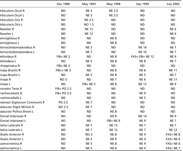
absence) and also the slow rate of disease progres-sion only affecting inferior motoneurons. Chronic reinnervation was found in cranial nerve muscles
(Orbicularis Oculi, Orbicularis Oris, Masseter, Genio
-glossus and S t e r n o c l e i d o m a s t o i d e u s), cervical mus-cles (Infraspinatus, Deltoideus, Biceps Brachii, Triceps, Pronator Teres, Extensor Digitorum Commu -nis, Abductor Digiti M i n imi, Abductor Pollicis Brevis, I Dorsal Interossei) and lumbosacral muscles (Va s t u s Lateralis, Tibialis Anterior and Vastus Lateralis) . Fibrillation potentials (active dennervation) were found in right I n f r a s p i n atus, Deltoideus, Biceps Bra chii, Triceps, Pronator Teres and Brachioradialis mus -c l e s) in the first ENMG study (1989). These findings could not be attributed to the cervical trauma at that time. After 1993, when motor neuron disea-se was suspected, fibrillation potentials were found only in left Tr i c e p s, left I Dorsal Interossei and right
Table 2. Electromyography (needle examination).
Dec 1989 May 1993 May 1998 Sep 1999 July 2003
Orbicularis Oculi R ND NE 3 NE 3.5 ND ND
Orbicularis Oculi L ND NE 3 NE 3.5 ND ND
Orbicularis Oris R ND NE 2.5 ND ND ND
Orbicularis Oris L ND NO 1.5 ND ND ND
Masseter R ND NE 12 ND ND NE 6
Masseter L ND NE 12 ND ND NE 6
Genioglossus R ND ND NE 8 ND ND
Genioglossus L ND ND NE 8 ND ND
Sternocleidomastoideus R ND NE 3 ND NE 16 NE 7
Sternocleidomastoideus L ND NE 7 ND NE 10 NE 7
Deltoideus R FIB+ NE 2 ND NE 8 FAS+ FIB+ NE 12 NE 9
Deltoideus L ND NE 5 NE 8 NE 8 NE 7
Infraspinatus R FIB+ NE 2 ND ND ND ND
Biceps Brachii R FIB++ NE 3 ND NE 8 NE 8 NE 11
Biceps Brachii L ND NE 5 NE 8 NE 5 NE 7
Triceps R NO 3 ND NE 7 NE 6 NE 17
Triceps L ND FIB+ NE 5 NE 8 NE 12 NE 9
Pronator Teres R FIB+ PO 2.5 ND ND ND ND
Brachioradialis R FIB+ PO 2.5 ND ND NE 9 ND
Brachioradialis L ND ND ND NE 5 ND
Extensor Digitorum Communis R PO 2.5 NE 7 ND ND ND
Abductor Digiti Minimi R NO 2.5 NE 7 ND ND ND
Abductor Pollicis Brevis L ND ND NE 8 ND ND
I Dorsal Interossei R ND ND NE 8 NE 14 NE 9
I Dorsal Interossei L ND ND FIB+ NE 9 NE 9 NE 7
Vastus Lateralis R ND NE 7 NE 15 NE 7 NE 7
Vastus Lateralis L ND NE 7 NE 12 NE 7 NE 12
Tibialis Anterior R ND NO 2 NE 8 NE 4 FAS+ NE 8
Tibialis Anterior L ND NE 3 NE 4 NE 4 FAS+ NE 6
Gastrocnemius R ND NE 5 NE 8 NE 4 FAS+ NE 6
Gastrocnemius L ND NE 5 NE 8 NE 9 FAS+ NE 7
Deltoideus muscles. Fasciculation potentials were found only in right Deltoideus and bilateral Gas
-trocnemius/Tibialis Anteriormuscles. We did not
find complex repetitive discharges as emphasized by others2. We concluded that the needle
exami-nation main finding was chronic reinnervation not precisely related to clinical picture reflecting very slow motor neuron degeneration with effective reinnervation. Sensory nerve conduction revealed low amplitude or absent SNAP and seemed to have an axonal loss progression. An interesting find-ing was the asymmetry of the findfind-ings in some ner-ves (e.g. no response in right and low amplitude in left for median and radial nerves). In clinical prac-tice significantly reduced SNAP amplitude in a pa-tient with clinical features consistent with M N D disease requires an explanation and any other cau-se, such as Kennedy’s diseacau-se, should be considered8.
Kennedy’s disease is considered as a slow pro-gressive form of MND symmetrically involving bul-bar and spinal motor neurons associated with testi-cular atrophy and gynecomastia2,4. The age of
on-set is earlier than for most MND being between 45-50 years2 , 4. but both disorders may not have
clas-sic findings and may be misdiagnosed4. The CAG
expansion repeat may be related to the age of on-set, being earlier when there are longer lengths o f r e p e a t s4. Although the electrophysiological findi n g s
are similar to those of MND, greater symmetry of findings and clear bulbar abnormalities are help-ful in distinguishing the disorders. Some patients have low amplitude/absent SNAP without sensory symptoms either negative or positive. Needle exam-ination shows large motor unit action potentials a n d scattered fibrillation potentials4. Early muscle c r a m p s ,
fasciculations, hand tremor, elevated CK, dysarthria, dysphagia and weakness in face, tongue and prox-imal limb muscles, are also described to be freq u e n t2 , 9.
Kennedy’s disease was studied in the province of Reggio Emilia in Northern Italy from 1980-1997. The mean incidence was 0.19 cases/100,000 for t h e male population; the average age at onset was 44.8 ± 10.1 and the average survival period was 27.3 ± 2.3 years. Whereas the incidence rate of Kennedy’s disease was 16 times lower than that of ALS, the incidence rate of progressive bulbar palsy in the m a l e population is only slightly higher than Kennedy’s disease10. Because of the presence of sporadic
cas-es or non-evident familial cascas-es, it is appropriate to consider this diagnostic possibility in making a d i a g-nosis of ALS in patients in whom lower motor neu-ron dysfunction or bulbar onset predominates1 0 , 1 1.
Our patient is a 55-year-old, presenting a unila-teral vocal fold and pharyngeal paresis, clinically reversible after a few months probably because of an active dennervation bout followed by an eff ic i e n t reinnervation; cramps were not a main complaint; fasciculations were evident in clinical examination and scattered electrophysiologically; CK was mildly elevated. In spite of the asymmetry of the initial symptoms, further clinical follow-up had revealed a symmetric complaint. Different from literature, t h e needle examination had showed a diffuse chron-ic reinnervation in cranial nerves muscles and proxi-mal/distal upper and lower limbs. In Brazil, the f i r s t two cases were reported by Seefeld et al.6; the s e
n-sory nerve conduction was normal, the muscles s t u-died in needle examination were restricted to low-er and upplow-er limbs and no molecular genetic study was done at that time (1995). In the other hand, there were elevated CK, gynecomastia, cramps, t r e-mor and slow progression of dennervation on low-er motoneurons, typical findings in Kennedy´s dise-ase. After that Kaimen-Maciel et al.7reported t h r e e
cases and in only one they could find abnormal elec-tromyography (spinal, medulla and pons motoneu-rons) without any details; nerve conduction was re-ferred as normal. All three cases had gynecomas-tia and abnormal molecular genetic study.
Expansions of unstable trinucleotide (CAG) repeats cause at least 15 inherited neurological diseases of which Kennedy’s disease was the first described1 2. It
also includes oculopharyngeal muscular dystrophy and myotonic dystrophy. Because of the signs of an-drogen insensitivity, the anan-drogen receptor became the candidate gene for Kennedy’s disease after the disease was mapped in the region of the X chromo-some in which this gene is located. The first exon of the androgen receptor contains a polymorphic CAG repeat that normally encodes 11 to 33 glutamines. In patients with Kennedy’s disease, this CAG repeat is expanded to encode a lengthened polyglutamine tract of 38 to 62 residues. Patients with longer polyg-lutamine expansions tend to present symptoms at an earlier age1 2. The mutated protein has an
expand-ed polyglutamine tract, forms intranuclear aggrega-tes and mediaaggrega-tes neurodegeneration through a tox-ic gain-of-function mechanism1 2.
slight asymmetry; third, a sub-clinical or subtle in-volvement of proximal/distal muscles of both upper and lower limbs; fourth, a probable evolution with bouts of acute dennervation, followed by an effi-cient reinnervation.
REFERENCES
1. Antonini G, Gragnani F, Romaniello A, et al. Sensory involvement in spinal-bulbar muscular atrophy (Kennedy’s disease). Muscle Nerve 2000;23:252-258.
2. Meriggioli MN, Rowin J, Sanders DB. Distinguishing clinical and elec-trodiagnostic features of X-linked bulbospinal neuronopathy. Muscle Nerve 1999;22:1693-1697.
3. Serratrice G, Pellissier JF, Pouget J. Neuronopathie bulbo-spinale liee a l’X: syndrome de Kennedy. Rev Neurol (Paris) 1988;144:756-758. 4. Daube JR. Electrodiagnostic studies in amyotrophic lateral sclerosis and
other motor neuron disorders. AAEM Minimonograph #18. Muscle Ner-ve 2000;23:1488-1502.
5. Nagashima T, Seko K, Hirose K, et al. Familial bulbo-spinal muscular a t rophy associated with testicular atrophy and sensory neuropathy (Ken-n e d y - A l t e r-Su(Ken-ng sy(Ken-ndrome): autopsy case report of two brothers. J Neurol Sci 1988;87:141-152.
6. Seefeld M, Cunha FM, Ferraz LE, Scola RH, Werneck LC. Doença de Kennedy: relato de dois casos. Arq Neuropsiquiatr 1995;53:471-474. 7. Kaimen-Maciel DR, Medeiros M, et al. Atrofia muscular bulbo espinal
recessiva ligada ao cromossomo X (doença de Kennedy): estudo de uma família. Arq Neuropsiquiatr 1998;56:639-645.
8. Eisen A, Swash M. Clinical neurophysiology of ALS (review). Clin Neurophysiol 2001;112:2190-2201.
9. Szabo A, Mechler F. A K e n n e d y - s z i n d roma-bulbospinalis izomatro p h i a . Ideggyogy Sz 2002;55:323-329.
10. Guidetti D, Sabadini R, Ferlini A, Torrente I. Epidemiological survey of X-linked bulbar and spinal muscular atrophy, or Kennedy disease, in the province of Reggio Emilia, Italy. Eur J Epidemiol 2001;17:587-591. 11. Wilde J, Moss T, Thrush D. X-linked bulbo-spinal neuronopathy: a family study of three patients. J Neurol Neurosurg Psychiatry 1987; 50:279-284.