1Pontifícia Universidade Católica do Rio Grande do Sul (PUCRS), Porto Alegre RS, Brazil: 1Interventional Neuroradiology Service, Hospital São Lucas PUCRS; 2Professor of Neurology, School of Medicine, PUCRS. Eduardo Raupp was the recipient of a scholarship awarded by Conselho de Aperfeiçoamento de Pessoal de Ensino Superior (CAPES).
Received 20 February 2004, received in final form 21 July 2004. Accepted 18 September 2004.
Dr. Eduardo F. Raupp - Rua André Puente 200/301 - 90035-150 Porto Alegre RS - Brasil. E-mail: [email protected]
DOES TREATMENT WITH N-BUTYL CYANOACRYLATE
EMBOLIZATION PROTECT AGAINST HEMORRHAGE
IN CEREBRAL ARTERIOVENOUS MALFORMATIONS?
Eduardo Floriani Raupp
1, Jefferson Fernandes
2ABSTRACT - Objective:To assess the role of this procedure to prevent hemorrhage in cerebral arteriovenous malformations (cAVM). Method:Between 1992 and 2000, we studied 104 patients submitted to emboliza-tion as the main treatment. Patients were followed until hemorrhage or death. Results:Follow-up ranged from 1.6 months to 8 years. The most frequent presentations were hemorrhage (50%) and seizures (38%). In addition, 40% were small (<30 mm); 56% were medium (30-60 mm). Obliteration was ≤1/3 in 11% of the cases; from 1/3 to ≤2/3 in 49%; >2/3 in 36%; complete in 5%. The risk of death was 1%/year, and of bleed-ing, 5.4%/year. Presentation with hemorrhage and low obliteration rate were the main factors associated with hemorrhage. Conclusion:cAVM embolization provides limited protection against hemorrhage with obliteration rates below 2/3. Presentation with hemorrhage is the main factor for predicting hemorrhage. KEY WORDS: therapeutic embolization, intracranial hemorrhage, bucrylate, intracranial arteriovenous malformation.
Embolização com N-butil cianoacrilato confere proteção contra hemorragia em malformações arteriovenosas cerebrais?
RESUMO - Objetivo:Avaliar o papel deste procedimento na prevenção de hemorragia em casos de malforma-ção arteriovenosa (MAV) cerebrais. Método:Entre 1992 e 2000, estudamos 104 pacientes submetidos a embolização como tratamento principal. Os pacientes foram seguidos até a ocorrência de hemorragia ou morte. Resultados:O período de seguimento variou de 1,6 mês a 8 anos. As apresentações mais freqüentes foram hemorragia (50%) e convulsões (38%). Além disso, 40% das lesões eram pequenas (<30 mm); 56% eram médias (30-60 mm). O grau de obliteração foi ≤1/3 em 11% dos casos; 1/3 a ≤2/3 em 49%; >2/3 em 36%; completa em 5%. O risco de morte foi 1%/ano, e de sangramento, 5,4%/ano. Apresentação com hemorra-gia e baixo grau de obliteração foram os principais fatores associados com hemorrahemorra-gia. Conclusão:A em-bolização em MAVs cerebrais confere proteção limitada contra hemorragia, com graus de obliteração abaixo de 2/3. Apresentação com hemorragia é o principal fator preditivo de hemorragia.
PALAVRAS-CHAVE: embolização terapêutica, hemorragia intracraniana, bucrilato, malformação arterioveno-sa intracraniana.
Cerebral arteriovenous malformations (cAVM) are characterized by a plexus of histologically undif-ferentiated vessels in which veins and arteries are directly connected without the usual intervening capillary network. These congenital lesions usual-ly affect otherwise healthy individuals, and are most often diagnosed by the age of 40 years. The usual symptoms are hemorrhage (about 50% of the cases), seizures (30%), headaches (20%), progres-sive neurological deficit (5%), and other less com-mon presentations (5%)1. The risk of hemorrhage varies from 2 to 4% per year, and may reach 18%
after a first bleeding episode1-4. Hemorrhage is usu-ally associated with 10 to 15% morbidity in cAVM, and this rate increases after each recurrence4-6. The mortality rate in patients with untreated cAVM has been estimated at 1% per year7.
per-fusion pressure, and presence of aneurysms2,3,10-13. Some investigators claim that presentation with sei-zures may be a protective factor against hemor-rhage in untreated cAVMs.1,3,14 Hemorrhage can only be prevented by total obliteration or excision of the lesion, since residual pathologic vessels may cause bleeding8,15. Several studies have also report-ed that embolization in cAVM may be useful in the management of patients presenting hard-to-con-trol seizures, progressive neurological deficit, and headaches, and to improve cerebral perfusion8,9,16. In addition, embolization has been thought to be useful in the elimination of aneurysms, which are independent risk factors for hemorrhage8,12,16-19. However, there are no conclusive data concerning the benefits of this procedure. In addition, since embolization is usually performed as a preopera-tive measure20, there is a dearth of data concern-ing the effects of embolization alone on preven-tion of hemorrhage in cAVM.
Therefore, the objective of the present paper was to assess the role of embolization to prevent hemorrhage and the association of this procedure with several known potential risk factors in a pop-ulation of patients submitted to embolization alo-ne, or submitted to surgery more than two months after the performance of embolization.
METHOD
The present study was carried out from January 1992 to June 2000. We studied a cohort of 104 patients who a) were submitted to embolization only because this was the indicated treatment; b) had an indication for em-bolization followed by surgery or radiosurgery but aban-doned this initial protocol and were submitted to em-bolization only; or c) underwent surgery more than two months after embolization. All presented plexiform pial cAVM. No control group was established for ethical rea-sons (it would be unethical not to treat patients at risk for hemorrhage). Patients who had been previously submitted to surgery or radiation therapy were exclud-ed. The study was approved by the appropriate Bioethics Research Committees. All patients provided written informed consent.
Data were collected for each patient starting with the first embolization session until the occurrence of ei-ther hemorrhage or death or until their last clinical eva-luation. Patients who did not present either outcome until the last clinical evaluation were censored from the analy-sis. Demographic data (sex and age) and information about clinical presentation were collected during anam-nesis or from the patient’s medical chart. We also collec-ted data about cAVM topography, size, angiographic aspects (venous drainage and association with aneurysms)
and percentage of obliteration after the last emboliza-tion session.
All embolizations were carried by the same profes-sional (E.F.R.) and employed catheters (mostly flow-di-rected) with or without a microguidewire system. For all procedures, a mixture of NBCA (Histoacryl B, Braun, Mel-sugen AG, Germany) and lipiodol (Lipiodol UF, Guerget, France) without tantalum was used. Until 1995, pedicu-lar injection and the full-column injection technique were used. After that, intranidal injection with induced hypo-tension was used whenever possible (considering the whole study period, pedicular injection was used in 29 patients, intranidal injection in 62, and in 13 patients both proce-dures were used).
Since the assessment of cAVM obliteration is subjec-tive (based on visual inspection), we divided each lesion into three-thirds, and patients were classified into four categories according to the rate of obliteration achieved:
≤1/3; >1/3 to ≤2/3; >2/3; or complete.
Statistical analysis –Cumulative incidence, density of incidence and their confidence intervals were calcula-ted for the occurrence of death or hemorrhage. In addi-tion, Kaplan-Meier curves were plotted, and measures of association between the outcomes of interest and po-tential risk factors were calculated, as well as relative risk (RR) and confidence intervals. The chi-square test was used to determine the significance of results. Cox proportion-al hazards regression modeling was used to evproportion-aluate the effect of risk factors selected on the bivariate analysis on the occurrence of hemorrhage. The significance leve-ls were α=0.05 for the bivariate analysis and α=0.10 for the multivariate analysis. Data were processed and analy-zed using the SPSS 9.0 software.
RESULTS
Of the 104 patients assessed, 57% were male, and age (mean ± standard deviation) was 30±13 years. The most common clinical presentation was hemorrhage (50%), followed by seizure (38%), headache (9%), and other (3%). Lesion topogra-phy was frontal in 29% of cases, parietal in 23%, temporal in 19%, deep in 17%, infratentorial in 7%, and occipital in 5%. In relation to size, most lesions were small (<30 mm) and medium (30-60 mm), with 40% and 56%, respectively.
considering only the 98 patients who underwent partial embolization, the density incidence was 5.6 (non significant).
Twenty patients presented 23 aneurysms; 16 of these aneurysms (70%) were intranidal. The ra-te of oblira-teration achieved with embolization was
≤1/3 in 11% of the cases; from 1/3 to ≤2/3 in 49%; >2/3 in 36%; and complete in 5%. During the cour-se of treatment with embolization, there were 18 cases (22.5%) of early complications (up to 72 hours after the procedure), distributed as follows: three cases (17%) of hemorrhagic complications that did not require surgery; 11 cases (61%) of ischemic
com-Fig 1. Occurrence of hemorrhage following embolization in cAVM patients.
plications; and four (22%) cases of seizures. Most of these complications were transient, but in three cas-es they rcas-esulted in permanent neurological deficit. Figure 1 shows that the rate of occurrence of hemorrhagic events was relatively constant along the first four years. From that point on, the hemor-rhage rate decreased significantly.
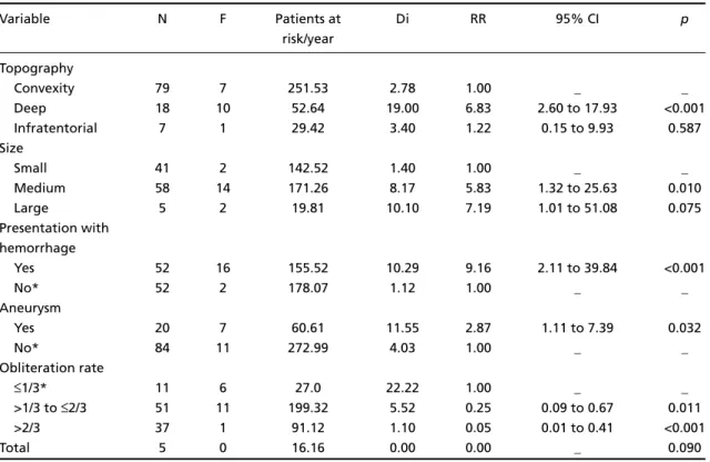
Table 1 shows that the factors that were most significantly associated with post-embolization in the bivariate analysis were deep topography, medi-um or large cAVMs, presentation with hemorrha-ge, presence of aneurysm, and low obliteration ra-te. Figure 2 shows the association between death or hemorrhage and presentation with hemorrha-ge, topography and obliteration rate.
The Cox proportional hazards regression mod-el (Table 2) confirmed the results of the bivariate analysis concerning low obliteration rate and pres-entation with hemorrhage, which remained as high risk factors. However, the relative risk of deep topography was significantly reduced, from high to moderate. Moreover, the presence of aneurysm (moderate relative risk on bivariate analysis) had its effect fully explained (RR≅1.0) after adjustment for the variables included in the multivariate analysis.
DISCUSSION
The prevention of hemorrhage is the main goal of treating cAVMs. Currently, cAVMs are usually treated with embolization followed by surgery or complementary obliteration with radiosurgery8. According to Spetzler and Martin, preoperative em-bolization of small and superficial cAVMs located in surgically accessible noneloquent locations brings little benefit21. However, it is important to note that
Fig 2. Occurrence of hemorrhage following embolization in cAVM patients according to A) presentation with (squares) or without (circles) hemorrhage; B) convexity (squares), infratentorial (triangles), or deep (circles) topography; and C) obliteration rate ≥2/3 (triangles); >1.3 or <2/3 (squares); or ≤1/3 (circles).
it is exactly in small lesions that embolization has the highest rates of cure8,22.
Luessenhop16observed that partial embolization may reduce the risk of hemorrhage by reducing in-tranidal pressure. However, the reports found in
the literature so far are inconclusive concerning the protective role of partial embolization7,12,16,18,23, since any residue of malformed vessels has the poten-tial to cause future bleeding, no matter what per-centage of the lesion was obliterated15,18,22,23. Partial embolization has also been thought to increase the risk of cAVM rupture, even in patients without a pre-vious hemorrhagic event8,10.
In the present cohort, the 5% yearly rate of bleeding recurrence was not significantly different from the 4% found by Ondra et al.6in a 24-year follow-up study of untreated cAVMs. The 1% mor-tality rate in our series was also similar to the rates reported by other authors for the natural history of cAVMs7. In addition, we observed that seizures were indeed a factor of protection against hemor-rhage, and that the factors that were most signifi-cantly associated with subsequent hemorrhage we-re pwe-resentation with hemorrhage and low oblitera-tion rate. The associaoblitera-tion between deep topogra-phy and hemorrhage was still present on multivaria-te analysis, although the power of the association was reduced.
Presentation with hemorrhage is generally accep-ted to be a strong predictor of bleeding recurrence in untreated cAVMs1,3,4,22. Some authors, however,
Table 1. Bivariate analysis of the correlation between hemorrhage after embolization in cAVM patients and sever-al variables
Variable N F Patients at Di RR 95% CI p
risk/year
Topography
Convexity 79 7 251.53 2.78 1.00 _ _
Deep 18 10 52.64 19.00 6.83 2.60 to 17.93 <0.001
Infratentorial 7 1 29.42 3.40 1.22 0.15 to 9.93 0.587
Size
Small 41 2 142.52 1.40 1.00 _ _
Medium 58 14 171.26 8.17 5.83 1.32 to 25.63 0.010
Large 5 2 19.81 10.10 7.19 1.01 to 51.08 0.075
Presentation with hemorrhage
Yes 52 16 155.52 10.29 9.16 2.11 to 39.84 <0.001
No* 52 2 178.07 1.12 1.00 _ _
Aneurysm
Yes 20 7 60.61 11.55 2.87 1.11 to 7.39 0.032
No* 84 11 272.99 4.03 1.00 _ _
Obliteration rate
≤1/3* 11 6 27.0 22.22 1.00 _ _
>1/3 to ≤2/3 51 11 199.32 5.52 0.25 0.09 to 0.67 0.011
>2/3 37 1 91.12 1.10 0.05 0.01 to 0.41 <0.001
Total 5 0 16.16 0.00 0.00 _ 0.090
* Reference category
Table 2. Risk of hemorrhage following embolization in cAVM patients associated with several factors adjusted by multivari-ate Cox proportional hazards modeling.
Variable RR 90% CI p
Obliteration rate
≤1/3* 1.00 _ _
>1/3 to ≤2/3 0.36 0.15 to 0.87 0.055 >2/3 0.08 0.01 to 0.46 0.019 Topography
Convexity 1.00 _ _
Deep 2.72 1.10 to 6.77 0.069
Infratentorial 1.04 0.17 to 6.31 0.971 Presentation with
hemorrhage
Yes 5.00 1.31 to 19.00 0.047
No* 1.00 _ _
Presence of aneurysm
Yes 0.94 0.37 to 2.41 0.921
No* _ _ _
have not observed this association18. On the other hand, presentation with seizures has been thought to be a factor of protection against bleeding1,14. In our series, out of 39 patients with seizures, only two had subsequent hemorrhage. In fact, careful analy-sis of angiographic studies for these cases showed evidence of accidental occlusion of drainage veins during NBCA embolization without immediate clin-ical effect. Therefore, in agreement with the litera-ture, our results support the notion that nonhem-orrhagic patients presenting with seizures are protec-ted against later bleeding. Since the only two cases of hemorrhage in this group probably resulted from accidental occlusion of draining veins, we believe that partial embolization does not have any negative in-fluence on the protective role of seizures.
Concerning obliteration rate, the multivariate analysis clearly shows that lesions with less than 2/3 obliteration had proportionally higher rebleed-ing rates. Gobin did not observe this association in partially embolized patients who later underwent radiosurgery, but this might be explained by the fact that in that study a multivariate technique was not used for data analysis22. Our patients with less than 1/3 obliteration bled even more often than ex-pected had the lesion been untreated. Szikora24also observed that patients with a low degree of oblit-eration had significantly higher hemorrhage rates than those expected for untreated lesions. For oblit-eration rates higher than 2/3, we observed a reble-eding rate that was significantly lower than that expected in untreated lesions. None of the cAVMs with full angiographic occlusion hemorrhaged du-ring follow-up, which is in agreement with other reports25. The higher bleeding rate in patients with a low degree of obliteration may be explained by intranidal hemodynamic changes with increased blood flow to regions prone to rupture. According to Pellettieri, partial obliteration may expose an occult compartment of the lesion that is not adapt-ed to hyperflow and may consequently rupture23. Findings concerning the topography of cAVM indicate that deep lesions are usually associated with presentation with hemorrhage, having a mo-re aggmo-ressive clinical course and a tendency towards recurring hemorrhages (recurrence rate may reach 30%)2,20. After adjustment for several relevant fac-tors, the association between deep lesion topog-raphy and occurrence of subsequent hemorrhage was confirmed (when compared to other loca-tions), although the risk was reduced. This may be explained by a higher perfusion pressure, short arte-rial pedicles and venous drainage in deep lesions10,26.
Associated aneurysms, especially intranidal, are also considered a factor of increased risk for hem-orrhage in untreated cAVMs2,12,27,28. The occlusion of aneurysms has been thought to confer relative protection2,12,29. but this was not what we observed in the present study, in which patients presented he-morrhage even after aneurysm occlusion. However, after controlling the effects of presentation with hemorrhage (Cox regression model), the relative risk associated with aneurysms practically disappea-red. This suggests that in patients with cAVM, he-morrhage at presentation has a more important role in the occurrence of subsequent hemorrhage than the presence of an intranidal aneurysm. Thus, it seems that occlusion of intranidal aneurysms in patients presenting with hemorrhage does not have an impact on subsequent bleeding. This may be a result of factors associated with rupture, which have not yet been completely explained. Also, in-tranidal aneurysms may be the consequence, and not the cause, of hemorrhage at presentation. Other authors have already followed a similar line of reasoning2,29. We also agree with Redekop et al.28 and Stein, apud Turjman et al.27in that the angio-graphic diagnosis of intranidal aneurysm is subjec-tive and operator-dependent. Therefore, we again highlight the role of presentation with hemorrha-ge as the factor of highest relevance in the occur-rence of subsequent hemorrhagic events.
REFERENCES
1. Crawford PM, West CR, Chadwick DW, Shaw MDM. Arteriovenous malformations of the brain: natural history in unoperated patients. J Neurol Neurosurg Psychiatry. 1986;49:1-10.
2. Marks MP, Lane B, Steinberg GK, Chang PJ. Hemorrhage in intracere-bral arteriovenous malformations: angiographic determinants. Radio-logy. 1990;176:807-813.
3. Mast H, Young WL, Koennecke HC, et al. Risk of spontaneous haem-orrhage after diagnosis of cerebral arteriovenous malformations. Lancet 1997;350:1065-1068.
4. Hartmann A, Mast H, Mohr JP, et al. Morbidity of intracranial hemor-rhage in patients with cerebral arteriovenous malformations. Stroke 1998;29:931-934.
5. Graf CJ, Perret GE, Torner JC. Bleeding from cerebral arteriovenous mal-formations as part of their natural history. J Neurosurg. 1983;58:331-337. 6. Ondra SL, Troupp H, George ED, Schwab K. The natural history of symp-tomatic arteriovenous malformations of the brain: a 24-year follow-up assessment. J Neurosurg. 1990;73:387-391.
7. Viñuela F, Dion J, Lylyk P, Duckwiler G. Update on interventional neu-roradiology. Neuroimaging Clin N Am. 1992;2:279-289.
8. Wikholm G. Role of transarterial embolization in the management of cerebral arteriovenous malformations. Acta Radiologica 1996;37 (Suppl.):S3-S25.
9. Al-Yamany M, Terbrugge KG, Willinsky R, Montanera W, Tymianski, Wallace MC. Palliative embolisation of brain arteriovenous malformations with progressive neurological deficit. Interv Neuroradiol 2000;6:177-183. 10. Spetzler RF, Hargraves RW, McCormick PW, Zabranski JM, Flom RA, Zimmerman RS. Relationship of perfusion pressure and size to risk of hemorrhage from arteriovenous malformations. J Neurosurg. 1992; 76:918-923.
11. Kader A, Young WL, Pile-Spellman J, et al. The influence of hemodynam-ic and anatomhemodynam-ic factors on hemorrhage from cerebral arteriovenous mal-formations. Neurosurgery 1994;34:801-808.
12. Lasjaunias P, Piske R, TerBrugge K, Willinsky R. Cerebral arteriovenous malformations (C.AVM) and associated arterial aneurysms (AA). Acta Neurochir (Wien) 1988;91:29-36.
13. Mansmann U, Meisel J, Brock M, Rodesch G, Alvarez H, Lasjaunias P. Factors associated with intracranial hemorrhage in cases of cerebral arte-riovenous malformation. Neurosurgery. 2000;46:272-281.
14. Forster DMC, Steiner L, Hakanson S. Arterionevenous malformations of the brain: a long-term clinical study. J Neurosurg 1972;37:562-570. 15. Guo WY, Karlsson B, Ericson K, Lindqvist M. Even the smallest
rem-nant of an AVM constitutes a risk of further bleeding: case report. Acta Neurochir (Wien) 1993;121:212-215.
16. Luessenhop AJ, Mujica PH. Embolization of segments of the circle of Willis and adjacent branches for management of certain inoperable cere-bral arteriovenous malformations. J Neurosurg. 1981;54:573-582. 17. Marks MP, Lane B, Steinberg GK, Sniper GJ. Intranidal aneurysms in
cerebral arteriovenous malformations: evaluation and endovascular treatment. Radiology 1992;183:355-360.
18. Lundqvist C, Wikholm G, Svendsen. Embolization of cerebral arteri-ovenous malformations: Part II. Clinical aspects on complications and late outcome. Neurosurgery 1996;39:460-469.
19. Luessenhop AJ, Presper JH. Surgical embolization of cerebral arteriove-nous malformations through internal carotid and vertebral arteries. J Neurosurg. 1975;42:443-451.
20. Lawton MT, Hamilton MG, Spetzler RF. Multimodality treatment of deep arteriovenous malformations: thalamus, basal ganglia, and brain stem. Neurosurgery 1995;37:29-36.
21. Spetzler RF, Martin NA. A proposed grading system for arteriovenous malformations. J Neurosurg 1986;65:476-483.
22. Gobin YP, Laurent A, Merienne L, et al. Treatment of brain arteriove-nous malformations by embolization and radiosurgery. J Neurosurg 1996;85:19-28.
23. Pellettieri L, Svendsen P, Wikholm G, Carlsson CA. Hidden compart-ments in AVMs-A new concept. Acta Radiol 1997;38:2-7.
24. Szikora I, Barath K, Vadja J, Czirják S, Futó J, Nyáry I. Bleeding frequen-cy following embolization of brain AVMs. Neuroradiology 1999; 41(Suppl):S45.
25. Wikholm G, Lundqvist C, Svendsen. Transarterial embolization of cerebral arteriovenous malformations. Improvement with experience. Am J Neuroradiol 1995;16:1811-1817.
26. Batjer H, Samson D. Arteriovenous malformations of the posterior fos-sa: clinical presentation, diagnostic evaluation, and surgical treatment. J Neurosurg 1986;64:849-856.
27. Turjman F, Massoud TF, Viñuela F, Sayre JW, Guglielmi G, Duckwiler G. Correlation of the angioarchitectural features of cerebral arteriove-nous with clinical presentation of hemorrhage. Neurosurgery. 1995;37:856-862.
28. Redekop G, TerBrugge K, Montanera W, Willinsky R. Arterial aneurysms associated with cerebral arteriovenous malformations: classification, inci-dence, and risk of hemorrhage. J Neurosurg 1998;89:539-546. 29. Monaco RG, Rodesh G, Alvarez H, Iizuka Y, Hui F, Lasjaunias P.