University of Taubaté, São Paulo SP, Brazil (UT), Faculty of the Medicine of the University of São Paulo, São Paulo SP, Brazil (FMUSP) and Biomedical Science Institute of the University of São Paulo São Paulo SP, Brazil (BSI): 1MD, Master in Neurology, Medicine Department, UT; 2Biochemistry, Phd, Department of Immunology, BSI; 3Biochemistry, Department of Immunology, Phd, BSI; 4MD, Phd, Department of Neurology, FMUSP; 5MD, Phd, Department of Neurology, FMUSP.
Received 4 March 2004, received in final form 4 May 2004. Accepted 9 June 2004.
Dr. Ronaldo Abraham - Rua Dr. Souza Alves 364 - 12020-030 Taubaté SP - Brasil. E-mail: [email protected]
TAENIA ANTIGENS DETECTION IN THE
CEREBROSPINAL FLUID OF PATIENTS WITH
NEUROCYSTICERCOSIS AND ITS RELATIONSHIP
WITH CLINICAL ACTIVITY OF THE DISEASE
Ronaldo Abraham
1, Alessandra Xavier Pardini
2, Adelaide José Vaz
3,
José Antonio Livramento
4, Luís dos Ramos Machado
5ABSTRACT - Objective:(1) To determine the concentration of Taeniaantigens in the cerebrospinal fluid (CSF) of patients with neurocysticercosis (NC); (2) to establish its relationship with clinical activity of the disease and with classical variables of CSF. Method: A CSF examination was performed in one sample from 36 patients with definitive diagnosis of NC, including: quantitative and cytomorphological study, bio-chemical tests, immunological reactions for cysticercosis and Taenia antigens. The antibodies for antigens detection were obtained from the larval form of Taenia crassiceps, ORF strain. After intraperitoneal pas-sage through female mice, a group of rabbits was immunized with vesicular fluid antigens. Results: The
Taeniaantigen was detected in CSF from 17 patients (47.2%), especially in those patients with epileptic symptoms in the last 12 months. Conclusion: Taenia antigens presence in CSF have significant relation-ship with clinically active forms of NC, being a more sensitive marker than the classic eosinophil presence.
KEY WORDS: neurocysticercosis, Taeniaantigens, neurocysticercosis clinical activity.
Dosagem de antígenos de Taeniano líquido cefalorraquidiano em pacientes com neurocisticer-cose e sua relação com a atividade clínica da doença
RESUMO -Objetivo: (1) Determinar a concentração de antígenos de Taeniano líquido cefalorraquidiano (LCR) em doentes com neurocisticercose; (2) estudar sua relação com a atividade clínica da doença e com as variáveis clássicas do LCR. Método: Em 36 pacientes com diagnóstico definido de neurocisticercose foi realizado exame do LCR, com estudo citológico e citomorfológico, exame bioquímico, reações imunoló-gicas para cisticercose e detecção de antígenos de Taenia. Os anticorpos para detecção desses antígenos foram obtidos a partir da forma larvar da Taenia crassiceps, cepa ORF. Após a inoculação e proliferação intraperitoneal dessa forma larvária em ratas, foi imunizado um grupo de coelhos com seu líquido vesi-cular. Resultados: Em 17 pacientes (47,2%) foi detectado antígeno de Taenia, especialmente naqueles com manifestação epiléptica nos últimos 12 meses. Conclusão: A detecção de antígeno de Taenia guar-da relação significativa com a vigência de formas clinicamente ativas, sendo, nestas formas, marcador mais sensível que a eosinofilorraquia.
PALAVRAS-CHAVE: neurocisticercose, antígenos de Taenia, atividade clínica da neurocisticercose.
Neurocysticercosis (NC) is defined as the infec-tion of the central nervous system caused by Cysti-cercus cellulosae, the larval stage of Taenia solium1,2 ,
acquired mainly by ingesting eggs of Taenia soli-umhidden in food, especially vegetables and fruits. Despite being considered an eradicable disease3,
it remains a public health challenge for most devel-oping countries4, representing an important
fac-tor in the genesis of epilepsy5,6. NC probably
ex-plains the high tropical countries ranges of active epilepsy, reaching almost twice the level of devel-oped countries7. In the last two decades NC became
an emerging problem in the United States of Ame-rica, where thousands of cases per year are now being reported8,9. In Southern California NC
neurosur-gical admissions, and reflects the importance of im-migrants as carriers of the disease10. In our
coun-try some regions are more affected than others, but the whole country is considered endemic for the disease11. At Ribeirão Preto City, São Paulo
State, Brazil, an estimated prevalence of 71.8 cas-es per 100.000 inhabitants was dcas-escribed1.
The clinical picture of NC is dominated by epilep-tic seizures, but a wide range of neurological symp-toms can occur12-15. Epileptic seizures occur more
often at the transitional stage of the cysts, but can also occur at the calcified stage, the so-called inac-tive form16-18. In a recent consensus proposing
diag-nostic criteria for NC, several images were emphasi-zed and classified as absolute, major and minor cri-teria19. Neuroimaging is strongly applied in the
diagnosis for NC, permitting visualization of the parasite in its different stages20-22. Examination of
cerebrospinal fluid (CSF) may be a valuable diag-nostic tool, providing sensitive information about the inflammatory process and activity of NC23-27.
Re-cently, a methodology able to detect anti-Taenia
antigens was developed, using highly purified an-tibodies against Taenia antigens, showing high sensibility and specificity28.
The purpose of this study is: (1) to determine concentration of anti-Taeniaantigens in cerebros-pinal fluid of patients with neurocysticercosis; (2) to establish its relationship with clinical activity of the disease and with classical variables of CSF.
METHOD
Between July 2002 and March 2003, 36 patients with definitive diagnosis of NC according to consensus diag-nostic criteria19, were attended at the Outpatient Clinic
of Infectious Diseases of the Neurological Clinics of the Hospital of the School of Medicine of University of São Paulo, and at the Outpatient Neurological Clinic of the Hospital of Taubaté, University of Taubaté. The study was developed according to ethical rules in research involv-ing human beinvolv-ings practiced at the Hospital of the School of Medicine of University of São Paulo, and submitted to analysis and approval of the Ethical Comission for Re-search Projects Analysis of that Hospital, under the re-search protocol number 132/03, according to resolution number 196/96 from Health National Council.
Patients were included in this study after signing a consent declaration. Concerning age, 9 patients (25%) were between 21 and 30 years old; 14 patients (38.8%) were between 31 and 40 years old, while 10 patients (27.7%) were between 41 and 50 years old. Only 3 patients (8.3%) were older than 51. Twenty three pa-tients (63.8%) were male. There was a predominance of white patients (30 patients, 83.3%), against 6 negro
pa-tients (16.7%). Thirty papa-tients (83.3%) originated from São Paulo State, while 6 patients (16.7%) came from the States of Minas Gerais and Bahia, three cases each.
Almost all patients (97.2%) presented epilepsy. Patients were classified in six groups, as regards clinical presen-tation and its temporal occurrence: (a) epileptic form, symptomatic in the last twelve months - 17 patients (47.2%); (b) epileptic form, asymptomatic in the last twel-ve months - 14 patients (38.9%); (c) epilepsy plus increa-sed intracranial pressure - 2 patients (5.5%); (d) epilepsy plus cerebrovascular involvement - 1 patient (2.8%); (e) epilepsy plus optic neuritis - 1 patient (2.8%); (f) headache plus psychic desorder - 1 patient (2.8%). All the patients with epilepsy were receiving antiepileptic drugs, even those asymptomatic in the last twelve months.
As regards magnetic resonance imaging, patients ex-hibited at the time of inclusion in the study the follow-ing findfollow-ings: (a) multiple cystic lesions with contrast enhancement in at least one of the lesions in 17 patients (47.3%); (b) multiple cystic lesions with no definite con-trast enhancement in one patient (2.8%); (c) single cys-tic lesion with contrast enhancement in 6 patients (16.6%); (d) multiple nodular lesions in 7 patients (19.4%); (e) sin-gle nodular lesion in 3 patients (8.3%); (f) multiple nodu-lar lesions with hydrocephalus in one patient (2.8%); (g) multiple parenchymal calcifications in one patient (2.8%). A CSF sample was collected by lumbar puncture in sitting position, in order to perform global leukocyte count, cytomorphological profile, biochemical tests (total protein content, adenosine-deaminase activity, protein electrophoresis), IgG class antibodies research for syphilis, toxoplasmosis and cysticercosis (comple-ment fixation test, indirect immunofluorescence, passive hemaglutination and enzyme-linked immunosorbent assay), besides cysticercus antigen research.
Antigens were detected in CSF samples by enzyme-lin-ked immunosorbent assay (ELISA) using polyclonal sera of rabbit anti-Taenia soliumcysticerci and anti-Taenia crassi-cepscysticerci vesicular fluid, as described by Pardini et al28.
A blood sample was also collected from all patients in order to perform a immunoblotting assay for cys-ticercosis.
RESULTS
CSF findings are shown at Table 1.
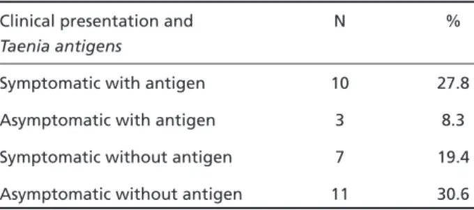
By comparing the clinical presentation of epilep-tic form with Taenia antigen detection, we observed a significant increase in the symptomatic group in the last twelve months as compared to the asympto-matic group (Tables 2 and 3).
DISCUSSION
NC is a disease with multiple clinical presenta-tions12,13and variable evolution profile largely
de-pending on immunological features. The relation-ship between host and parasite is complex. Immune evasion mechanism, besides different levels of local immunodepression, allows a longer and pacif-ic parasite survival within the central nervous sys-tem without producing significant inflammatory reaction9. Usually, clinical activity takes place when
cyst degeneration begins. Often multiple cysts in different phases of evolution coexist in the same patient making clinical management more difficult.
Correct diagnosis per seis not sufficient to de-termine severity, adequate therapeutic regimen and prognosis. It is necessary to know whether the disease is active: (1) under image criteria (cysts with-out enhancement) and (2) under immunological and clinical criteria. While diagnostic procedures are quite developed, disease activity criteria are poor and based almost exclusively on neuroimag-ing16,17. Besides specific anti-Taeniaantibodies which
may persist for long time in CSF, the detection of
Taeniaantigens may be related to the acute phase of the inflammatory activity. This inflammatory activity is closely related with NC clinical activity.
Patients included in this study present peculiar clinical picture, with an absolute preponderance of epileptic form, possibly due to the strict applica-tion of diagnostic criteria defined by the recent con-sensus on NC diagnosis19. These criteria for
defini-tive diagnosis of NC virtually excluded all patients with non-epileptic clinical manifestations, includ-ing the most severe hypertensive forms. The pa-tients here included were often without epileptic crisis in the last 12 months. A few patients had oth-er neurological manifestations. Despite this bias, our patients match the age, gender and race distri-bution referred in the literature. It means that it is a representative population, what allows us to validate the results.
CSF examination shows classical variables for the diagnosis of NC known for several decades:
pleocy-tosis, eosinophilorraquia and the presence of speci-fic antibodies23-25. This last topic has become a very
sensible and specific parameter for the diagnosis, thanks to new techniques introduced in the clini-cal practice, like the enzyme-linked immunosorbant assay and immunoblotting.
In this group of patients, pleocytosis occurred in 16.7% of the cases, eosinophilorraquia in 30.6% and presence of specific antibodies in 75% of the cases; the complete syndrome occurred in only fi-ve patients (13.9%). Neutrophils were obserfi-ved in 63.9 % of the cases, and a high protein content in half of the patients. Nine patients (25%)
present-Table 1. CSF in patients with NC.
CSF N %
Pleocytosis 6 16.7
Presence of neutrophils 23 63.9
Presence of eosinophils 11 30.6
Protein increase 18 50.0
Gamma globulin increase 9 25.0
Positive complement fixation reaction 5 13.9 Positive indirect immunofluorescent test 24 66.7 Positive hemagglutination test 24 66.7 Positive enzyme-linked immunosorbent assay 27 75.0 Positive antigen detection 17 47.2
N, number of cases.
Table 2. Clinical activity of epileptic form in the last twelve months related to antigen detection.
Clinical presentation and N %
Taenia antigens
Symptomatic with antigen 10 27.8
Asymptomatic with antigen 3 8.3
Symptomatic without antigen 7 19.4 Asymptomatic without antigen 11 30.6
N, number of cases.
Table 3. Clinical activity related to antigen detection in patients with NC.
Variable p S/NS
Clinical activity (all patients) vs.antigen detected 0.01 S Epileptic seizures in the last twelve months vs. antigen detected 0.02 S
ed high levels of gamma globulins, but in only one with oligoclonal distribution. These results in-dicate the occurrence of non-cicatricial NC, with poor inflammatory reaction and immunorelease of specific antibodies.
Nowadays, the diagnosis for NC is greatly relat-ed to neuroimaging19 . NC is one of the rare
dis-eases where image morphology permits etiologi-cal diagnostic, as if we could see the parasite. Neu-roimaging also permits follow-up of different phas-es of parasite, from the vphas-esicular until the calcified stages21,22. Nevertheless, neuroimaging
informa-tion is morphological in nature, not funcinforma-tional. The data obtained from neuroimaging are not always proportional to the severity of the disease. Pa-tients with multiple lesions may present asympto-matic, while patients with few images can be pro-foundly ill. Sotelo et al.18tried to establish clinical
activity criteria in order not to treat cicatricial forms of the disease, and predominantly morphological criteria have been adopted. Since then, presence of intact cysts in the brain parenchyma has been a frequent reference to “active forms of NC”. Such reference does not seem reasonable, since inflam-matory activity and clinical manifestations are absent at that time.
The concept of disease activity in patients with the NC diagnosis is relevant, and is not yet well es-tablished. Discrepancies between clinical presenta-tion and image often turn therapeutic decisions dif-ficult. There are no criteria to confirm whether the disease, rather than the image, is active or not.
In the most severe forms of NC, pleocytosis with presence of eosinophils in the CSF is one of the activ-ity criteria related to cyst rupture and consequent antigen release at the nervous system. If we admit that antigen release is related to inflammatory activity, and that inflammatory activity is related to the clinical activity of the disease, we can test the hypothesis that Taeniaantigen detection with inflammatory activity in the CSF is correlated with NC immunological active phase.
Taenia antigen was detected in part of the pa-tients (47.2%), all of them with definitive NC. It excludes the universality of the phenomenon. There is a non- casual statistic relationship between Taenia
antigen and the occurrence of clinically active NC (p = 0.02) among patients with the epileptic form. So, it can be considered a clinical activity marker of the disease, at least in the epileptic form.
Taeniaantigen detection was not statistically re-lated to: (1) pleocytosis; (2) presence of neutrophils; (3) elevated protein content; (4) elevated gamma globulin fraction; (5) presence of specific
anti-Taeniaantibodies (ELISA). These tests did not show concordance with antigen dosage related to its fre-quency (McNemar test) neither to the quantitative variation (regression tests). Nevertheless, the pres-ence of eosinophils is related in a significant way to the occurrence of Taeniaantigen (p=0.006). The-se two determinations must translate the same phe-nomenon but eosinophilorraquia is significantly less sensitive.
We conclude that, in epileptic form of NC, Taenia
antigen dosage may be able to give suitable infor-mation about disease activity, in a more sensitive way than any other classical variable of CSF.
REFERENCES
1. Takayanagui OM. Prevenção da neurocisticercose (CD ROM - Curso Pré-Congresso XX Congresso Brasileiro de Neurologia - setembro/2002, Florianópolis, SC).
2. White AC Jr. Neurocysticercosis: update on epidemiology, pathogen-esis, diagnosis and management. Ann Rev Med 2000;51:187-206. 3. Schantz PM, Cruz M, Sarti E, Pawlowski Z. Potential eradicability of
tae-niasis and cysticercosis. Bull Pan Am Health Organ 1993;27:397-403. 4. Román G, Sotelo J, Del Brutto O, et al. A proposal to declare
neurocys-ticercosis an international reportable disease. Bull World Health Organ 2000;78:399-403.
Table 5. Presence of antibodies (ELISA) related to antigen detec-tion.
Variable r p S/NS
Reactive ELISA and antigen - ~ 0.80 NS (McNemar)
Reactive ELISA and antigen 0.30 0.08 NS (regression analysis)
r, Pearson correlation coefficient; p, associated probability; S, statisti-cally significant value; NS, statististatisti-cally not-significant value.
Table 4. Clinical variables of cerebrospinal fluid related to anti-gen detection: probability study.
Variable p S/NS
Number of cells x antigen 0.052 NS
Neutrophils x antigen 0.55 NS
Eosinophils x antigen 0.006 S
Increased protein content x antigen 0.74 NS Increased gamma globulin fraction x 0.18 NS antigen
ELISA reactive x antigen 0.10 NS
5. Del Brutto OH, Santibañez R, Noboa CA, Aguirre R, Diaz E, Alarcón TA. Epilepsy due to neurocysticercosis: analysis of 203 patients. Neurology 1992;42:389-392.
6. Manreza MLG. Epilepsia e neurocisticercose. In Guerreiro CAM, Guerreiro MM, Cendes F, Lopes-Cendes I (eds). Epilepsia. São Paulo: Lemos Editorial, 2000:255-264.
7. Bittencourt PRM, Adamolekum B, Bharucha N, et al. Epilepsy in the tropics: I. Epidemiology, socioeconomic risk factors, and etiology. Epilepsia 1996;37:1121-1127.
8. Shandera WX, White AC Jr, Chen JC, Diaz P, Armstrong R. Neuro-cysticercosis in Houston, Texas: a report of 112 cases. Medicine 1994;73: 37-52.
9. Carpio A. Neurocysticercosis: an update. Lancet Infect Dis 2002;2:751-762. 10. Schantz PM, Moore AC, Muñoz JL, et al. Neurocysticercosis in an orthodox Jewish community in New York City. N Engl J Med 1992;327:692-695.
11. Agapejev S. Epidemiologia e importância do estudo da neurocisticer-cose em nosso meio (CD ROM - Curso Pré-Congresso XX Congresso Brasileiro de Neurologia - setembro/2002, Florianópolis, SC). 12. Wittig EO. Neurocisticercose: formas clínicas e aspectos
anátomo-patológicos. In Machado LR, Livramento JA, Spina-França A, Nóbrega JPS (eds). Neuroinfecção 96. São Paulo: Clínica Neurológica HC/FMUSP, 1996:193-204.
13. Takayanagui OM, Jardim E. Aspectos clínicos da neurocisticercose: análise de 500 casos. Arq Neuropsiquiatr 1983;41:50-63.
14. Barinagarrementeria F, Cantú C. Frequency of cerebral arteritis in sub-arachnoid cysticercosis: an angiographic study. Stroke 1998;29:123-125. 15. Coelho CMF. Neurocisticercose: crises epilépticas, neuroimagem e líquido cefalorraqueano; estudo de 62 casos. Tese (doutorado) Universidade de São Paulo. São Paulo, 2002.
16. Carpio A, Placencia M, Santillán F, Escobar A. A proposal for classifi-cation of neurocysticercosis. Can J Neurol Sci 1994;21:43-47. 17. Salgado P, Rojas R, Sotelo J. Cysticercosis: Clinical classification based
on imaging studies. Arch Intern Med 1997;157:1991-1997.
18. Sotelo J, Guerrero J, Rubio F. A new classification based on active and inactive forms. Arch Intern Med 1988;148:442-445.
19. Del Brutto OH, Rajshektar V, White AC Jr, et al. Proposed diagnostic criteria for neurocysticercosis. Neurology 2001;57:177-183.
20. Machado LR, Nóbrega JPS, Barros NG, Livramento J A, Baccheschi LA, Spina-França A. Computed tomography in neurocysticercosis: a 10-year long evolution analysis of 100 patients with an appraisal of a new clas-sification. Arq Neuropsiquiatr 1990;48:414-418.
21. Kramer LD, Locke GE, Byrd SE, Daryabagi J. Cerebral cysticercosis: doc-umentation of natural history with CT. Radiology 1989;171:459-462. 22. Dumas JL, Visy JM, Belin C, Gaston A, Goldlust D, Dumas M.
Paren-chymal neurocysticercosis: follow-up and staging by MRI. Neurora-diology 1997;39:12-18.
23. Livramento JA, Machado LR, Spina-França A. Sinalização do líquido cefalorraqueano em doenças inflamatórias crônicas do sistema ner-voso central. Arq Neuropsiquiatr 1986;44:351-358.
24. Machado LR. Líquido cefalorraqueano e neurocisticercose: aspectos evolutivos da resposta inflamatória celular. Arq Neuropsiquiatr 1987;45:353-363.
25. Spina-França A, Livramento J A, Machado LR. Cysticercosis of central nervous system and CSF: immunodiagnosis of 1573 patients in 63 years (1929-1992). Arq Neuropsiquiatr 1993;51:16-20.
26. Bueno EC. Diagnóstico imunológico da neurocisticercose (CD ROM -Curso Pré-Congresso XX Congresso Brasileiro de Neurologia - setem-bro/2002, Florianópolis, SC).
27. Bueno EC, Vaz AJ, Machado LR, Livramento JA, Mielli SR. Specific
Taenia crassicepsand Taenia soliumantigenic peptides for neurocysticer-cosis immunodiagnosis using serum samples. J Clin Microbiol 2000;38:146-151.