THE EXPRESSION OF NGFr AND PGP 9.5 IN
LEPROSY REACTIONAL CUTANEOUS LESIONS
An assessment of the nerve fiber
status using immunostaining
Sérgio Luiz Gomes Antunes
1,2, Yong Liang
3, José Augusto da Costa Neri
1,
Mary Haak-Frendscho
4, Olle Johansson
3ABSTRACT - The effects of reactional episodes on the cutaneous nerve fibers of leprosy patients was assessed in six patients (three with reversal reactions and three with erythema nodosum leprosum). Cryosections of cutaneous biopsy of reactional lesions taken during the episode and of another sample during the remission period were immunostained with anti-NGFr and anti-PGP 9.5 (indirect immunofluorescence). We found no significant statistical difference in the number of NGFr- and PGP 9.5-positive fibers between the reactional and post-reactional groups. A significant difference was detected between the number of NGFr and PGP 9.5-stained fibers inside of the reactional group of biopsy cryosections but this difference was ascribed to the distinct aspects of the nerve fibers displayed whether stained with anti-NGFr or with anti-PGP 9.5; NGFr-positive branches looked larger and so interpreted as containing more fibers. In addition, a substantial number NGFr-positive fibers were PGP 9.5-negative. No differences in the number of stained fibers among the distinct cutaneous regions examined (epidermis + upper dermis, mid and deep dermis) was detected. In conclusion, the number of PGP- and NGFr-positive fibers were not significantly different in the reactional and post-reactional biopsies in the present study. NGFr-staining of the nerve fibers is different from their PGP-imunoreactivity and the evaluation of the nerve fiber status on an innervated target organ should be carried out choosing markers for both components of nerve fibers (Schwann cells and axons).
KEY WORDS: nerve growth factor receptor, protein gene product 9.5, leprosy, neurotrophic factor.
Abbreviations: NGFr: nerve growth factor receptor; PGP 9.5: protein gene product; BB: borderline borderline patient; BL: borderline lepromatous patient; ENL: erythema nodosum leprosum; LL: lepromatous lepromatous patient; MDT: multidrug therapy; TIR: type I reactions; TIIR: type II reactions.
Expressão de NGF Expressão de NGFExpressão de NGF
Expressão de NGFExpressão de NGFr e PGP 9.5 nas lesões cutâneas reacionais da hanseníase: uma avaliação do status dasr e PGP 9.5 nas lesões cutâneas reacionais da hanseníase: uma avaliação do status dasr e PGP 9.5 nas lesões cutâneas reacionais da hanseníase: uma avaliação do status dasr e PGP 9.5 nas lesões cutâneas reacionais da hanseníase: uma avaliação do status dasr e PGP 9.5 nas lesões cutâneas reacionais da hanseníase: uma avaliação do status das fibras nervosas utilizando imunomarcação
fibras nervosas utilizando imunomarcaçãofibras nervosas utilizando imunomarcação fibras nervosas utilizando imunomarcaçãofibras nervosas utilizando imunomarcação
RESUMO - O efeito das reações hansenianas sobre a inervação cutânea de pacientes hansenianos foi avaliado em seis pacientes (três com reação reversa e três com eritema nodoso leprosum). Cortes congelados de biópsias de lesões cutâneas reacionais colhidas na ocasião da reação e de biópsias colhidas após a remissão do quadro reacional na mesma região ocupada previamente pela lesão foram marcados pela técnica de imunofluorescência indireta utilizando os anticorpos anti-NGFr e anti-PGP 9.5. Não foi encontrada diferença significativa na quantificação de fibras positivas para NGFr e para PGP 9.5 entre as biópsias colhidas durante a reação e as biópsias colhidas no período de remissão. Entretanto, no grupo de biópsias da reação houve uma significativa diferença entre a quantidade de fibras NGFr-positivas e as fibras imunomarcadas para PGP 9.5. Essa diferença contudo foi atribuída aos diferentes aspectos que a mesma fibra pode assumir quando marcadas com NGFr ou com PGP 9..5 separadamente. O presente estudo também mostrou que a avaliação das condições das fibras nervosas de um órgão deve ser realizada com marcadores para o axônio e para células de Schwann.
PALAVRAS-CHAVE: receptor do fator de crescimento neuronal, produto protéico do gen 9.5, lepra, fator neurotrópico.
Abbreviações: NGFr: nerve growth factor receptor; PGP 9.5: protein gene product; BB: borderline borderline patient; BL: borderline lepromatous patient; ENL: erythema nodosum leprosum; LL: lepromatous lepromatous patient; MDT: multidrug therapy; TIR: type I reactions; TIIR: type II reactions.
1Oswaldo Cruz Institute, Laboratory of Leprosy, Rio de Janeiro RJ Brazil; 2Iguaçu University, Nova Iguaçu PR Brazil; 3The Experimental
Dermatology Unit, Department of Neuroscience, Karolinska Institute, Stockholm, Sweden; 4Promega Corporation, Madison, WI, USA
Received 20 October 2002, received in final form 20 January 2003. Accepted 27 January 2003.
Leprosy reactions are acute recurrent clinical epi-sodes which occur during the chronic course of the disease. There are two types of leprosy reaction: type I (TIR) and type II (TIIR; erythema nodosum leprosum = ENL, or multiform erythema) reactions1. TIR is
cha-racterized by the appearance of new lesions or reinfiltration of old ones, accompanied by neural symptoms and it is interpreted as a shift of the im-munological status towards the tuberculoid pole (up-grading) or towards the lepromatous pole (down-grading)1. It occurs on the borderline patients of the
immunopathologic spectrum of the disease2. The
emergence of immature tuberculoid granulomas, in-creased lymphocytic infiltration, edema and vascular congestion with bacillary clearing on a borderline lepromatous infiltrate are the histopathological fea-tures of the TIR episode. TIIR (ENL) is clinically charac-terized by the appearance of subcutaneous nodules and general symptoms such as fever, malaise, ocu-lar manifestations, edema and orchitis. On the histo-pathology, TIIR exhibits a characteristic neutrophilic infiltration upon a lepromatous cell infiltrate in the dermal-hypodermal boundary. Edema and nuclear fragmentation generally also occurs1. The triggering
mechanism and the exact meaning of reactions have not been clarified yet, however exacerbation of the immune response has been detected during reactional episodes3-7. Leprosy reactions increase the
morbidity of the disease due to nerve damage and impairment of nerve function8. These undesirable
effects may even occur after the conclusion of multidrug therapy5. Paresthesia, hypoesthesia and
paresia become progressively more intense if treat-ment is not promptly instituted1. Despite the
de-crease on the prevalence of leprosy in the world after the institution of the multidrug therapy, reactions continue to occur and may cause severe nerve da-mage to the patient under treatment or even after its conclusion.
The comprehension of leprosy pathogeny de-pends on an integrated knowledge of the patho-biology of the peripheral nervous system and the study on neuron survival, degeneration and regene-ration which has remarkably progressed in the recent decade is within this large theme9. These
pathobio-logical processes have been studied at molecular le-vels and so, the participation of neurotrophilic fac-tors, cytoskeletal filaments, signaling molecules, membrane receptors, and degrading enzymes have been disclosed10. Neurotrophic factors or
neurotro-phins are represented by the NGF family and other family of molecules which bind to membrane receptors, mediating their effects. NGF receptors
dis-play two types of affinity: the high-affinity receptors or tropomyosin-related kinase receptors (trk A, trk B and trk C); each of them binding preferentially to a member of the nerve growth factor family) and the low-affinity receptor p75, which may enhance the affinity of tkr for NGF but does not mediate directly any neurotrophic effect11. Low-affinity NGFr (p75)
may also directly mediate NGF-induced Schwann cell migration12, the development of
calcitonin-gene-rela-ted-peptide- and substance P-containing fibers13 and
it also plays a role in NGF-induced apoptosis14. P75
NGFr is present in non-neuronal cells but its biological activity on them is unknown15. The expression of
ner-ve growth factor receptors by Schwann cells corres-ponds to its non-myelinating status which may be constitutive or induced in the regenerative process and the loss of NGFr, NCAM, GFAP and L1 expression corresponds to the achievement of a remyelinating activity on regenerating axons16.
Neurotrophism in leprosy has been studied by Anand et al.17, who detected depletion of NGF in
the skin of leprosy patients using enzyme-linked im-munoassay. Facer et al18,19 related the lack of NGF
neurotrophism to the decreased nerve function and decreased expression of an axonal sodium channel. Antunes et al,20 have also compared between NGFr
and PGP expression in the early macular cutaneous lesions of leprosy. Attempts to detect neurotrophic disturbances examining NGFr immunohistochemical expression were also carried out in peripheral neu-ropathies other than leprosy19, and in amyotrophic
lateral sclerosis21. On the other hand, protein gene
product 9.5 (PGP 9.5) is widely utilized as a neuronal marker in studies of the peripheral nerve patholo-gies22,23. These studies have shown the extent of
peripheral nerve damage in target organs, in diseases in which the peripheral nervous system was involved. In the present study we evaluated the expression of NGFr (p75) and protein gene product 9.5 (PGP 9.5) in skin biopsies taken from patients under the reactional episodes and in the remission period. Re-garding the two selected markers, NGFr is predomi-nantly expressed by Schwann cells and on sym-pathetic and sensory neural crest-derived axons9 in
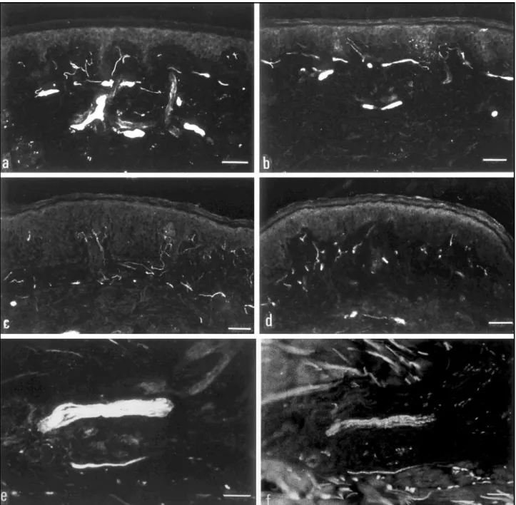
Figs a-f: NGFr immunoreactive fibers in a reactional (a) and in a post-reactional biopsy (b). The number and pattern of positive fibers were not significantly different in both groups. c-d: PGP 9.5 immunohistochemical expression in reactional (c) and post-reactional biopsies (d). The intensity and the pattern of immunoreactivity is not distinct between both sections. Note that the intensity of the immunoreactivity as well as the thickness of the positive fibers are lower than those shown in figures a and b, stained with anti-NGFr. This occurs due to the NGFr expression on perineurium and on Schwann cells together. e-f: NGFr (e) and PGP staining (f) of a nerve branch surrounded by inflammatory infiltrate (same field). Note that the NGFr-positive fibers are more intensely stained and also thicker than that shown with PGP staining.
are implicated in neural degeneration and regenera-tion considering that these two processes undoub-tedly occur in leprosy24.
METHOD
Six leprosy patients were selected for this study. Their clinical data are showed in Table 1. These patients had the diagnosis of leprosy confirmed in the leprosy outpatient service of the Oswaldo Cruz Institute. Leprosy patients are
The selected patients were submitted to a first skin biopsy during the reactional episode and a second one during the remission period. The patients were considered to be in remission after the reactional lesions had decreased either in number or in degree of infiltration and all the general reactional symptoms had subsided. Both biopsies (reactional and post-reactional) were serially taken from close skin sites of the patient. The specific treatment for each kind of reaction (prednisone 1mg/kg of body weight/
day for TIR and thalidomide 200 mg/day for TIIR) was con-veniently instituted for each patient.
The cutaneous biopsies were taken with a 6-mm punch and were fixed in a 4% paraformaldehyde solution con-taining 14% of saturated picric acid for two hours at 4oC,
rinsed four times in 0.1 M phosphate buffer with 10% sucrose added, stored in liquid nitrogen, and sectioned on a Microm cryostat (Heidelberg, Germany). The sections were thawed onto pre-coated glass slides (Super
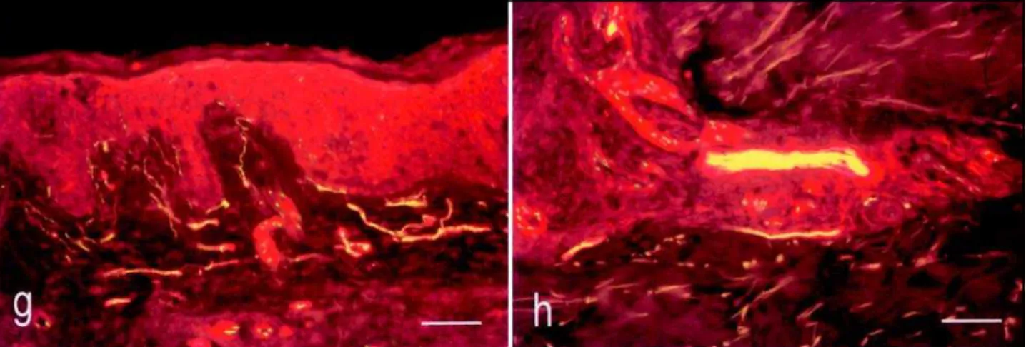
Figs g-h: Double staining of NGFr and PGP showing the nerve fibers in the subepidermal region and a branch in the deep dermis. Green color corresponds to the PGP-positivity alone and reddish color to the positivity. Yellowish color is the result of simultaneous NGFr-and PGP-positivity.
Table 1. Clinical data of the patients.
Patient Age Sex Clinical MDT Reaction Skin lesion Sensorial Paresthesia Paresia General Time between
form impairment symptoms reaction and
remission ERS 29 M LL MB TIIR Diffuse Shins, knees, No Intrinsic No 5 months
infiltration ankles muscles of the hands
HJ 21 M BL MB TIIR Plaques/ No No No No 1 month
nodules
JSS 16 M BL MB TIIR Plaque Hand, feet Forearms, Intrinsic Edema of 4 months Infiltration feet muscles of the ankles
of the ears the hands
MPC 63 M BL MB TIR Plaques Shin, right Feet Intrinsic Edema of 1 month (diffuse) foot the muscles of ankles
hands
MV 63 M BB MB TIR Foveolar Soles Right No No 1 month
plaques elbow
(diffuse)
ZAC 59 F BL MB TIR Plaques Feet No No No 5 months
(diffuse)
Frost®Plus, Menzel-Gläser, Braunschweig, Germany). The
other half was processed for routine diagnostic procedures (hematoxylin-eosin and Wade staining).
Two non-adjacent sections for each biopsy were picked for the staining and counting of positive fibers and both biopsies (reactional and post-reactional) of each patient were processed in parallel for the immunostaining of NGFr and PGP. Double-staining with indirect immunofluores-cence methods were utilized.
The primary antibodies selected for this study were rabbit anti-NGFr (Chemicon USA) and mouse anti-human PGP (UltraClone Cambridge) diluted in 0.01 M phosphate buffer containing 0.3% Triton X-100, in which the sections were incubated overnight at 4oC in a humid chamber
follo-wed by the incubation of fluorescein isothyocyanate (FITC)-conjugated donkey anti-mouse (1:160, Jackson Im-munoResearch Laboratories, West Grove, PA, USA) toge-ther with LRSC-conjugated donkey anti-chicken (1:160, Jackson ImmunoResearch Laboratories, West Grove, PA, USA). Both were diluted in 0.01 M phosphate buffer con-taining 0.3% Triton X-100.
The sections were incubated with both primary anti-bodies overnight at 40C in a humid atmosphere and control
sections were incubated with corresponding normal sera instead, this was followed by the incubation with both second antibodies. The observation was performed using different excitation lights with a photomicroscope (Nikon,
Tokyo, Japan). The immunohistochemical expression of NGFr and PGP markers were compared in both types of biopsies. NGFr- and the PGP-stained nerve fibers were counted separately on the same field using the appropriate filter for the respective fluorochrome used. The frame utilized for taking microphotographs was employed as a standard field for counting nerve fibers. The immunoreactive nerve fibers inside six frames per section were counted. The fra-mes were placed on the section according to distribution of two on the upper dermis, two on the middermis and two on the deep dermis. The objective lens utilized on the counting was the 20X objective matched with a 10X ocu-lar. The nerve endings in the subepidermal region as well as the ones surrounding anexial structures, and dermal vessels were counted as individual fibers; average thick nerve branches on the mid dermis were estimated to have about forty-five fibers and the thicker branches in the deep dermis exhibiting the presence of perineurium were mated to contain about seventy-five nerve fibers. This esti-mation was based on a previous ultrastructural visualiza-tion of cross secvisualiza-tions of dermal nerve branches performed in other study25.
The results of nerve fibers quantification were analyzed with the Statistica (Statsoft Inc. USA) software using the Wilcoxon’s non-parametric paired analysis, Mann Whitney U and ANOVA tests.
RESULTS
NGFr- and PGP- immunoreactive fibers were ob-served in all the reactional and post-reactional biop-sies examined. The higher density of terminal fibers were exhibited on the subepidermal regions (Figs a, b, c, d) surrounding the anexial structures, microves-sels, and among the smooth muscle cells of arrector-pillus muscles. Thick branches with NGFr-positive perineurial boundaries were seen in the mid and deep dermal regions (Fig a). No NGFr-immunoreac-tive fibers were seen in the epidermis but only PGP-positive fibers.
The number of NGFr- and of PGP- immunoreactive nerve fibers in the whole skin and per region of the skin sections, in the reactional biopsies were not significantly different from those in the post-reac-tional group. (Table 2) (Fig a-b, c-d).
The great majority of fibers were both NGFr- and PGP 9.5-positive, but they look more intensely stai-ned and thicker when staistai-ned with anti-NGFr (Fig e-f). This more evident expression of NGFr was due to the immunoreactivity of perineurium, Schwann cells and axons together; the less intense PGP immuno-reactivity instead, was ascribed to single axonal ex-pression of this protein. In addition, few NGFr-stained fibers were PGP 9.5-negative in the same field.
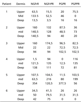
Table 2. Number of NGFr- and PGP-positive nerve fibers in reactional biopsies (NGFrR and PGPR) and in post-reactional biopsies (NGFrPR and PGPPR) (6 frames/section, Magnif.: 200X). Patient Dermis NGFrR NGFrPR PGPR PGPPR
1 Upper 63,5 15,5 20 75,5
Mid 133.5 52,5 46 0
Deep 13,5 3,5 16 16
2 Upper 160 132 87,5 36
mid 140,5 128 48,5 73
Deep 140,5 94 40 20
3 Upper 160 174,5 96,5 82,5
Mid 22 22 72,5 72,5
Deep 94 94 102,5 102,5
4 Upper 1,5 94 0 116
mid 121,5 135 12,5 135
Deep 138 111 72 81
5 Upper 107,5 104,5 11,5 103,5
mid 63,5 216 80 199
Deep 354 120,5 377,5 56
6 Upper 34,5 41,5 26 26
mid 50 79,5 31,5 31,5
As NGFr-positive branches look larger than the PGP-stained ones, we considered the former as con-taining more fibers than the latter, finding then, a higher number of NGFr- than of PGP-immunoreactive fibers inside of reactional but not in the post-reac-tional group (Table 2). This result however, will be commented and interpreted in the following section of this article.
No significant differences in the number of PGP 9.5- and of NGFr-immunoreactive fibers were found among the distinct cutaneous section regions counted inside each group (Table 2).
DISCUSSION
Basically we have found two results in this study: 1) the number of NGFr- and PGP-positive fibers were not significantly different in the reactional and in the post-reactional biopsies, 2) the total number of NGFr-positive fibers in the whole skin sections was significantly higher than that of PGP-immunoreactive ones in reactional but not in the post-reactional group of biopsies.
The first finding should be considered cautiously since comparison of samples with higher number of patients in each group could unveil a significant dif-ference in the number of positive fibers. Nevertheless, we expected to find a higher number of NGFr- and PGP-positive fibers in the post-reactional group due to a supposed nerve regeneration which is reported to occur in leprosy patients’ nerve trunks26. Miko
however, this author remarks that the regeneration observed in the nerve trunks was not effective because the restoration of nerve function was not significantly achieved in the lapse of time (2 to 40 years) between the diagnosis of the patients and the beginning of his investigation. This implies that regenerating fibers, which are observed in the trunk may not reach the target organs, whick is the skin in the case of this study.
Expression of NGFr p75 on Schwann cells corres-ponds to a non-myelinating phenotype of this cell and also both neural crest-derived sensory and sym-pathetic neurons normally express this receptor9.
Regeneration of nerve fibers induces increased NGFr expression on the axons and the reexpression of NGFr on denervated Schwann cells. The expected conse-quences of a leprosy reactional episode on the NGFr-immunoreactivity of peripheral nerve fibers therefore would be a decrease of this expression followed by an increased reexpression of this receptor by the Sch-wann cells and by the axons. In the present study we could not disclose any effect of the reaction on
the number of NGFr and PGP 9.5-immunostained nerve fibers. This finding however, doesn’t rule out other changes in molecular expression of nerve fibers.
Unfortunately it was not possible in this study to compare the number of the positive fibers in reac-tional biopsies with those of the pre-reacreac-tional state. This procedure could disclose a possible decrease of nerve fibers during reactional episode and a recovery of the their previous number in the remission stage.
The higher number of NGFr-positive fibers than that of PGP-positive ones in the same specimen of the reactional group of biopsies may be partially ex-plained by the fact that distinct structures of the nerve fiber express the chosen markers. Therefore, NGFr is expressed on the Schwann cells, on perineu-rial cells and on the axons27,28 while PGP 9.5 is only
an axonal marker. This distinct pattern of expression confers to a bundle of fibers distinct appearances depending on wheter they were stained for NGFr or for PGP. NGFr-positive branches seem to be larger and more evident because of the axons, Schwann cells embracing them and the perineurial cells toge-ther express this receptor strengthening the intensity and thickening the area of the nerve branch immu-noreactivity. PGP-positive fibers are thinner and com-paratively less evident because only axons are stai-ned. The estimation of the number of the nerve fibers based on the thickness of the positive nerve branches may mislead us to the conclusion in respect of the comparison of NGFr and PGP immunoreactivity in the same biopsy specimen. In addition, few of NGFr-positive fibers were PGP-negative, suggesting a selec-tive molecular change of the fiber or axonal degeneration leaving surviving NGFr-positive Schwann cells. Using the methods of the present study, it was not possible to discern which of these two hypothesis correspond to reality. Immunoelec-tronmicroscopy with both anti-NGFr and PGP 9.5 would enlighten this point. Therefore, we could not state with the methods used in present study that there is really a higher number of NGFr- than of PGP-positive fibers in either group of biopsies.
The results of this investigation show that morpho-logical and functional evaluation of nerve fibers should be carried out with markers for both Schwann cells and axons and also allow a reinterpretation of our previous findings20 in which we reported a decreased
not accounting the Schwann cell component of nerve fiber expressing NGFr. Under the light of Liang et al reports and also of the present study, we can state that PGP-negative fiber may retain its NGFr-positivity because of NGFr-expressing Schwann cells.
In conclusion, the number of PGP- and NGFr-posi-tive fibers was not significantly different in the reac-tional and post-reacreac-tional biopsies in the present study. NGFr expression is different from PGP positivity on the nerve fibers and the evaluation of the nerve fiber status on an innervated target organ should be carried out choosing markers for both components of nerve fibers.
Acknowledgements Acknowledgements Acknowledgements Acknowledgements
Acknowledgements - This study was supported by grants from the Fundação de Amparo à Pesquisa do Estado do Rio de Janeiro (FAPERJ), and from the Cancer and Allergy Foundation in Sweden, as well as by funds from the Oswaldo Cruz Institute and from the Karolinska Institute. We also express our thanks to Haroldo Matos, Department of Computer Science, State University of Rio de Janeiro, Brazil for his expert advice in statistical analysis. Ms Agnetha Bonnevier and Ms Eva-Karin Johansson are gratefully acknowledged for skilful technical and secretarial assistance.
REFERENCES
1. Bryceson A, Pfaltzgraff RE. Immunological complications: reactions. In Leprosy. Medicine in the tropics. Edingburgh: Churchill Livingstone, 1990:115-126.
2. Sehgal VN, Srivastava G, Sundharam JA. Immunology of reactions in leprosy: current status. Int J Dermatol 1988;27:157-162.
3. Yamamura M, Uyemura K, Deans RJ, et al. Defining protective responses to pathogens: cytokine profiles in leprosy lesions. Science 1992;254:277-282.
4. Sarno EN, Grau GE, Vieira LMM, Nery JA. Serum levels of tumor necrosis factor-alpha and interleukin-1b during leprosy reactional states. Clin Exp Immunol 1991;84:103-108.
5. Lienhardt C, Fine PEM. Type I reaction, neuritis, and disability in leprosy. Lepr Rev 1994;65:9-33.
6. Uyemura K, Band H, Ohmen J, Brenner MB, Rea TH, Modlin RL. γδ T cells in leprosy lesions. Curr Top Microbiol Immunol 1991;173:203-207. 7. Moraes MO, Sarno EN, Almeida AS, et al. Cytokine m RNA expression in leprosy: a possible role for interferon-γ and interleukin-12 in reactions (RR and ENL). Scand J Immunol1999;50:541-549.
8. Thacker AK, Chandra S, Mukhija RD, Sarkari NB. Electro-physiological evaluation of nerves during reactions in leprosy. J Neurol 1996;243:530-535.
9. Stoll G, Müller HW. Nerve injury, axonal regeneration and neural regeneration: basic insights. Brain Pathol 1999;9:313-325.
10. Zoghbi HY, Gage FH, Choi DW. Neurobiology of disease. Curr Opinion Neurobiol 2000;10:655-660.
11. Meakin SO, Shooter EM The nerve growth factor family of receptors. Trends Neurosci 1992;15:323-331.
12. Anton ES, Weskamp G, Reichardt LF, Matthew WD. Nerve growth factor and its low affinity receptor promote Schwann cell migration. Proc Natl Acad Sci USA 1994;91:2795-2799.
13. Lindsay RM, Harmar AJ. Nerve growth factor regulates expression of neuropeptide genes in adult sensory neurons. Nature 1989;337:362-364. 14. Frade JM, Rodriguez-Tebar, Barde Y-A. Induction of cell death by endogenous nerve growth factor through its p75 receptor. Nature 1996;383:166-168.
15. Hermann JL, Menter DG, Hamada J, Marchetti D, Nakajima M, Nicolson GL. Mediation of NGF stimulated extracellular matrix invasion by the human melanoma low affinity p75 neurotrophin receptor: melanoma p75 functions independently on trk A. Mol Biol Cell 1993;4: 1205-1216. 16. Marlini R. Expression and functional roles of neural surface molecules and extracellular matrix components during developent and regeneration of peripheral nerves. J Neurocytol 1994;23:1-28. 17. Anand P, Pandya S, Ladiwala U, Singhal B, Sinicropi DV,
Williams-Chestnut RE. Depletion of nerve growth factor in leprosy. Lancet 1994;344:129-130
18. Facer P, Mann D, Mathur R, et al. Do nerve growth factor-related mechanisms contribute to loss of cutaneous nociception in leprosy? Pain 2000;85:231-238.
19. Facer P, Mathur R, Pandya SS, Ladiwala U, Singhal BS, Anand P. Correlation of quantitative tests of nerve and target organ dysfunction with skin immunohistology in leprosy. Brain 1998;121:2239-2247. 20. Antunes SLGA, Sarno EN, Holmqvist G, Johansson O. A comparison
of the expression of NGFr with PGP 9.5 and NSE in the cutaneous lesions of early leprosy patients using immunohistochemistry. Int J Lepr 1997;65:357-365.
21. Aquilonius SM, Ashmark H, Ebendal T, Gillberg PG. No re-expression of high affinity nerve growth factor binding sites in spinal motor neurons in amyotrophic lateral sclerosis. Eur Neurol 1992;32:216-218. 22. Johansson O, Hilliges M, Ståhle-Bäckdahl M. Intraepidermal neuron-specific enolase (NSE)-immunoreactive nerve fibres: evidence for sprouting in uremic patients on maintenance hemodialysis. Neurosci Lett 1989;99:281-286. 23. Karanth SS, Springall DR, Lucas S, et al. Changes in nerves and
neuropeptides in skin from 100 leprosy patients investigated by immunocytochemistry. J Pathol 1989;157:15-26
24. Shetty VP, Antia NH. Pathology of nerve damage in leprosy. In Shetty VP, Antia NH. The peripheral nerve in leprosy and other neuropathies. Calcutta Oxford Univ Press, 1997;79-138.
25. Antunes SLG, Sarno EN. Ultrastructural study of dermal nerves in the cutaneous lesions of early leprosy patients. Hansenologia Interna-tionalis 1997;21:14-21.
26. Miko TL, Gschmeissner SE, le Maitre C, Kinfu Y, Kazen R, Pereira JH. Regeneration at the predilective damage sites of nerve trunks in treated leprosy.Lepr Rev1993;64:330-337
27. Liang Y, Johansson O. Light and electron microscopic demonstration of the p75 nerve growth factor receptor in normal human cutaneous nerve fibers: new vistas. J Invest Dermatol 1998;111:114-118. 28. Liang Y, Marcusson JA, Johansson O. Light and electron microscopic