290 291
290 291
Effect of conventional TENS on pain and electromyographic
activity of masticatory muscles in TMD patients
Efeito da TENS convencional sobre a dor e a atividade
eletromiográfica dos músculos mastigatórios em pacientes
com DTM
Delaine Rodrigues*
Anamaria Oliveira Siriani** Fausto Bérzin***
ABSTRACT: Temporomandibular disorders (TMD) are characterized by several signs and symptoms, such as pain and changes in the electrical activity of masticatory muscles. Considering that transcutaneous electrical nerve stimulation (TENS) is a resource indicated to promote analgesia, the objective of this study was to evaluate the effect of TENS on pain and electromyographic (EMG) activity of the jaw elevator muscles in TMD patients. This study evaluated 35 female volunteers: 19 TMD patients (mean age = 23.04 ± 3.5) and 16 normal subjects (mean age = 23.3 ± 3.0). Transcutaneous electrical nerve stimulation (conventional mode, 150 Hz) was applied once to each group for 45 minutes. Surface electromyography (gain of 100 times and 1 kHz sampling frequency) and the visual analogue scale (VAS) were applied before and immediately after TENS application. Both VAS data and root mean square (RMS) values were analyzed using Student's t-test. The TMD group, compared to the control group, showed higher EMG activity of the jaw elevator muscles at rest. No difference was observed between the groups regarding maximum voluntary clenching (MVC). In TMD patients, TENS reduced both pain and EMG activity of the anterior portion of the temporal muscle, increasing the activity of the masseter muscles during MVC. It is possible to conclude that a single TENS application is effective in pain reduction. However, it does not act homogeneously on the features of the electric activity of the muscles evaluated.
DESCRIPTORS: Temporomandibular joint; Electromyography; Masticatory muscles; Transcutaneous electric nerve stimulation; Facial pain.
RESUMO: A desordem temporomandibular (DTM) é caracterizada por diversos sinais e sintomas, como dor e al-teração do sinal eletromiográfico dos músculos da mastigação. Considerando que a estimulação elétrica nervosa transcutânea (TENS) é um recurso indicado para promover analgesia, o objetivo deste trabalho foi avaliar o efeito da TENS na dor e na atividade eletromiográfica (EMG) dos músculos elevadores da mandíbula em indivíduos com DTM. Foram selecionados 35 voluntários do sexo feminino, 19 portadores de DTM (média de 23,04 anos ± 3,5) e 16 normais (média de 23,3 anos ± 3,0). A TENS (modo convencional, 150 Hz) foi aplicada uma vez em ambos os gru-pos, por 45 min. O exame EMG de superfície (ganho de 100 vezes e freqüência de amostragem de 1 kHz) e a escala visual analógica (EVA) foram realizados antes e imediatamente depois da aplicação da TENS. Os dados da EVA e os valores da “root mean square” (RMS) foram analisados pelo teste t de Student. Observou-se que, na situação de repouso, os indivíduos com DTM apresentam um aumento da atividade EMG dos músculos elevadores da mandí-bula em relação aos do grupo controle. Na contração voluntária máxima (CVM), não foram observadas diferenças entre os grupos. Nos portadores de DTM, a TENS reduziu a dor, a atividade EMG da porção anterior do músculo temporal e aumentou a atividade dos músculos masseteres durante a CVM. É possível concluir que apenas uma aplicação da TENS é efetiva para reduzir a dor, porém não atua de forma homogênea sobre as características da atividade elétrica dos músculos avaliados.
DESCRITORES: Articulação temporomandibular; Eletromiografia; Músculos mastigatórios; Estimulação elétrica transcutânea do nervo; Dor facial.
* PhD, Professor, Undergraduate and Graduate Courses in Physiotherapy, Methodist University of Piracicaba.
** PhD, Professor, Graduate Course in Orthopedics, Traumatology and Rehabilitation, School of Medicine of Ribeirão Preto, Uni-versity of São Paulo.
*** Head Professor, Department of Morphology, School of Dentistry of Piracicaba, State University of Campinas.
INTRODUCTION
Temporomandibular disorders (TMD) are
290 291
290 291
in the electromyographic signs of masticatory muscles.
According to some studies3,13,17, TMD subjects
present increased electromyographic (EMG) activ-ity of masticatory muscles with the jaw at rest, i. e., closed lips with no teeth contact, and lower level of clenching electric activation. Some authors11,16,19
also reported that, during mastication, subjects with TMD present activity in jaw-closing muscles (masseter and temporal muscles) during the open-ing phase, when they should be relaxed.
Cooper et al.2 (1991) and Lous et al.8 (1970)
stated that, among the masticatory muscles, the anterior portion of temporal muscles shows the highest electromyographic activity (hyperactivity) when the jaw is at rest position. However, this hy-peractivity of masticatory muscles at rest has been widely discussed. Some authors believe it is an exaggeration to consider muscular hyperactivity alterations from 1 to 2% of the maximum produc-tion of electrical activity of a muscle10.
Different researches have found different results in myoelectric activity during clenching. Naeije, Hansson12 (1986) and Rodrigues15 (2000)
did not observe statistically significant differences in masticatory muscle EMG activity between con-trol and TMD volunteers.
Many studies have demonstrated the effect of some therapeutic resources that would reduce pain and reestablish function of the masticatory muscles. Under this aspect, transcutaneous elec-tric nerve stimulation (TENS) deserves special at-tention because it presents no side effects and enables both pain reduction5 and alteration in
EMG activity16.
Works on the effects of TENS on masticatory muscles show that it is an effective resource in the relief of pain and in the decrease of EMG ac-tivity of masticatory muscles at rest1,2,6,21, in the
reduction of myoelectric activity of the jaw-closing muscles during the opening phase of the jaw in TMD patients16, and in the increase of
muscu-lar electrical activity recorded during clenching in TMD patients1,2. On the other hand, recent
stud-ies15 did not present the same results.
Thus, the objective of the present work was to evaluate the effect of a single TENS application in conventional mode (high-frequency and sensorial threshold) on pain and on the amplitude of masti-catory muscle EMG records of TMD patients.
MATERIALS AND METHODS
Thirty five female volunteers (n = 35) partici-pated in this study: nineteen TMD patients, aged 19 to 33 (mean age = 23.04, SD = ± 3.5 years), and sixteen clinically normal subjects aged 19 to 31 (mean age = 23.3, SD = ± 3.0 years). In the TMD group, volunteers presented:
1. pain in the temporal and masseter muscles during functional activities for at least one year and a maximum of 5 years;
2. occlusal parafunction (clenching and/or grind-ing) for at least one year;
3. pain and/or muscular fatigue when waking up;
4. no functional limitation.
The control group included subjects with no occlusal parafunction, no joint trauma, no joint and/or muscular pain during functional activities, no muscular fatigue and/or pain when waking up. Patients with dental loss, systemic disease which could affect TMJ or cervical joints, e. g. rheumatoid arthritis, osteoarthrosis, osteoporosis, diabetes, and patients taking analgesic, muscle relaxant or anti-inflammatory drugs were excluded, because such drugs could influence the results.
Volunteers were selected after functional evaluation (personal data, anamnesis, background facts, visual inspection, muscular palpation, and functional exam), and dental evaluation, consid-ering oral and dental conditions. All volunteers signed the formal participation form submitted to and approved by the Human Research Ethics Committee, School of Dentistry of Piracicaba, State University of Campinas.
Surface electromyography was recorded at the anterior temporal and masseter muscles using surface differential electrodes of silver bars 10 mm apart, 10 mm long, 2 mm wide, gain of 100 times, input impedance of 10 GΩ and common mode of rejection ratio (CMRR) of 130 dB (Lynx Tecnologia Eletrônica Ltda., São Paulo, SP, Brazil). The EMG signals were analogically amplified with gain of 100 X, filtered (10-509 Hz bandpass Butterworth filter, second order filter) and sampled by 12 bits A/D covert board (model CAD 12/46, Lynx Tec-nologia Eletrônica Ltda., São Paulo, SP, Brazil) with 1 kHz frequency. The signals were digitally bandpass filtered (10-500 Hz).
292 293
292 293
(3 x 4 cm) (Bio-logic Systems Corp., Mundelein, IL, Chicago) was also used as a reference electrode to reduce noise acquisition, fixed on the right wrist of the volunteer.
The volunteers remained comfortably sat on a chair with their bodies lined up and their backs at rest (Frankfurt plan parallel to the ground), eyes open, and arms resting on inferior limbs.
Three five-second recordings of the surface electromyography signals were performed during each of the following situations: (1) rest: subject with mandible at rest, teeth separated, and lips to-gether; (2) isometric contraction: subjects clenched their teeth. During clenching, the researcher is-sued a verbal command for maximum clenching while volunteers clenched Parafilm M (American National Can, Chicago, IL). The material was placed bilaterally between upper and lower teeth, from second premolars to second molars. Previ-ous training was given to familiarize subjects with the experimental procedure. Before the removal of surface electromyography electrodes, marks were drawn on volunteers' skin to indicate where to replace them after electrical stimulation.
Transcutaneous electrical nerve stimulation was delivered by a transcutaneous electrical stim-ulator (model Dualpex 961, Quark Produtos Médi-cos, Piracicaba, SP, Brazil) of two channels and four circular self-adhesive percutaneous electrodes measuring 42 mm (ValuTrode Fallbrook, Califor-nia, USA). TENS was applied using frequency of 150 Hz; pulse width of 20 µs; intensity (mA) ex-clusively set at the subject’s sensorial threshold; modulation up to 50% of variation frequency; qua-dratic biphasic symmetrical pulse and length of application of 45 minutes.
The volunteers were stimulated in dorsal de-cubitus, adequately positioned with a roll under their knees. The percutaneous electrodes of electri-cal stimulation were placed on the masseter and on the anterior portion of the temporal muscle bilaterally. The channels were distributed so that one channel would stimulate the masseter and the other, the temporal, due to their morphological differences and distinct intensity quantities for a given limit of electrical excitability. After the period of application of the electrical stimulation, record-ing electrodes were repositioned and the EMG sig-nal was recorded as described previously.
Pain evaluation was performed using a visual analogue scale (VAS), which was applied before and immediately after electrical stimulation in both studied groups through different recording forms,
so that subjects could not observe what had been written before the application. The intensity of pain was recorded in centimeters, measured from the beginning of the scale (no pain) to the point estab-lished by the volunteer. These data showed regular distribution and were compared using Student’s t-test, at 5% significance level for the values before and after the application of electrical stimulation only for the TMD group. The value obtained in the scale for the control group was zero, that is, they did not present pain before or after the ap-plication.
The masseter and the anterior portion of the temporal muscle EMG amplitudes were analyzed bilaterally by root mean square (RMS) values. The RMS values at rest and at clenching obtained before and after TENS application were analyzed using paired Student’s t-test. RMS values of the experi-mental groups were also compared using Student’s t-test. For all analyses, the values p ≤ 0.05 and p ≤ 0.01 were considered significant and highly significant, respectively.
RESULTS
Pain intensity – VAS mean values
The TENS application promoted significant (p = 0.001) pain relief just for the TMD group (Ta-ble 1).
EMG amplitude – RMS values
The RMS values of both groups recorded with the jaw at rest before TENS are shown in Table 2. It can be observed that EMG activity at rest is higher in the TMD group than in the control one (p ≤ 0.05).
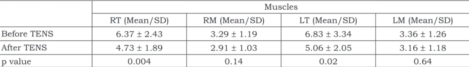
The comparison of RMS values of the TMD group recorded with the jaw at rest before and af-ter TENS are shown in Table 3. It can be observed that after TENS there was a significant decrease (p ≤ 0.05) in EMG activity of the anterior portion
TABLE 1 - Mean values and standard deviation of the
VAS in the TMD group (n = 19) before and after TENS application. Values in cm.
TENS treatment
Before TENS After TENS
Pain intensity 5.75 ± 1.41 1.32 ± 1.05*
292 293
292 293
of the temporal muscle bilaterally in the group with TMD, whereas no significant difference was observed for the right and left masseter muscles.
The RMS values of the control and TMD groups recorded during clenching before the TENS appli-cation are shown in Table 4. There is no
signifi-cant difference between TMD and control groups (p > 0.05).
The comparison of clenching RMS values of the TMD group before and after TENS is shown in Table 5. It can be seen that after TENS there was significant increase in the EMG activity of the masseter muscles in the TMD group, whereas no TABLE 2 - Mean values and standard deviation (SD) of EMG activity recorded with the jaw at rest in TMD (n = 19)
and control (n = 16) groups before TENS application. Values in µvolts. Muscles
RT (Mean/SD) RM (Mean/SD) LT (Mean/SD) LM (Mean/SD)
TMD group 6.37 ± 2.43 3.29 ± 1.19 6.83 ± 3.34 3.36 ± 1.26
Control group 2.47 ± 0.56 2.39 ± 0.55 2.74 ± 0.64 2.67 ± 0.71
p value 0.0001 0.025 0.0005 0.0536
RT and LT: anterior portion of the right and left temporal muscles. RM and LM: right and left masseters. EMG activity: electromyo-graphic activity. TENS: transcutaneous electrical nerve stimulation. TMD: temporomandibular disorders.
TABLE 3 - Mean values and standard deviation (SD) of EMG activity recorded with the jaw at rest in the TMD group
(n = 19) before and after TENS application. Values in µvolts.
Muscles
RT (Mean/SD) RM (Mean/SD) LT (Mean/SD) LM (Mean/SD)
Before TENS 6.37 ± 2.43 3.29 ± 1.19 6.83 ± 3.34 3.36 ± 1.26
After TENS 4.73 ± 1.89 2.91 ± 1.03 5.06 ± 2.05 3.16 ± 1.18
p value 0.004 0.14 0.02 0.64
RT and LT: anterior portion of the right and left temporal muscles. RM and LM: right and left masseters. EMG activity: electromyo-graphic activity. TENS: transcutaneous electrical nerve stimulation. TMD: temporomandibular disorders.
TABLE 4 - Mean values and standard deviation (SD) of EMG activity recorded during clenching in TMD (n = 19)
and control (n = 16) groups before TENS application. Values in µvolts. Muscles
RT (Mean/SD) RM (Mean/SD) LT (Mean/SD) LM (Mean/SD)
TMD group 116.37 ± 31.58 145.79 ± 39.34 157.58 ± 41.15 158.99 ± 34.48
Control group 123.66 ± 34.99 168.16 ± 69.62 167.48 ± 50.39 167.55 ± 50.52
p value 0.74 0.21 0.07 0.57
RT and LT: anterior portion of the right and left temporal muscles. RM and LM: right and left masseters. EMG activity: electromyo-graphic activity. TENS: transcutaneous electrical nerve stimulation. TMD: temporomandibular disorders.
TABLE 5 - Mean values and standard deviation (SD) of EMG activity recorded during clenching in the TMD group
(n = 19) before and after TENS application. Values in µvolts.
Muscles
RT (Mean/SD) RM (Mean/SD) LT (Mean/SD) LM (Mean/SD)
Before TENS 116.37 ± 31.58 145.79 ± 39.34 157.58 ± 41.15 158.99 ± 34.48
After TENS 115.68 ± 31.10 190.68 ± 48.05 161.07 ± 46.32 171.07 ± 46.32
p value 0.8483 0.0001 0.687 0.0392
294 295
294 295
significant difference was observed for the anterior portion of the temporal muscle.
DISCUSSION
The results of this study showed that, at rest, TMD subjects show higher myoelectric activity of the jaw elevator muscles than the control group. This is more evident in the anterior portion of the temporal muscle. No significant differences were observed between the evaluated groups dur-ing clenchdur-ing. We could also observe that TENS promoted pain relief with concurrent reduction in myoelectric activity in the anterior portion of the temporal muscle at rest, and an increase of that activity in the masseter muscles during clenching in the experimental groups.
Several authors reported that TMD subjects present hyperactivity of the masticatory muscles with the jaw at rest and this may cause ische mia, muscular fatigue, and pain7,14,22. Lund et al.9
(1991), Lund, Sessle10 (2000) and Stohler et al.20
(1996) criticize the use of the word “hyperactiv-ity” meaning increased activity in the masticatory muscles with the jaw at rest in TMD subjects. According to Lund, Sessle10 (2000), the so-called
muscular resting “hyperactivity” of the jaw eleva-tor muscles ranges from 1 to 2% of the maximum activity recorded using electromyography during maximum voluntary clenching.
According to the authors mentioned above, the word “hyperactivity” at rest is not adequate. To receive this denomination, the activity records should contain values between 9 and 10 % of the maximum activity recorded during maximum vol-untary clenching, period during which, according to Finsterer4 (2001), we can identify the typical
in-terference pattern of the surface electromyographic record.
On the other hand, the results obtained in this work and in clinical experience show that altera-tions in electromyographic signals of TMD sub-jects must be considered. Although more studies are necessary in order to determine their causes, the increase in electromyographic amplitude of the jaw elevator muscles at rest is probably due to sensorial-motor interactions, of which pain, a symptom present in most TMD subjects evaluated, can modify the generation of action potentials and, finally, the amplitude of myoelectric activity.
The temporal muscle, among the jaw eleva-tors, is where manifestations of increased electro-myographic activity were more frequently reported
in cases of temporomandibular disorder. Data of the present work are in agreement with this find-ing and are in accordance with Lous et al.8 (1970)
and Cooper et al.2 (1991).
The reduction in myoelectric activity of the jaw elevator muscles in records with mandibular rest as a result of TENS, as observed in the pres-ent study, was previously described by other au-thors1,2,6,21 studying TMD subjects as well.
It should be pointed out that only the anterior portion of the left and right temporal muscles pre-sented significant reduction of electromyographic activity, showing that TENS did not cause the same result for all studied muscles. These data are in accordance with the findings of Kamyszek et al.6 (2001), who, through the use of 30- to
40-minute ultralow frequency stimulation, observed a reduction in electromyographic activity at rest, but a non-homogenous effect in the masticatory muscles studied.
On the other hand, data from the present work disagree with the findings of Cooper et al.2 (1991),
who, as a result of a 60- to 90-minute low fre-quency TENS application, obtained uniform effect in the decrease of electromyographic activity at rest and increase of that activity during isometric contraction of the jaw elevator muscles.
We believe that to obtain the same TENS effect in all the studied muscles, both at rest and dur-ing clenchdur-ing, further studies are necessary with variation in the stimulation frequency for a period of over 45 minutes, in order to determine the influ-ence of these variables in the observed results.
As to clenching, the results of the present work showed that there was no significant differ-ence between RMS values of the control and TMD groups.
Data from the present work are in agreement with the findings of Naeije, Hansson12 (1986), who
did not find significant differences in electromyo-graphic activity recorded during isometric contrac-tion of subjects clinically normal or with pathologi-cal conditions. However, our data differ from those of Sheikholeslam et al.18 (1980), who evaluated the
masseter muscles and the anterior portion of the temporal muscle of 39 TMD subjects, aged 14 to 70, and of 45 non-TMD subjects, aged 20 to 32, by surface electromyography. As to the symptoms, TMD subjects presented pain in the masticatory muscles, sensitivity to touch in the temporoman-dibular joint, and limited opening width.
294 295
294 295
and, according to Naeije, Hansson12 (1986),
sub-jects with arthrogenic disorders show lower elec-tromyographic activity than myogenic temporo-mandibular disorder subjects.
Lund et al.9 (1991) reported that, in situations
of chronic muscular pain, muscular contraction ability is reduced due to the decrease in activity of the agonist muscles and the increase in activity of the antagonist muscles. The results of this work disagree with the statements regarding no signifi-cant difference observed in clenching myoelectric activity of the jaw elevator muscles compared with the control group. Besides, the suprahyoid mus-cles, which are antagonistic to the temporal and masseter muscles, were not evaluated.
As to clenching electromyographic activity, we can see that the related literature is contro-versial. Some authors reported decrease in the electromyographic activity in pain and others did not. Thus, future studies should use samples with
similar disorders as well as the records of the jaw depressor muscles.
CONCLUSION
In the conditions of this experiment, it was assumed that TMD subjects present higher elec-tromyographic activity with the jaw at rest than clinically normal subjects, and that such situation is not defined as a causal factor of temporoman-dibular disorders, but as one of the symptoms of the syndrome. A single conventional TENS applica-tion for 45 minutes is efficacious for the relief of pain. However, in general, it does not provide the same effect on the electromyographic activity of the studied muscles, which were recorded both at rest and during isometric contraction. Thus, further studies with longer application and/or different stimulation frequencies are necessary.
REFERENCES
1. Cooper BC. The role of bioelectronic instrumentation in the documentation and management of temporomandibular disorders. Oral Surg Oral Med Oral Pathol Oral Radiol Endod 1997;83:91-100.
2. Cooper BC, Cooper DL, Lucente FE. Electromyography of masticatory muscles in craniomandibular disorders. Laryngoscope 1991;101:150-7.
3. Dahlström L. Electromyographic studies of cranioman-dibular disorders: a review of the literature. J Oral Rehabil 1989;16:1-20.
4. Finsterer J. EMG-interference pattern analysis. J Electro-myogr Kinesiol 2001;11:231-46.
5. Johnson MI, Ashton CH, Thompson JW. The clinical use of TENS. J Orthop Med 1992;14:3-12.
6. Kamyszek G, Ketcham R, Garcia R Jr, Radke J. Electro-myographic evidence of reduced muscle activity when ULF-TENS is applied to the Vth and VIIth cranial nerves. Cranio
2001;19:162-8.
7. Laskin DM. Etiology of the pain-dysfunction syndrome. J Am Dent Assoc 1969;79:147-53.
8. Lous I, Sheikholeslam A, Moller E. Postural activity in sub-jects with functional disorders of the chewing apparatus. Scand J Dent Res 1970;78:404-10.
9. Lund JP, Donga R, Widmer CG, Stohler CS. The pain-ad-aptation model: a discussion of the relationship between chronic musculoskeletal pain and motor activity. Can J Physiol Pharmacol 1991;69:683-94.
10. Lund JP, Sessle BJ. Mecanismos neurofisiológicos. In: Zarb GA, Carlsson GE, Sessle BJ, Mohl ND. Disfunções da articulação temporomandibular e dos músculos da masti-gação. 2ª ed. São Paulo: Santos; 2000. p. 188-207. 11. Moller E, Sheikholeslam A, Lous I. Response of
eleva-tor activity during mastication to treatment of functional disorders. Scand J Dent Res 1984;92:64-83.
12. Naeije M, Hansson TL. Electromyographic screening of myogenous and arthrogenous TMJ dysfunction patients. J Oral Rehabil 1986;13:433-41.
13. Pinho JC, Caldas FM, Mora MJ, Santana-Penin U. Electromyographic activity in patients with temporoman-dibular disorders. J Oral Rehabil 2000;27:985-90. 14. Ramfjord SP. Bruxism, a clinical and
electromyo-graphic study. J Am Dent Assoc 1961;62:21-44.
15. Rodrigues D. Efeito da estimulação elétrica nervosa transcutânea na atividade elétrica do m. masseter e da porção anterior do m. temporal em indivíduos portadores de desordem craniomandibular – análise eletromiográfica [Dissertação de Mestrado]. São Carlos: Universidade Fe-deral de São Carlos; 2000.
16. Rodrigues D, Oliveira AS, Bérzin F. Effect of TENS on the activation pattern of the masticatory muscles in TMD patients. Braz J Oral Sci 2004;3:510-5.
17. Schroeder H, Siegmund H, Santibanez G, Kluge A. Causes and signs of temporomandibular joint pain and dysfunction: an electromyographical investigation. J Oral Rehabil 1991;18:301-10.
18. Sheikholeslam A, Moller E, Lous I. Pain, tenderness and strength of human mandibular elevators. Scand J Dent Res 1980;88:60-6.
19. Stohler CS, Ashton-Miller JA, Carlson DS. The effects of pain from the mandibular joint and muscles on masticatory motor behaviour in man. Arch Oral Biol 1988;33:175-82. 20. Stohler CS, Zhang X, Lund JP. The effect of
experi-mental jaw muscle pain on postural muscle activity. Pain 1996;66:215-21.
21. Treacy K. Awareness/relaxation training and trans-cutaneous electrical neural stimulation in the treatment of bruxism. J Oral Rehabil 1999;26:280-7.
22. Ueda HM, Kato M, Saifuddin M, Tabe H, Yamaguchi K, Tanne K. Differences in the fatigue of masticatory and neck muscles between male and female. J Oral Rehabil 2002;29:575-82.