36e
Rev Bras Med Esporte _ Vol. 13, Nº 1 – Jan/Fev, 2007 1. Laboratório de Estudos Clínicos em Fisioterapia, Universidade deRi-beirão Preto, RiRi-beirão Preto.
Received in 21/6/05. Final version received in 7/ 12/05. Approved in 17/7/06. Correspondence to: Avenida do Café, 1.139, Vila Amélia – 14050-230 – Ribeirão Preto, SP. Tels.: (16) 3966-5406 / (16) 602-3079. E-mail: fta_ gabriel [email protected]
Electromyographic activity during one-legged
squatting under different foot positions
Gabriel Ribeiro1, Valdeci Carlos Dionísio1 and Gil Lúcio Almeida1
O
RIGINALA
RTICLEKeywords: Muscle. Weight-Bearing. Rehabilitation. Knee. ENGLISH VERSION
ABSTRACT
The specific aim of this study was to quantify muscle activity while performing squat with technique variations. Eight healthy volunteers performed squats under 5 types of foot positions: neu-tral position, under a 10o descending wedge, under a 10o
ascend-ing wedge, under a 10o medial wedge and under a 10o lateral
wedge. It was evaluated electromyographic data of vastus media-lis oblique, vastus lateramedia-lis, rectus femoris, biceps femoris, lateral gastrocnemius and tibialis anterior muscles, using One-Way ANO-VA. The EMG integrated values was not significantly different across the 5 foot positions. The results of this study suggest that differ-ent foot positions during one-legged squat do not cause changes in muscle recruitment.
INTRODUCTION
Squat exercises have been widely used in the last years in train-ing and rehabilitation programs of the lower limb. The application of variations of this exercise with the aim to observe different pat-terns of muscular activity, has been very usual(1-7). The
understand-ing of the pattern of the muscular activity durunderstand-ing squat may in-crease the selection of prevention, rehabilitation and sports training programs of the lower limb(8).
The use of orthoses(6), different situations(2,4,7) and feet
stanc-es(3), as well as the application of different degrees of rotation of
the lower extremity(1,4-5) were some of the strategies used in order
to trigger selective muscular recruitment during squat.
During single-legged squat, the electromyographic behavior of the quadriceps musculature was studied associated to different foot stances in the medium-lateral direction(3). In this study, no
dif-ferences were observed in the electromyographic behavior (EMG) of two uniarticular units(3): vastus medialis oblique and vastus
later-alis muscles. On the other hand, Hertel et al.(6) found differences in
the EMG activity pattern in this very task using an orthosis for cor-rection of foot pronation and supination. Nevertheless, the trunk positioning and the angular velocity of the knee were not controlled in any of these studies, which could influence the muscular re-cruitment pattern(9-12).
We hypothesize that with the control of the angular velocity of the knee and the sagittal and frontal trunk dislocation, we could observe a different muscular activity pattern during the single-legged squat associated to different foot stances on the medium-lateral and antero-posterior positions.
Since single-legged squat is frequently applied in rehabilitation activities(13-14) and in tests used in the pre-participation physical
exams(15-16), this study may contribute for the understanding of this
task, besides aiding in the elaboration of rehabilitation programs.
Thus, the aim of the present study was to quantify the muscular activity during single-legged squats with variations in the technique and with control of 2 kinematic parameters –angular velocity of the knee and trunk dislocation.
METHODOLOGY
Subjects
Eight healthy and right-handed subjects (four male, four female) age range 18-24 years participated in the study. The medical histo-ry of all subjects was evaluated and later the ones who did not present any organic deficiency, surgery or back/lower limbs pain were selected. The descriptive statistics of these subjects is found in table 1.
TABLE 1
Descriptive statistics for men (n = 4), women (n = 4) and total sample (n = 8)*
Age (years) Height (cm) Weight (Kg)
Men 19.7 ± 1.0 1.7 ± 0.0 69.6 ± 0.90
Women 22.7 ± 0.7 1.6 ± 0.0 54.7 ± 1.80
Total sample 21.2 ± 0.8 1.6 ± 0.0 62.1 ± 2.97
* Values expressed as mean ± EPM.
Prior to the experiment, all subjects read and signed a clarified consent form approved by the Committee of Ethics in Research of the Ribeirão Preto University.
Instruments
Registration of the electromyographic data (EMG)
Prior to the data collection, the site of each electrode was shaved and cleaned with alcohol in order to facilitate its adherence as well as the conduction of EMG signals. Bipolar surface electrodes (10 x 1 mm) DelSYS (model DE2.2L) were positioned with a 3 cm dis-tance between them. The procedures described by Bevilaqua-Gros-si et al.(17). were followed for the electrodes placement on the
vas-tus medialis oblique muscle (VMO). For the straight femoral (SF), vastus lateralis (VL), biceps femoral (BF), gastrocnemius lateralis (GL) and anterior tibialis (AT), the electrodes were placed accord-ing to the procedures applied by Basmajian and Blumenstein(18). In
all collections, the site was identified and prepared by the same researcher. The EMG signals were amplified (x 2000), filtered (band pass 20-450 Hz) and registered. The data were digitalized at 12 bits, collected by an IBM-PC computer at 1000 Hz and expressed in millivolts (mV). The EMG signals were not normalized since the comparisons are done within the same muscle, and not between different muscles.
Registration of the kinematic data
Rev Bras Med Esporte _ Vol. 13, Nº 1 – Jan/Fev, 2007
37e
were attached to the center of the shoulder, hip, knee and anklejoints (lateral extremity of the acromio; femoral major trochanter; femoral lateral epicondyle and lateral malleolus) (figure 1). The in-frared signal emission of these points was with 100 Hz frequency by a tridimensional optical system OPTOTRAK 3020.
Data processing
The electromyographic signals (EMG) and the registrations of the X, Y and Z points were synchronized by a ODAU II – Optotrak Dates Acquisition Unit II synchronizer and later mathematically treat-ed by a code in MatLab (Math Works Inc., version 6.0). The linear dislocation of the points, the dislocation of the knee and angular velocity were calculated in this code. Moreover, the EMG signals of each muscle were quantified. All squat tasks which had linear dislocation longer than three centimeters in the antero-posterior and latero-lateral positions were discarded with the purpose to re-duce the interferences derived from each subject’s balance. The linear dislocation of the mark on the acromio was used as refer-ence for the performance of this procedure.
The electromyographic signals were calculated in the descend-ing phase of the squat, usdescend-ing as reference for the calculation the angular velocity of the knee. The beginning of the movement was considered when the initial velocity reached 5% of the velocity peak and the end of the movement when the final velocity reached 5% of the velocity peak. During the movement two phases were considered (acceleration and desacceleration). The acceleration phase was considered between the beginning of the movement and the velocity peak and the desacceleration phase was consid-ered between the velocity peak and the end of the movement.
The velocity of the knee was used for the quantification of the electromyographic signals of the vastus medialis oblique (VMO); straight femoral (SF), vastus lateralis (VL); biceps femoral (BF); gas-trocnemius lateralis (GL) and anterior tibial (AT) muscles. The elec-tromyographic signals (EMG) were integrated in the acceleration and desacceleration phases of the movement. Mean of the EMG of each muscle was calculated for each kind of squat for each sub-ject.
Data analysis
All data used in the statistical analysis were submitted to the Kolmogorov-Smirnov test in order to verify whether they present-ed a normal distribution. All analyzpresent-ed data presentpresent-ed normal distri-bution. The one-way (ANOVA) variance analysis was used in order to individually test the effect of the type of squat over the elec-tromyographic signal of each muscle. The same analysis was ap-plied for the comparison of dislocation, angular velocity of the knee and trunk dislocation in the different types of squat. In all evalua-tions the significance level considered was lower or equal to 5% (p ≤ 0.05).
RESULTS
Dislocation and angular velocity
The breadth of the angular dislocation of the knee and the knee joint angular velocity were not statistically different (table 2) in the five types of squats performed in different foot stances (NP; PF; DF; S and P). The values of the angular dislocation are expressed in figure 2.
Electromyographic analysis
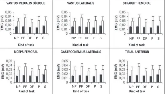
The statistical analysis did not show effect of the different types of single-legged squat over the electromyographic activity (EMG) of the muscles analyzed in the acceleration and desacceleration phases (table 3). The EMG of the analyzed muscles in the acceler-Figure 1 – – – – – Graphic representation of the stances of the individuals after
single-legged squat. NP = neutral position; DF = dorsal flexion; PF = plan-tar flexion; S = supination; P = pronation.
Position of the marks – LED (Light Emis-sion Diode)
1. shoulder – acromio 2. hip – femoral major trochanter 3. knee – femoral lateral epicondyle 4. ankle – lateral malleolus
Procedure
Single-legged squat with foot on Neutral Position (NP)
Each subject was placed standing, with the right limb on the center of a square (80 x 80 cm) drawn on the ground. Each partic-ipant was asked to keep the left knee flexed approximately 60o.
The subject kept the right knee in total extension and the upper limbs extended and raised to the front with a 90o shoulder flexion
angle. A glass board was placed in the front of the participant with marks setting the targets. These marks would delimit the initial and final positions of the movement and represent a breadth of 35o to 40o of knee flexion, having as reference the arm dislocation.
From the initial position, when receiving the verbal command, the subject performed the single-legged squat by knee flexion. The subjects were motivated to perform each movement the fastest as possible. When reaching the target, the subject would keep the position until receiving another verbal command to return to the initial position. Only the descending phase of the squat was ana-lyzed. A series of 10 squats was performed. Figure 1 represents the graphic form of the single-legged squat with foot in neutral position.
Single-legged squats with foot on Plantar Flexion (PF); Dorsal Flexion (DF), Supination (S) and Pronation (P) stances
For the performance of right foot squats in the PF, DF, S and P positions the same procedure described above was performed, associated with the use of a wooden platform (55 x 55 cm). This platform with 10o inclination was set in order to position the foot in
10o of PF; 10o of DF; 10o of S and 10o of P prior to the beginning of
the squat. The choice of a platform with 10o inclination was based
on the study by Hung and Gross(3). Figure 1 graphic represents the
single-legged squat with the foot on plantar flexion.
TABLE 2
(ANOVA) variance analysis – Kinematic parameters
F Freedom degrees p
Angular dislocation of the knee 1.27 (4, 28) < 0.30
Angular velocity of the knee 0.85 (4, 28) < 0.50
NP DF
38e
Rev Bras Med Esporte _ Vol. 13, Nº 1 – Jan/Fev, 2007 ation and desacceleration phases of the single-legged squat isrep-resented in figure 3.
DISCUSSION
The outlining of the study was mainly directed in order to pro-vide information concerning the influence of foot stances over the electromyographic activity of uniarticular and biarticular muscles during single-legged squat. Single-legged squat is a task of com-plex weight discharge where several muscles act together.(16).
Moreover, single-legged squat is a task applied in the rehabilitation of the muscle-skeletal system(13) and is related to sports activities;
being important to athletes’ dribbling, jumping and balancing(15). It
has been more recently suggested that such task, if performed with the foot positioned in 25-degree decline, could be beneficial to subjects recovering from patellar tendinopathy(14).
Despite the relevance of the single-legged squat in the athletic and rehabilitation activities, few works investigated the electromyo-graphic behavior of the main muscles involved in the single-legged squat. Hung and Gross(3) investigated the influence of the foot
medial and lateral inclination over the EMG activity during single-legged squat. They did not find difference in the vastus medialis oblique and vastus lateralis ratio, using a 10o board for foot
inclina-tion(3). These results are contrary to the ones observed by Hertel et
al.(6) who found an increase in the EMG activity of the vastus
medi-alis and the gluteus medius during the squat with utilization of orth-eses for correction of the turn-out in the medium-lateral position. These authors(6) did not observe correlation with the orthesis
incli-nation position, that is, regardless the postural correction, the use of the orthesis determined increase of the electromyographic ac-tivity of the vastus medialis and the gluteus medius.
Our results did not show differences in the EMG activity during the tasks proposed, corroborating thus the results by Hung and Gross(3). The difference may be given to the fact that in our studies
a 10o platform was used, which caused alterations of the foot
stanc-es as a whole, while Hertel et al.(6) used an orthesis only on the
back of the foot (inside the shoe) which caused a local alteration of one region of the foot. Besides that, differences in the task perfor-mance where Hertel et al.(6) did not control the trunk position or
the angular velocity of the knee, could have influenced the results. Richardson and Bullock(9) verified that the increase of the
flex-ion-extension movement of the knee caused the EMG activity of the straight femoral and the hamstrings to increase, while the EMG activity of the vastus medialis and the vastus lateralis did not suf-fer alteration. Hagood et al.(10) find a great increase of the EMG
activity antagonist of the quadriceps and hamstrings applying an increase of the joint velocity of the knee. In our study we moni-tored the angular velocity of the knee in the five types of performed squat, except for the influence of velocity over the EMG. Accord-ing to Henry et al.(11) the EMG activity of the thigh muscles is
influ-enced by postural dislocations in the lateral and antero-posterior direction. Within this context, another study(12) verified EMG
alter-ations of the soleum muscle during squats performance over un-steady surface. These authors also related the EMG alterations of the soleum to its role in the postural control. We also performed the control of the trunk dislocation with the aim to decrease the effect of the postural control over the EMG.
TABLE 3
(ANOVA) variance analysis – Electromyographic activity (EMG) in the acceleration (AP) and desacceleration phases (DP) Muscles F Freedom p Muscles F Freedom p
(FA) degrees (DF) degrees
VMO 2.09 (4, 28) < 0.10 VMO 1.33 (4, 28) < 0.28
SF 1.24 (4, 28) < 0.31 SF 0.61 (4, 28) < 0.65
VL 2.18 (4, 28) < 0.09 VL 1.41 (4, 28) < 0.25
BF 1.11 (4, 28) < 0.37 BF 0.14 (4, 28) < 0.96
GL 1.28 (4, 28) < 0.30 GL 1.19 (4, 28) < 0.33
AT 4.59 (4, 28) < 0.50 AT 1.61 (4, 28) < 0.20
Figure 3 – – – – – Electromyographic activity (EMG) of the muscles analyzed during the different types of single-legged squat. The black bars represent the EMG in the acceleration phase and the white bars the EMG in the desacceleration phase of the movement. All values were expressed by means and Standard error. Figure 2 – – – – – Dislocation and angular velocity of the knee joint during the
different types of single-legged squat. All values were expressed by means and standard error.
Dislocation
Type of squat
V
elocity
Type of squat
VASTUS MEDIALIS OBLIQUE VASTUS LATERALIS STRAIGHT FEMORAL
BICEPS FEMORAL GASTROCNEMIUS LATERALIS TIBIAL ANTERIOR
Kind of task Kind of task Kind of task
Kind of task Kind of task Kind of task
NP PF DF P S NP PF DF P S NP PF DF P S
NP PF DF P S NP PF DF P S
Rev Bras Med Esporte _ Vol. 13, Nº 1 – Jan/Fev, 2007
39e
Clinically speaking, a protocol applying single-legged squatexer-cises associated to the foot stances on a platform with 25o
inclina-tion was able to cause clinical improvement in athletes with patel-lar tendinopathy(11). Compared with our study, this higher inclination
(25o) kept the ankle in plantar flexion, which could favor a different
muscular recruitment pattern of the quadriceps, justifying thus the improvement of the inflammation. Further studies exploring the kinematic, kinetic and electromyographic behavior are needed in order to understand the positive results in the patellar tendinopathic treatment.
CONCLUSION
Our results suggest that alterations in foot stance during single-legged squat, according to the described methodology, do not cause alterations in the muscular recruitment pattern of the main mus-cles involved in the task.
All the authors declared there is not any potential conflict of inter-ests regarding this article.
REFERENCES
1. Ninos JC, Irrgang JJ, Burdett R, Weiss JR. Electromyographic analysis of the squat performed in self-selected lower extremity neutral rotation and 30 de-grees of lower extremity turn-out from the self-selected neutral position. J Or-thop Sports Phys Ther. 1997;25:307-15.
2. Anderson R, Courtney C, Carmeli E. EMG analysis of the vastus medialis / vastus lateralis muscles utilizing the unloaded narrow- and wide-stance squats. J Sport Rehabil. 1998;7:236-47.
3. Hung YJ, Gross MT. Effect of foot position on eletromyographic activity of the vastus medialis oblique and vastus lateralis during lower extremity weight-bear-ing activities. J Orthop Sports Phys Ther. 1999;29:93-102.
4. Escamilla RF, Flesig GS, Zheng N, Lander JE, Barrentine SW, Andrews JR, et al. Effects of technique variations on knee biomechanics during the squat and leg press. Med Sci Sports Exerc. 2001;33:1552-66.
5. Lam PL, Ng GYF. Activation of the quadriceps muscle during semisquatting with different hip and knee positions in patients with anterior knee pain. Am J Phys Med Rehabil. 2001;80:804-8.
6. Hertel J, Sloss BR, Earl JE. Effect of foot orthotics on quadriceps and gluteus medius electromyographic activity during selected exercises. Arch Phys Med Rehabil. 2005;86:26-30.
7. McCaw ST, Melrose DR. Stance width and bar load effects on leg muscle activ-ity during the parallel squat. Med Sci Sports Exerc. 1999;31:428-36.
8. Escamilla RF. Knee biomechanics of the dynamic squat exercise. Med Sci Sports Exerc. 2001;33:127-41.
9. Richardson C, Bullock MI. Changes in muscle activity during fast, alternating flexion-extension movements of the knee. Scand J Rehabil Med. 1986;18:51-8.
10. Hagood S, Solomonow M, Baratta R, Zhou BH, D’Ambrosia R. The effect of joint velocity on the contribution of the antagonist musculature to knee stiffness and laxity. Am J Sports Med. 1990;18:182-7.
11. Henry SM, Fung J, Horak FB. Control of stance during lateral and anterior / pos-terior surface translations. IEEE Trans Rehabil Eng. 1998;6:32-42.
12. Anderson K, Behm DG. Trunk muscle activity increases with unstable squat move-ments. Can J Appl Physiol. 2005;30:33-45.
13. Ng GY, Cheng JM. The effects of patellar taping on pain and neuromuscular performance in subjects with patellofemoral pain syndrome. Clin Rehabil. 2002; 16:821-7.
14. Young MA, Cook JL, Purdam CR, Kiss ZS, Alfredson H. Eccentric decline squat protocol offers superior results at 12 months compared with traditional eccen-tric protocol for patellar tendinopathy in volleyball players. Br J Sports Med. 2005;39:102-5.
15. Zeller BL, McCrory JL, Kibler WB, Tymothy UL. Differences in kinematics and electromyographic activity between men and women during single-legged squat. Am J Sports Med. 2003;31:449-58.
16. Tagesson S, Öberg B, Kvist J. Passive and dynamic translation in the knee is not influenced by knee exercises in healthy individuals. Scand J Med Sci Sports. 2005;15:139-47.
17. Bevilaqua-Grossi D, Felicio LR, Simões R, et al. Avaliação eletromiográfica dos músculos estabilizadores da patela durante exercício isométrico de agachamen-to em indivíduos com síndrome da dor femoropatelar. Rev Bras Med Esporte. 2005;11:159-63.