Regadas FSP, Figueiredo WR, Nogueira MAA, Bezerra CRS, Sousa PC. Role of bowel preparation on colocolonic anastomosis: experimental study in dogs. J Coloproctol, 2012;32(4): 359-364.
AbstrACt: The aim of the present study was to evaluate the eficacy of colocolonic anastomosis with and without preoperative bowel preparation. Methods: The study compared 42 female dogs (Canis familiaris), divided into 2 groups of 21 animals: Group I (control) – submitted to bowel preparation – and Group II (study) – without previous bowel preparation –. All animals were submitted to laparotomy with sectioning of the descending colon and primary anastomosis using polypropylene thread. Following euthanasia on the 21st postopera-tive day (POD), a second laparotomy was performed to evaluate the anastomosis with regard to complications, intra-abdominal adhesions and anastomotic burst pressure. results: One animal from each group (4.5%) died. The death in Group I occurred on seventh POD due to anastomotic dehiscence. The death in Group II occurred on tenth POD due to deep incisional infection at the surgical site and complete dehiscence of the abdominal wall. The groups did not differ signiicantly with regard to adhesion grade or anastomotic burst pressure (one specimen burst in each group) (p>0.05). Conclusion: Colocolonic anastomosis without previous bowel preparation was shown to be safe and eficacious, suggesting it is not an indispensable procedure in colorectal anastomosis surgery.
Keywords: colorectal surgery; postoperative complications; anastomotic leak.
resuMo: Esse estudo avaliou a eicácia da anastomose colocólica sem preparo intestinal prévio comparando-a com a anastomose realizada com preparo. Método: Foram utilizados 42 animais (Canis familiares) fêmeas distribuídos em 2 grupos com 21 animais em cada: Grupo I (controle) – com preparo intestinal – e Grupo II (estudo) – sem preparo intestinal prévio –. Os animais de ambos os grupos foram submetidos à laparotomia com secção do cólon descendente e à anastomose primária com io de polipropileno, bem como à euta -násia no 21º dia de pós-operatório com laparotomia e à avaliação da anastomose colocólica quanto à presença de complicações, grau de aderências intestinais e pressão de ruptura da anastomose. resultados: Ocorreu um (4,5%) óbito em cada grupo, sendo o do Grupo I no
sétimo dia pós-operatório em decorrência da deiscência da anastomose colocólica e o do Grupo II no décimo dia de pós-operatório por causa de infecção em sítio cirúrgico com deiscência total da parede abdominal. Não foi observada diferença estatisticamente signiicante no grau de aderências intestinais tampouco no teste de pressão de ruptura entre os grupos (um espécime sofreu ruptura em casa grupo) (p>0,05). Conclusão: A anastomose colocólica sem preparo intestinal apresentou a mesma segurança e eicácia da anastomose realizada
com preparo prévio, sugerindo não ser indispensável na cirurgia colorretal com anastomose.
Palavras-chave: cirurgia colorretal; complicações pós-operatórias; fístula anastomótica.
role of bowel preparation on colocolonic anastomosis:
experimental study in dogs
Francisco Sérgio Pinheiro Regadas1, Welligton Ribeiro Figueiredo2,
Miguel Augusto Arcoverde Nogueira3, Carlos Renato Sales Bezerra4, Péricles Cerqueira de Sousa5
1Full Professor at Faculty of Medicine, Universidade Federal do Ceará (UFC) – Fortaleza (CE), Brazil. 2Digestive Tract
Surgeon, Master student in Surgery, UFC – Fortaleza (CE), Brazil. 3Coloproctologist, Doctoral candidate in Surgery,
UFC – Fortaleza (CE), Brazil. 4Digestive Tract Surgeon, Doctoral candidate in Surgery, UFC – Fortaleza (CE), Brazil. 5Digestive Tract Surgeon, Master student in Surgery, UFC – Fortaleza (CE), Brazil.
Study carried out at Post-graduation program stricto sensu in Surgery, Surgery Department, School of Medicine, Universidade Federal do Ceará – Fortaleza (CE), Brazil.
Financing source: none.
Conlict of interest: nothing to declare.
INtroDuCtIoN
Colorectal surgery with primary anastomo-sis is associated with a range of mild to severe complications, from simple surgical site infection to anastomotic dehiscence and fistula. Thus, the postoperative recovery of patients submitted to colorectal surgery remains a major challenge for surgeons.
Mechanical bowel preparation of the colon prior to elective surgery was first proposed over a hundred years ago. It has since been used to re-duce or eliminate the fecal mass, thereby minimiz-ing infection and complications in perianastomotic tissues and ensuring more esthetic outcomes1.
Bowel preparation involves a set of proce-dures designed to completely remove fecal resi-dues and significantly reduce the bacterial flora in the colon with the least possible discomfort and risk for the patient2.
Thorough bowel cleansing is by most sur-geons considered one of the most important fac-tors in the prevention of complications3. In fact, since the days of Halsted, the presence of feces in-side the colon has been viewed as a major cause of anastomotic dehiscence4. Not surprisingly, many authors believe preoperative bowel preparation is essential in the prevention of infectious complica-tions following colorectal surgery5-8.
Based on the literature, it is difficult to de-termine exactly when preoperative bowel prepa-ration became a standard procedure in colorectal surgery, but the earliest studies on colon and rec-tum cleansing were carried out by Maunsell in the early 1890s9.
However, in the 1990s, the use of bowel prep-aration as an indispensable preoperative procedure
came into question, and criteria for when it ought
to be avoided was proposed10. In addition, a num-ber of studies documented the favorable evolution of patients submitted to emergency left colon re-section with primary anastomosis without previous colon preparation, raising doubts about its indis-pensability11. It has been shown that under certain circumstances mechanical bowel preparation can actually stimulate bacterial growth and transloca-tion, both of which favor the emergence of
sep-tic complications from colorectal surgery12. Other researchers believe mechanical colon preparation does not improve postoperative morbidity rates and may even increase the incidence of infectious complications, fistulas and hydroelectrolyte im-balance10. In a study with five years of follow-up, Fillmann, in 2001, reported lower fistula and in-fection rates among patients who were not submit-ted to preoperative bowel preparation13. Thus, to a number of authors, anastomosis may be performed safely without preoperative bowel preparation14,15.
The aim of the present study was to evalu-ate the efficacy of colocolonic anastomosis in dogs with and without preoperative bowel preparation.
MAterIALs AND MetHoDs
This study included 42 female dogs (Canis
fa-miliaris) weighing 8.4–16.9 kg, distributed at
ran-dom in two groups of 21 animals each:
• Group I (control) – animals with preoperative
bowel preparation;
• Group II (study) – animals not submitted to
preoperative bowel preparation.
The animals in Group I (control) were sub-mitted to preoperative bowel preparation with a 12% glycerin solution administered rectally one day before surgery.
Surgical technique — once the animals
were anesthetized, a digital rectal examination was performed individually to determine bowel preparation according to the classification pro-posed by O’Dweyr: excellent (absence of feces); good (presence of minimal fecal residue);
accept-able (presence of liquid feces); soiled (presence of
solid feces)16. The procedure consisted of a medi-an trmedi-ansumbilical laparotomy, identification of the descending colon at 20 cm from the anal margin, and colotomy with sectioning of the entire colon circumference. In both groups, the colotomy was closed manually with a continuous single-layer ex-tramucosal suture using polypropylene 3–0 thread.
(POD 21), the animals were euthanized and a sec-ond laparotomy was performed through the same incision. The anastomoses were evaluated with re-gard to integrity and the presence of fistulas and dehiscence, while the abdominal cavity was eval-uated for adhesions using the classification pro-posed by Knightly: 0=no adhesions, 1=single thin and easily separable adhesion, 2=less extensive but weak adhesions which withstand traction poor-ly, 3=extensive visceral adhesions extending to ab-dominal wall, 4=numerous extensive and visceral adhesions involving the mesentery, bowel, omen-tum and abdominal wall17. A 6 cm-colon segment centered on the anastomosis was resected and cau-terized at the proximal extremity using a urethral probe (#8.0). In order to determine the anastomot-ic bursting pressure, the proximal extremity of the colon segment was tied to a sphygmomanometer with two cotton threads (size 2–0) while the distal extremity was closed with Kelly forceps to prevent air from escaping (Figure 1).
Then the colon segment was inflated continu-ously with a manual bulb to a maximum pressure of 300 mmHg, or until the suture burst (Figure 2). The site of anastomotic disruption, if any, was ex-amined and the respective pressure was registered.
The weight, colon preparation, postoperative clinical evolution, intra-abdominal adhesion score and anastomotic bursting pressure were registered for all the animals, and the two groups were compared.
resuLts
One animal in each group (4.5%) died. The death in Group I (study) occurred on POD 7 due to anas-tomotic dehiscence. The death in Group II (control) occurred on POD 10 due to deep incisional infection at the surgical site and complete dehiscence of the ab-dominal wall with evisceration and intact anastomo-sis. The two groups did not differ with regard to de-hiscence (p>0.05). Likewise, the observed difference
in average weight did not reach statistical signiicance
(p>0.05).
According to the O’Dweyr classiication, bowel
preparation was considered good in 70% and excellent in 30% of the animals in Group II.
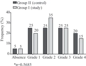
The distribution of the animals according to in-tra-abdominal adhesion grade is shown in Graph 1. In Group I, adhesions were predominantly grade 2 (35%) and grade 3 (25%), while the distribution
Figure 1. System used to test the bursting strength of colon
was more homogenous in Group II (grades 1, 2 and 3=25%; grade 4=20%). However, the difference was
not statistically signiicant (p=0.5685). (Figure 3) When testing the bursting strength by inlation at
up to 300 mmHg, one colon segment from each group (5.0%) was disrupted, one at 270 mmHg (Group I) and one at 220 mmHg (Group II). The difference was not
statistically signiicant (p>0.05) (Figure 4).
DIsCussIoN
Female dogs were used in this study because they are easy to obtain and handle, their bowels are relatively similar to human bowels, and the size of the pelvic cavity is appropriate to test the procedure18. In addition, canine and human bowels also feature
rela-tively similar intestinal microlora, blood supply and
descending colon anatomy16.
On the average, the animals in Group I weighed more than the animals in Group II, but the difference
was not statistically signiicant. Thus, body weight
cannot be considered a determining factor in our sam-ple.
The bowel preparation of the animals in Group I was successful, indicating that bowel cleansing with 12% glycerin solution 24 hours prior to surgery is
ef-icient in dogs19.
There is still some discussion about the most appropriate way to perform anastomosis,
wheth-er to use a continuous single-laywheth-er suture or sepa-rate stitches, and which thread to use. The method a dopted in this study (continuous single-layer ex-tramucosal suture) is simple, swift and inexpensive. It is associated with impermeability and low levels of
tissue inlammation, and is considered as safe as sep -arate stitches20,21. Monoilament thread is preferred
to multiilament thread which is known to favor the development of infections and inlammatory reac -tions14. The polypropylene thread used in this study
is monoilament, has high tensile strength and is as
-sociated with very little inlammatory reaction22. The mortality rate was similar in the two groups. Both the observed deaths were the result of infectious complications. One animal in Group I presented anas-tomotic dehiscence evolving towards peritonitis and died on POD 7. Another animal in Group II presented deep incisional infection at the surgical site leading to complete dehiscence of the abdominal wall and died on POD 10. These results suggest that mechani-cal bowel preparation prior to surgery does not reduce mortality in dogs submitted to colorectal anastomosis surgery14,15,23.
The grade of adhesions is an indirect measure
of anastomotic complications and, consequently, of wound healing. Based on Knightly’s classiication
of adhesions17, no signiicant differences between the groups were observed, indicating that
mechani-cal bowel preparation did not inluence the devel
-Figure 4. Disruption of anastomosis submitted to inlation at up
to 300 mmHg.
No disruption Disruption 0
20 40 60 80
100 95 95*
5 5
Group II (control) Group I (study)
*p=0,9714 F re q u en cy ( % )
Absence Grade 1 Grade 2 Grade 3 Grade 4 0
10 20 30 40
Group II (control) Group I (study)
5 5 25 20 25 35 *p=0,5685 25 25 20 15 F re q u en cy ( % )
reFereNCes
1. Hares MM, Alexander-Williams J. The effect of bowel preparation on colonic surgery. World J Surg 1982;2(6):175-81.
2. Habr-Gama A, Gama-Rodrigues JJ, Teixeira MG, Alves PRA, Ventura TCM, Quintanilha AG, et al. Preparo intestinal pela ingestão de manitol a 10%. Rev Bras Coloproct 1981;1(2):84-94.
3. Güenaga KF, Matos D, Wille-Jørgensen P. Preoperative mechanical bowel preparation in elective colorectal surgery. An update of systematic review of the literature and meta-analysis. J Coloproctol 2012;1(32):7-17.
4. Ravo B, Metwally N, Castera P, Polasnky PJ, Ger R. The importance of intraluminal anastomotic fecal contact and peritonitis in colonic anastomotic leakages. An experimental study. Dis Colon Rectum 1988;1(31):868-71.
5. Rosenberg IL, Graham NG, Dombal FT, Goligher JC. Preparation of the intestine in patients undergoing major large-bowel surgery, mainly for neoplasmas of the colon and rectum. Br J Surg 1971;58(4):266-9.
6. Irvin TT, Goligher JC. Aetiology of disruption of intestinal anastomoses. Br J Surg 1973;60(6):461-4.
7. Buffara JR, Brenner S, Souza FJ, Marchesini JB, Malaia O. Infecção em cirurgia colorretal. Estudo retrospectivo de 621 casos. Rev Bras Colo-Proct 1988;3(8):94-7.
8. Pitrez FAB. Pré e pós-operatório em cirurgia geral e especializada. 2a ed. Porto Alegre: Artmed; 2003. 266 p. 9. Graney MJ, Graney CM. Colorectal surgery from antiquity
to the modern era. Dis Colon Rectum 1980;23(6):432-41. 10. Fillmann EEP, Fillmann LS, Fillmann HS. Cirurgia
colorretal eletiva sem preparo. In: Habr-Gama A, Barone B. Atualização em coloproctologia. São Paulo: Sociedade Brasileira de Coloproctologia e Associação Latino-americana de Coloproctologia; 1995. p. 269-71. 11. Koruth NM, Krukowski ZH, Youngson GG, Hendry WS,
Logie JR, Jones PF, et al. Intra-operative colonic irrigation in
the management of left-sided large bowel emergencies. Br J Surg 1985;72(9):708-11.
12. Valarini R, Lemos R, Quintana LFC, Cordova LF, Cabrera PFA, Repka JD, et al. Estudo da translocação bacteriana após sutura primária do colo com e sem limpeza mecânica: trabalho experimental em cães. Rev Bras Coloproct 1998;18(1):22-9. 13. Fillmann LS, Perondi F, Fillmann HS, Fillmann EEP. Cirurgia
eletiva para o câncer colo-retal sem preparo mecânico da luz intestinal: Análise após 5 anos de acompanhamento. Rev Bras Coloproct 2001;4(21):246-9.
14. Torres Neto JR, Fakhouri R, Menezes MVA, Santos JS, Prudente ACL, Monteiro JTS, et al. Estudo Histomorfométrico de Anastomoses Primárias de Cólon em Coelhos, Com e Sem Preparo Intestinal. Rev Bras Coloproct 2007;4(27):384-90. 15. Scabini S, Rimini E, Romairone E, Scordamaglia R, Damiani
G, Pertile D, et al. Colon and rectal surgery for câncer without mechanical bowel preparation: onecenter ramdomized prospective trial. World J Surg Oncol 2010;8:35.
16. O’Dwyer PJ, Conway MC, McDermott EW, O’Higgins NJ. Effect of mechanical bowel preparation on anastomotic integrity following low anterior resection in dog. Br J Surg 1989;76(7):756-8.
17. Knigthly JJ, Agostino D, Cliffton EE. The effect of ibrinolisyn and heparin on the formation of peritoneal adhesions. Surgery 1962;52(4):250-8.
18. Regadas SMM, Regadas FSP, Rodrigues LV, Carvalho MCGS, Regadas Filho FSP. Modelo experimental de sutura manual em cólon de cão por vídeo-laparoscopia. Acta Cir Bras 2005;20(4):323-8.
19. Bezerra CRS. Fechamento do coto distal do cólon sigmóide comparando sutura contínua com lacre plástico. Estudo experimental em cães. [dissertation]. Fortaleza (CE): Universidade Federal do Ceará; 2010.
20. Regadas FSP, Castro Filho HF, Nicodemo AM, Morano JCOD, Sampaio ZS. Estudo comparativo entre sutura contínua e separada em anastomose cólica. Estudo experimental em ratos. Acta Cir Bras 1990;4:141-5.
opment of adhesions. This inding contradicts the
notion that preoperative colon cleansing reduces the risk of contamination of the peritoneum and perian-astomotic tissues, thereby minimizing the develop-ment and severity of adhesions2.
Studies on healing of intestinal sutures often em-ploy mechanical parameters such as burst tension
test-ing in which a bowel segment is distended with liquid
or air up to a predetermined pressure level, or until it bursts17,24. In this study, the bowel segments tested
were carefully centered on the anastomosis. Burst
pressure is an eficient parameter to evaluate the heal -ing of intestinal anastomoses provided the disruption occurs at the site of the anastomosis17,24. In our study, disruption occurred in one specimen from each group,
thus no signiicant difference was observed. In conclu -sion, colocolonic anastomosis without previous bowel
21. Figueiredo AF. Efeitos da suplementação nutricional com glicina e com glutamina na cicatrização colônica em coelhos [dissertation]. Belo Horizonte (MG): Universidade Federal de Minas Gerais; 2007.
22. Ribeiro FJC. Avaliação qualitativa e quantitativa da resposta inlamatória comparando a ação do io de polipropileno com o io de poligliconato em anastomoses realizadas em colon de ratos [dissertation]. Fortaleza (CE): Universidade Federal do Ceará; 1998.
23. Feres O, Santos Jr JCM, Andrade JI. The role of mechanical bowel preparation for colonic resection and anastomosis: an experimental study. Int J Colorectal Dis 2001;16(6):353-6.
24. Gonçalves CG. Cicatrização de anastomose colônica e nutrição pré-operatória em ratos desnutridos: estudo tensiométrico e de deposição de colágeno [dissertation]. Curitiba (PR): Universidade Federal do Paraná; 2005.
Correspondence to:
Francisco Sergio Pinheiro Regadas
Faculdade de Medicina da Universidade Federal do Ceará Avenida Atilano de Moura, 430, apto. 200