Rev Bras Cardiol Invasiva. 2012;20(4):427-30
© 2012 Sociedade Brasileira de Hemodinâmica e Cardiologia Intervencionista. Published by Elsevier Editora Ltda. All rights reserved.
Agenesis of the Inferior Vena Cava
Caroline Saltz Gensas
1, Leonardo Martins Pires
2, Marcelo Lapa Kruse
3, Tiago Luiz Luz Leiria
4,
Daniel Garcia Gomes
5, Gustavo Glotz de Lima
6ABSTRACT
Agenesis of the inferior vena cava is a rare malformation. Its most common cause is dysgenesis during embryogenesis, but it may also be related to intrauterine or perinatal thrombosis. It is usually asymptomatic, associated or not with other congenital malformations and may be related to increased risk of chronic venous insuficiency and deep vein thrombosis, especially in young individuals. Diagnosis is often incidental, during ab-dominal surgery or radiological procedures. We reported ive cases of agenesis of the inferior vena cava detected during electrophysiological procedures.
DESCRIPTORS: Electrophysiology. Cardiovascular abnormalities. Vena cava, inferior.
1 Medicine Academic at Universidade Federal de Ciências da Saúde
de Porto Alegre. Porto Alegre, RS, Brazil.
2 Master. Cardiologist Physician and Electrophysiologist at Instituto de
Cardiologia/Fundação Universitária de Cardiologia. Porto Alegre, RS, Brazil.
3 Doctor. Cardiologist Physician and Electrophysiologist at Instituto de
Cardiologia/Fundação Universitária de Cardiologia. Porto Alegre, RS, Brazil.
4 Doctor. Cardiologist Physician and Electrophysiologist at Instituto de
Cardiologia/Fundação Universitária de Cardiologia. Porto Alegre, RS, Brazil.
5 Cardiologist Physician and Trainee at Electrophysiologist Service
of Instituto de Cardiologia/Fundação Universitária de Cardiologia. Porto Alegre, RS, Brazil.
6 Doctor. Cardiologist Physician and Head of Electrophysiology Service
of Instituto de Cardiologia/Fundação Universitária de Cardiologia. Porto Alegre, RS, Brazil.
Correspondence to: Gustavo Glotz de Lima. Av. Princesa Isabel, 370 – Porto Alegre, RS, Brazil – CEP 90620-000
E-mail: gglima.pesquisa@gmail.com
Received on: 9/5/2012 • Accepted on: 11/15/2012 RESUMO
Agenesia da Veia Cava Inferior
Agenesia da veia cava inferior é uma malformação rara. Sua causa mais comum é a disgenesia durante a embriogênese, mas também pode estar relacionada a trombose intrauterina ou perinatal. Normalmente é assintomática, em associação, ou não, com outras malformações congênitas, e pode cursar com maior risco de insuiciência venosa crônica e trombose venosa profunda, especialmente em jovens. Seu diagnóstico frequentemente é acidental, durante cirurgias abdominais ou procedimentos radiológicos. Relatamos cinco casos de agene-sia da veia cava inferior detectada durante procedimentos eletroisiológicos.
DESCRITORES: Eletroisiologia. Anormalidades cardiovasculares. Veia cava inferior.
Case Report
T
he electrophysiological study is an invasive test used to diagnose heart rhythm and conduction disorders. The test can be used to measure atrioventricular conduction intervals, clarify arrhyth-mogenic mechanisms, and evaluate antiarrhythmic agent efficacy. Currently, it is used to identify and map re-entrant circuits and ectopic foci for subse-quent treatment with catheter ablation. Therefore, the electrophysiological test has diagnostic, therapeutic, and prognostic value.Access to the heart is usually attained through a femoral or subclavian venipuncture to insert a catheter into the heart chambers. Agenesis of the inferior vena
cava is generally an asymptomatic condition that may be unknown to the patient, and can occur in approxi-mately 0.5% of the population.1 During
electrophysio-logical procedures, catheter progression dificulties in the cephalad direction raise the possibility of this anomaly; therefore, manoeuvres are required to conirm the presence of agenesis.
Gensas et al.
Agenesis of the Inferior Vena Cava
Rev Bras Cardiol Invasiva. 2012;20(4):427-30
428
CASE REPORTS
Case 1
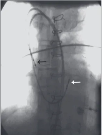
Male patient, 60 years-old, with a history of ischemic stroke in 2005 and acute myocardial infarction in 2007, prior to coronary artery bypass graft (CABG) surgery, and normal ventricular function. In February 2010, the patient was admitted to the Electrophysiology Labora-tory for investigation of syncope unrelated to exertion, with no premonitory symptoms; he was submitted to an electrophysiological study with right femoral vein puncture. During catheter insertion, there was dificulty in advancing the cephalad catheter. A venography was performed (Figure 1), which showed agenesis of the inferior vena cava and dilated hemiazygos vein, with venous drainage into the common trunk, the superior vena cava, and subsequently the right atrium (Figure 2). After the venography, an electrophysiological study was performed using a stimulation protocol (Figure 3), which showed normal sinus function and normal atrio-ventricular and intraatrio-ventricular conduction.
Case 2
Female patient, 12 years-old, with tetralogy of Fallot repair (systemic-pulmonary shunt performed in 2000) and a permanent pacemaker for sinus node dysfunction (implanted in 2000). In March 2012,
the patient was admitted to the Electrophysiology Laboratory to undergo an electrophysiological study for sudden death stratification. A right femoral veni-puncture was performed. Catheterization of the right chambers was attempted using fluoroscopy, without success. A pigtail catheter was inserted, and the venography was performed, which showed the ab-sence of the inferior vena cava and venous drainage through the hemiazygos vein to the superior vena cava. Ventricular pacing was not performed, due to the inability to insert the catheters.
Case 3
Female patient, 21 years of age, was referred to the Electrophysiology Laboratory to undergo an electro-physiological study in April of 2012 to evaluate frequent episodes of tachycardia. The patient had a history of syncope, and complex congenital heart disease repair had been performed 19 years earlier (double outlet right ventricle, pulmonary stenosis, atrial septal defect); she underwent a permanent pacemaker implant in 2004. During the examination, a femoral venipuncture was performed, and a right-chamber catheterization was attempted with the insertion of a multipolar electrode catheter, which was unsuccessful due to agenesis of the inferior vena cava. Programmed ventricular pacing was
Figure 1 – Angiography showing inferior vena cava agenesis (white arrow), with catheter progression through the hemiazygos vein (black arrow).
Figure 2 – Schematic graph showing dilated hemiazygos vein draining into the common venous trunk, superior vena cava, and subsequently, into the right atrium.
Hemiazygos Superior
vena cava
Gensas et al. Agenesis of the Inferior Vena Cava
Rev Bras Cardiol Invasiva. 2012;20(4):427-30
429
performed with a pacemaker, which showed a sinus rhythm and a complete atrioventricular block that did not induce sustained ventricular tachycardia.
Case 4
Female patient, 40 years of age, was referred to the Electrophysiology Laboratory for an electrophysiological study and ablation. A right femoral venipuncture was performed for catheter insertion. During catheter insertion, there was dificulty in advancing the cephalad catheter. An angiography of the venous system was performed, which showed an anomalous course of venous drainage into the lower half of the body, directed to the superior vena cava. The catheters were inserted into the heart cavity through the superior vena cava and positioned initially in the low right atrium, coronary sinus, and His bundle region. The atrial extrastimulation demonstrated dual atrioventricular nodal conduction and induced reproducible episodes of supraventricular tachycardia with concentric retrograde atrial activation. Slow nodal pathway ablation was performed successfully. However, postoperatively, the patient had deep vein thrombosis and pulmonary embolism of small intensity, with no associated symptoms. Anticoagulation was initiated, and the patient improved.
Case 5
Female patient, 62 years of age, was referred to the Electrophysiology Laboratory for an electro-physiological study and ablation due to a palpitation complaint. Three diagnostic electrode catheters were introduced into the right chamber; however, cathe-ter progression showed the presence of anomalous drainage into the lower half of the body to the supe-rior vena cava. During the examination, posteroseptal accessory bundle was diagnosed, with successful bundle ablation.
DISCUSSION
Venous system development during embryogenesis is a complex process, during which the development, regression, and anastomosis of three pairs of veins (posterior cardinal, subcardinal, and supracardinal) form the inferior vena cava.2,3 If the originally paired
structures are not joined between the sixth and eighth weeks of gestation, malformations can occur, such as a duplicated inferior vena cava, inferior vena cava agenesis, and the interruption of a certain segment (infrahepatic, prerenal, renal, or infrarenal), among others.3,4 Inferior vena cava malformations are present
in 0.07% to 8.7% of the population3 and may be
asymptomatic or associated with nonspecific symp-toms. However, they may also be associated with an increased risk of deep venous thrombosis, which is present in 5% of young individuals who have deep venous thrombosis.5
Agenesis (also called atresia or aplasia) of the inferior vena cava is a rare malformation that occurs in 0.005% to 1% of the population. Its most common cause is dysgenesis during embryogenesis (due to the simultaneous occurrence of a defect in the venous system of the three embryonic segments), but it can also occur due to in utero or perinatal thrombosis
(without embryological abnormalities).6 Since the
pa-tient shows no symptoms, the diagnosis of agenesis is often accidentally attained during abdominal surgery or radiological procedures;5 in these cases, collateral
circulation develops from the lumbar, azygos, and hemiazygos systems, which compensate for the mal-formed lower vena cava function.7 However, in the
event of insuficient collateralization, the slow blood low in the lower limbs and pelvis leads to venous stasis and to an increased propensity for thrombosis.8 Thus,
chronic venous insuficiency or deep vein thrombosis (especially in the iliac and femoral veins) may occur with a higher recurrence rate of thrombosis in these patients. Furthermore, comorbidities may be present, such as splenic abnormalities, intestinal malrotation, pulmonary dysgenesis, renal agenesis, dextrocardia, or other congenital heart diseases.5 Among the present
patients, two had other congenital heart diseases, and one patient developed DVT followed by pulmonary thromboembolism.
Gensas et al.
Agenesis of the Inferior Vena Cava
Rev Bras Cardiol Invasiva. 2012;20(4):427-30
430
Inferior vena cava agenesis should be suspected when young patients (up to 30 years-old) have idio-pathic deep venous thrombosis, especially bilate-rally in the iliac veins, and no risk factors (such as hypercoagulable states).8 However, it is important to
remember that, in the absence of other anomalies, agenesis can be asymptomatic due to collateral venous system development.7
In case of diagnostic suspicion, imaging examina-tions should be performed. The best imaging methods to diagnose inferior vena cava abnormalities are computed tomography and magnetic resonance angiography, since the diagnosis of inferior vena cava abnormalities solely by ultrasonography is considered to be dificult.1,2,5
Treat-ment should focus on the prevention of complications, especially thrombosis. For this reason, several authors have recommended the use of anticoagulants.7 The
patient must be advised to avoid other risk factors for thrombosis, such as the use of oral contraception and long periods of immobilization, as well as unnecessary surgical interventions.7
Patients referred for electrophysiological study or ablation may come to the electrophysiology laboratory without a prior diagnosis of inferior vena cava agenesis. Thus, the electrophysiologist should remember that these morphological alterations can occur, in order to avoid inadvertent complications of vascular access and catheter handling. For this purpose, other routes (e.g., the subclavian vein) may be used to access the venous system.
CONFLICT OF INTEREST
The authors declare that they have been granted sponsorship for scientiic events and equipment for the Electrophysiology Laboratory from St. Jude Medical and Biotronik.
REFERENCES
1. Onzi RR, Costa LF, Angnes RF, Domingues LA, Moraes PS, Armani Li, et al. Malformação de veia cava inferior e trom-bose venosa profunda: fator de risco de tromtrom-bose venosa em jovens. J Vasc Bras. 2007;6(2):186-9.
2. Iqbal J, Nagaraju E. Congenital absence of inferior vena cava and thrombosis: a case report. J Med Case Rep. 2008;2:46-9. 3. Cho BC, Choi HJ, Kang SM, Chang J, Lee SM, Yang DG, et
al. Congenital absence of inferior vena cava as a rare cause of pulmonary thromboembolism. Yonsei Med J. 2004;45(5):947-51. 4. Obernosterer A, Aschauer M, Schnedl W, Lipp RW. Anomalies
of the inferior vena cava in patients with iliac venous throm-bosis. Ann Intern Med. 2002;136(1):37-41.
5. Konopka CL, Salame M, Padulla GA, Muradás RR, Batistella JC. Agenesia de veia cava inferior associada à trombose venosa profunda. J Vasc Bras. 2010;9(3):196-9.
6. Lambert M, Marboeuf P, Midulla M, Trillot N, Beregi JP, Mounier-Vehier C, et al. Inferior vena cava agenesis and deep vein thrombosis: 10 patients and review of the literature. Vasc Med. 2010;15(6):451-9.
7. Felício ML, Martins AS, Andrade RR, Silva MAM. Ausência parcial de veia cava inferior associada à malformação intes-tinal. Rev Bras Cir Cardiovasc. 2007;22(3):362-4.