191
Radiol Bras. 2012 Jul/Ago;45(4):191–197
Neuroendocrine tumors of the lung: major radiologic
findings in a series of 22 histopathologically confirmed cases
*
Tumores neuroendócrinos do pulmão: principais achados radiológicos em uma série de 22 casos com confirmação anatomopatológica
Marcel Koenigkam Santos1, André Rodrigues Façanha Barreto2, Francisco Abaeté Chagas Neto3, Valdair Francisco Muglia4, Jorge Elias Júnior5
Objective: To describe key imaging findings in a series of cases of primary neuroendocrine tumors of the lung (NTLs), with emphasis on computed tomography changes. Materials and Methods: Imaging studies of 22 patients (12 men, mean age 60 years) with histopathologically confirmed diagnosis, evaluated in the authors’s institution during the last five years were retrospectively reviewed by two radiologists, with findings being consensually described focusing on changes observed at computed tomography. Results: The authors have described five typical carcinoids, three atypical carcinoids, three large-cell neuroendocrine carcinomas (LCNCs), and 11 small-cell lung cancers (SCLCs). Only one typical carci-noid presented the characteristic appearance of central endobronchial nodule with distal pulmonary atelectasis, while the others were pulmonary nodules or masses. The atypical carcinoids corresponded to peripheral heterogeneous masses. One out of the three LCNCs was a peripheral homogeneous mass, while the others were ill-defined and heterogeneous. The 11 SCLCs corresponded to central, infiltrating and heterogeneous masses with secondary pleuropulmonary changes. Calcifications were absent both in LGNCs and SCLCs. Metastases were found initially and also at follow-up of all the cases of LCNCs and SCLCs. Conclusion: Although some imaging features may be similar, radiologic findings consid-ered together with clinical information may play a relevant role in the differentiation of histological types of NTLs. Keywords: Computed tomography; Lung neoplasms; Neuroendocrine tumors.
Objetivo: Descrever os principais achados de imagem em uma série de casos de tumores neuroendócrinos primários do pulmão (TNPs), destacando as alterações na tomografia computadorizada. Materiais e Métodos: Exames de 22 pacientes (12 homens, idade média de 60 anos) avaliados nos últimos cinco anos em nosso serviço, com confirma-ção histopatológica, foram retrospectivamente revistos por dois médicos radiologistas e os achados foram descritos em consenso, focando as alterações tomográficas. Resultados: Descrevemos 5 carcinoides típicos, 3 carcinoides atípicos, 3 carcinomas neuroendócrinos de grandes células (CNGCs) e 11 cânceres pulmonares de pequenas células (CPPCs). Apenas um carcinoide típico apresentou aspecto característico de nódulo endobrônquico central com ate-lectasia pulmonar distal, enquanto os demais foram nódulos ou massas pulmonares. Os carcinoides atípicos eram massas pulmonares periféricas e heterogêneas. Um CNGC era massa periférica delimitada e homogênea, enquanto os demais eram mal delimitados e heterogêneos. Os 11 CPPCs eram massas centrais, infiltrativas e heterogêneas, com alterações pleuropulmonares secundárias. Calcificações estavam ausentes nos CNGCs e CPPCs. Metástases foram vistas inicialmente ou no seguimento de todos os CNGCs e CPPCs. Conclusão: Apesar de alguns aspectos semelhan-tes nos exames de imagem, os achados radiológicos, quando integrados às informações clínicas, podem constituir critérios importantes na diferenciação dos tipos histológicos de TNPs.
Unitermos: Tomografia computadorizada; Neoplasias pulmonares; Tumores neuroendócrinos. Abstract
Resumo
* Study developed at Centro de Ciências das Imagens e Física Médica (CCIFM) – Hospital das Clínicas da Faculdade de Medi-cina de Ribeirão Preto da Universidade de São Paulo (HCFMRP-USP), Ribeirão Preto, SP, Brazil.
1. PhD, MD, Radiologist, Assistant Physician at Hospital das Clínicas da Faculdade de Medicina de Ribeirão Preto da Univer-sidade de São Paulo (HCFMRP-USP). Fellow Post-doctoral de-gree, Department of Diagnostic and Interventional Radiology, Heidelberg University, Germany.
2. MD, Radiologist at Clínica Radius, Clínica São Carlos Ima-gem and Santa Casa de Misericórdia de Fortaleza, Fortaleza, CE, Brazil.
3. MD, Radiologist, Fellow of the Program of Health Sciences Applied to the Locomotor System – Faculdade de Medicina de
Koenigkam Santos M, Barreto ARF, Chagas Neto FA, Muglia VF, Elias Jr J. Neuroendocrine tumors of the lung: major radiologic findings in a series of 22 histopathologically confirmed cases. Radiol Bras. 2012 Jul/Ago;45(4):191–197.
Ribeirão Preto da Universidade de São Paulo (FMRP-USP), Ri-beirão Preto, SP, Brazil.
4. PhD, MD, Radiologist, Associate Professor at Division of Radiology, Department of Medical Practice – Faculdade de Me-dicina de Ribeirão Preto da Universidade de São Paulo (FMRP-USP), Ribeirão Preto, SP, Brazil.
5. Private Docent, MD, Radiologist, Associate Professor at Division of Radiology, Department of Medical Practice, Head of the Division of Radiology – Hospital das Clínicas da Faculdade de Medicina de Ribeirão Preto da Universidade de São Paulo (HCFMRP-USP), Ribeirão Preto, SP, Brazil.
Mailing Address: Dr. Marcel Koenigkam Santos. Avenida Ban-deirantes, 3900, Campus Universitário, Monte Alegre. Ribeirão Preto, SP, Brazil, 14048-900. E-mail: [email protected]
Received April 24, 2012. Accepted after revision June 22, 2012.
INTRODUCTION
with similar pathological features, and being capable of producing and secreting peptide hormones and neuroamines(2).
Since the early 1990’s, because of its clini-cal and histologiclini-cal characteristics, SCLC is classified as a neuroendocrine neopla-sia of the lung. Neuroendocrine tumors of the lung comprise 25% of all lung neo-plasms, with a wide spectrum of clinical behaviors, being currently classified into four types: typical carcinoid tumor (low malignancy grade), atypical carcinoid tu-mor (intermediate malignancy grade), large-cell neuroendocrine carcinomas (LCNC) and SCLC, these both with high malignancy grade, the latter representing most of the primary neuroendocrine lung lesions(3,4).
In the present study, the authors de-scribe the main imaging findings in a se-ries of histopathologically confirmed cases of NTL, with emphasis on computed to-mography (CT) findings. Also, the authors make a brief description of the main clini-cal data, including information on the evo-lution of the cases, correlating them with radiological and anatomopathological data.
of the clinical routine in the assessment of such patients, it exempted a term of free and informed consent in addition to the one obtained previously to the performance of the exams. Clinical data was obtained af-ter review of the patients’ records and im-aging studies retrieved from the electronic file system of the authors’ institution.
Images were reviewed by two radiolo-gists, and the findings were described in consensus. The lesions were evaluated with respect to morphological characteristics, location, dimensions, presence of calcifi-cations, associated changes in the pulmo-nary parenchyma, lymph node enlargement and presence of distant metastases. All the imaging studies stored in the electronic file system were reviewed, including plain ra-diographs and magnetic resonance imaging (MRI) studies, but the reviewers have par-ticularly focused on the description of CT findings, which is currently the most accu-rate radiological method and most com-monly utilized in the evaluation of lung tumors. In spite of not being related to the main objective of the present study, the post-treatment follow-up exams, whenever available, were also reviewed for
correla-toms, such as weight loss and malaise, were decribed with higher frequency in cases of LCNC and SCLC. Along the medical records review process, data confirming the presence of paraneoplastic syndrome due to ectopic production of hormones were not found for any of the patients. As regards histological type, the lesions of the 22 pa-tients included: five typical carcinoid le-sions, three atypical carcinoid lele-sions, three LCNCs and 11 SCLCs (Table 1).
Among the five patients with typical carcinoid lesions, only one (20%) had a well defined central endobronchial nodule with soft tissue density in association with distal lung atelectasis(5) (Figure 1). Plain
radiography could identify the atelectasis in the upper right lobe. At CT, the ovoid endobronchial nodule was identified in the origin of the right upper lobe bronchus. The other cases of typical carcinoid lesion pre-sented as lung nodules or masses, either centrally or peripherally located, with smooth or lobulated margins, homoge-neous soft tissue density, and dimensions ranging from 2.1 to 5.5 cm in the largest diameter (Figures 2 and 3). Calcifications were found in two lesions (40%) and were
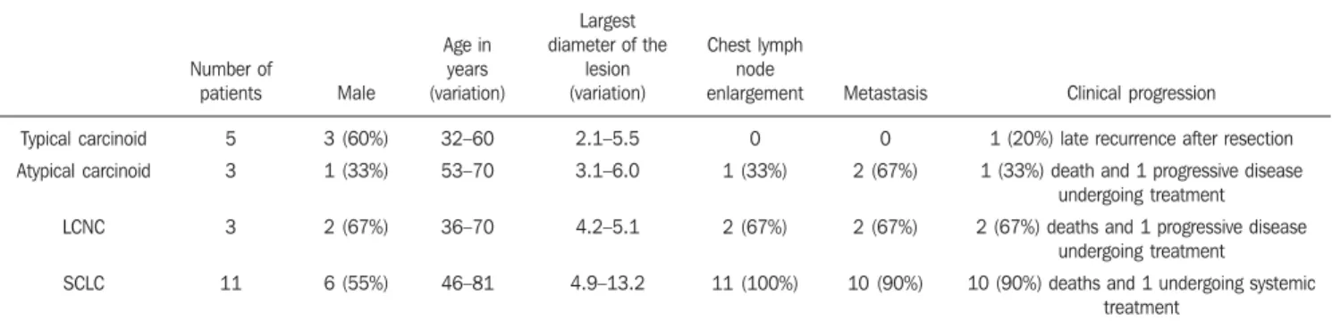
Table 1 Summary of clinical and radiological data of the 22 patients diagnosed with primary neuroendocrine tumor of the lung.
Typical carcinoid Atypical carcinoid LCNC SCLC Number of patients 5 3 3 11 Male 3 (60%) 1 (33%) 2 (67%) 6 (55%) Age in years (variation) 32–60 53–70 36–70 46–81 Largest diameter of the
lesion (variation) 2.1–5.5 3.1–6.0 4.2–5.1 4.9–13.2 Chest lymph node enlargement 0 1 (33%) 2 (67%) 11 (100%) Metastasis 0 2 (67%) 2 (67%) 10 (90%) Clinical progression
1 (20%) late recurrence after resection
1 (33%) death and 1 progressive disease undergoing treatment
2 (67%) deaths and 1 progressive disease undergoing treatment
10 (90%) deaths and 1 undergoing systemic treatment
Figure 1. Lesion corresponding to typical carcinoid tumor. Axial chest CT images after intravenous injection of iodinated contrast medium. On the lung window, one may identify the amputation of the upper right lobe bronchus (arrow on A) and the distal pulmonary atelectasis (C). On the mediastinal window, a well defined, ovoid endobronchial nodule is identified, with homogeneous attenuation (arrow on B).
more clearly seen at CT. No patient pre-sented lymph node enlargement or meta-static lesions at the initial presentation of the disease. Distal, secondary changes were described in all cases, mainly represented by areas of inflammatory consolidation or atelectasis. All five patients were submit-ted to surgical resection (either segmentec-tomy or lobecsegmentec-tomy) and only one patient
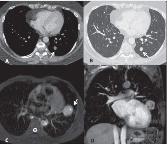
presented recurrence of the disease in the clinical follow-up, with mediastinal lymph node enlargement identified six years after diagnosis (Figure 3). One patient with a typical carcinoid lesion also underwent MRI, which demonstrated the presence of a well defined nodule in the left lower lobe with intermediate signal intensity on T1-weighted and hyperintense signal on
T2-weighed sequences, also with restriction in diffusion weighted imaging and prominent contrast-enhancement more noticeable in delayed phases and with homogeneous appearance (Figure 2).
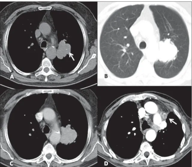
The three cases of atypical carcinoid tumors were identified both at plain radi-ography and CT, described as peripheral lung masses with lobulated or irregular
contours, with heterogeneous density and contrast-enhancement, dimensions ranging between 3.1 and 6.0 cm and with second-ary changes in the adjacent lung paren-chyma (Figure 4). One lesion presented nodular calcifications identified at plain radiography, but best characterized at CT. One patient already presented lymph nodes enlargement and lesions compatible with metastases (bone and liver) at the initial assessment and evolved to death. Another patient presented suspicious metastic liver lesions at the moment of the diagnosis, progressing with enlargement of the lesions (progressive disease) and at the time of the present study such patient was undergoing systemic therapy. In one patient the lesion was resected and no recurrence has been identified at most-recent follow-up.
Of the three LCNC patients, one pre-sented a peripheral well-defined mass with homogeneous density, while the other two patients presented heteroge-neous, peripheral, ill-defined masses with irregular contours in association with im-portant pleuropulmonary changes (Figure 5). The lesions dimensions ranged from 4.2 to 5.1 cm and calcifications were not found. The two patients presenting with heterogeneous lesions already presented lymph nodes enlargement and distant metastatic lesions at their initial assess-ment, and underwent non-surgical therapy, but evolved with progressive disease at follow-up and, later, death. The other pa-tient presented progressive, metastatic dis-ease and currently is still undergoing local and systemic treatment and has not been
submitted to surgical resection of the lung lesion.
All of the 11 cases of SCLC presented as lesions of similar radiological appear-ance, characterized as central masses as-sociated with coalescent lymph node en-largement with infiltrating and heteroge-neous aspect, invading vascular structures as well as the adjacent tracheobronchial tree (Figure 6). Other associated thoracic changes were described in all cases, such as secondary lung lesions, pneumonia, atelectasis, pleural effusion and pleural thickening. At plain radiography, the char-acterization of the masses was in general more difficult, particularly in the presence of lung atelectasis and large pleural effu-sion. Distant metastatic lesions were also found in ten patients (90%) at the moment
of diagnosis, mainly in bones and upper abdomen (liver and adrenal glands), al-ready seen at the first chest CT scan. In general, the lesions that could be measured were larger than 5.0 cm in their largest
diameter, some of them greater than 10.0 cm. Calcifications were not identified in any lesion. Like in the cases of LCNC, all SCLC patients were submitted to local and/or systemic therapy and evolved with
progressive disease and death few months after biopsy and histopathological diagno-sis, with the exception of one patient who remained under treatment at the moment of the present review.
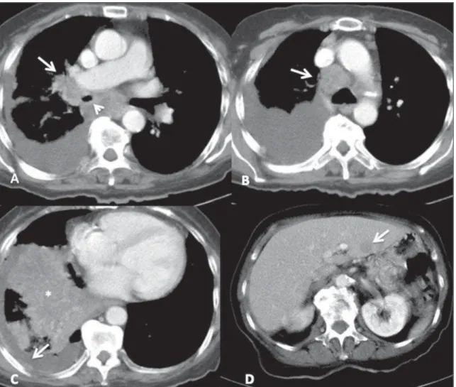
Figure 5. Large-cell neuroendocrine carcinoma. Axial chest CT images, with mediastinal window before (A) and after contrast injection (B), more images with lung window, with thick slices (C) and high resolution, thin sections (D). One observes a mass with partially defined limits in the upper left lobe, with hetero-geneous post-contrast enhancement (arrow on B), in association with secondary changes of the adjacent lung parenchyma, including interstitial thickening due to carcinomatous lymphangitis, best seen on high resolution images (arrow on D).
DISCUSSION
Neuroendocrine tumors of the lung originate in the bronchial mucosa and present similar histopathological character-istics, but present a wide clinical spectrum with behavior ranging from the relatively indolent and well differentiated typical carcinoid lesion to the most aggressive lung tumor, the SCLC(6). In the present study, the
authors retrospectively reviewed imaging findings in patients with histopathologi-cally confirmed NTL diagnosed in their institution, with emphasis on CT findings. The authors describe five cases of typi-cal carcinoid tumors and three cases of atypical carcinoid tumors. Carcinoid tu-mors occur most frequently in the gas-trointestinal tract (80% to 90%), with
bron-chial or lung carcinoid tumors being rep-resented by the typical carcinoid type in most cases (approximately 90%) and less frequently by the atypical carcinoid type(7).
Atypical carcinoid tumors are assciated with smoking, most commonly affecting male patients (2:1) and at older ages (59 years, on average), while typical carcinoid tumors lack established association with smoking, affecting younger patients of both genders. Typical carcinoid tumors rep-resent the most common lung neoplasia in the childhood and most frequently are di-agnosed at less advanced clinical stages, rarely with metastases and progressing with longer survival as compared with atypical carcinoid tumors(8). Among the presented
cases, only one case (20%) of typical car-cinoid tumor presented recurrence after
surgical resection of the primary lesion, while two patients (67%) with atypical tu-mors presented metastatic dissemination of the disease, and one (33%) progressed to death.
In the present study, the authors found only one case of central endobronchial nodule characteristic of typical carcinoid tumor, but atypical carcinoid tumors were found as larger and more heterogeneous peripheral masses, more frequently with metastases at diagnosis or follow-up, like-wise described in the literature. Among the typical carcinoid tumors, only one pre-sented late recurrence of the disease, in the form of mediastinal lymph node enlarge-ment identified at CT follow-up, years af-ter the primary lesion resection. According to the medical literature, imaging findings
of both typical and atypical carcinoid tu-mors are similarly described, being mainly found as well-defined nodules or masses, sometimes lobulated, and when elongated, with the longer axis parallel to the bronchi. Most commonly, the lesions are central, next to the source bronchi and present punctiform or diffuse calcifications iden-tified at CT in up to 30% of the cases. Car-cinoid tumors tend to be well vascularized, with prominent contrast enhancement, which also aids in the differentiation be-tween lesions and the commonly associated distal secondary changes (pneumonia, atectasis and bronchi with mucoid impac-tion)(9). Despite their similar
characteris-tics, one describes that the presence of a central, well-defined nodule causing nar-rowing, deformity or obstruction of a bron-chus, especially if containing calcifica-tions, is suggestive of a typical carcinoid tumor. On the other hand, atypical carci-noid tumors tend to be larger, more periph-eral (distal to the segmental bronchus) and more heterogeneous, besides presenting a higher incidence of metastatic lesions at the diagnostic imaging(10).
In the present study, the authors de-scribed three cases of LCNC, all of them presenting as peripheral heterogeneous masses associated with metatastic disease, and none of the lesions with calcifications. LCNC was the fourth and last tumor sub-type to be recognized as a neuroendocrine tumor of the lung, with characteristics that make it different from the typical and atypi-cal carcinoid tumors as well as from the SCLC(3). It is predominantly found in men
(2.5 times greater incidence than in women), above the age of 60, and in 60% of the cases in association with smoking. The prognosis for patients with LCNC is poorer than for patients with the other NSCLC types, with lower five-year survival rate(11).
However, the imaging findings are de-scribed as being similar to those of most adenocarcinomas and squamous cell carci-nomas, with predominance of nodules or peripheral masses, irregular or spiculated margins, heterogeneous density with foci of necrosis and less frequently with calci-fications (approximately 9%). Pleural thick-ening or effusion are also commonly de-scribed(12). Interestingly, in the present
re-view, one patient was a 36-year-old woman.
Two (67%) patients with LCNC progressed to death months after the diagnosis, despite of the treatment.
In the present study, the authors de-scribed 11 cases of patients with SCLC. Lesions were similar to those described as most characteristic in the medical literature, with no isolated findings of nodule or pe-ripheral mass. All SCLC patients aready presented extensive disease at the moment of diagnosis and 10 (90%) patients pro-gressed to death few months after diagno-sis. Small-cell lung cancer represents ap-proximately 20% of all bronchogenic car-cinomas, is the lung neoplasia with stron-ger association with smoking and affects individuas at mean age of 70 years(6). The
literature reports that, at the moment of diagnosis, the patient usually presents ad-vanced disease with metastasis, mainly to bones, liver, adrenal glands or brain(13). The
SCLC lesions are central in most cases and present in association with hilar and/or mediastinal lymph node enlargement, with a confluent aspect, many times forming large, infiltrating masses, and leading to the displacement or narrowing of the tracheo-bronchial tree and central vessels. Findings of atectasis, other lung lesions and pleural effusion are common. Calcifications are described as being present in up to 23% of cases. In the present study, lesions with calcifications were not found. More rarely, SCLC presents as a peripheral lung lesion without lymph node enlargement, and in such cases it may be confused with other types of bronchogenic carcinomas and with the other NTLs(1,14). Probably, the present
study’s sample of SCLC was underesti-mated, considering its prevalence and the characteristic of specialized reference hos-pital of the authors’ institution. The authors believe that such limitation of the present study was related to the fact that only cases with histopathological and immunohis-tochemical confirmation and radiological exams performed in their own institution were included.
In this series of cases, only one patient with lung neoplasia underwent MRI, with a typical carcinoid lesion presenting as a lung nodule. The role of MRI in the evalu-ation of lung neoplasms is increasing, par-ticularly in the staging of lesions and in the clinical follow-up of the treatment.
How-ever, little has been reported in the litera-ture(15) about MRI in the evaluation of
NTL, and its role, particularly regarding diagnostic accuracy in the different histo-logical types of NTL, still requires further investigation.
REFERENCES
1. Rosado-de-Christenson ML, Templeton PA, Moran CA. Bronchogenic carcinoma: radiologic pathologic correlation. Radiographics. 1994;14: 429–46.
2. Melmon KL, Sjoerdsma A, Mason DT. Distinc-tive clinical and therapeutic aspects of the syn-drome associated with bronchial carcinoid tu-mors. Am J Med. 1965;39:568–81.
3. Travis WD, Linnoila RI, Tsokos MG, et al. Neu-roendocrine tumors of the lung with proposed cri-teria for large-cell neuroendocrine carcinoma. An ultrastructural, immunohistochemical, and flow cytometric study of 35 cases. Am J Surg Pathol. 1991;15:529–53.
4. Travis WD, Colby TV, Corrin B, et al. Histologi-cal typing of lung and pleural tumors. 3rd ed. Ber-lin, Germany: Springer-Verlag; 1999. 5. Magid D, Siegelman SS, Eggleston JC, et al.
Pul-monary carcinoid tumors: CT assessment. J Comput Assist Tomogr. 1989;13:244–7. 6. Chong S, Lee KS, Chung MJ, et al.
Neuroendo-crine tumors of the lung: clinical, pathologic, and imaging findings. Radiographics. 2006;26:41– 57.
7. Modlin IM, Lye KD, Kidd M. A 5-decade analy-sis of 13,715 carcinoid tumors. Cancer. 2003;97: 934–59.
8. Ducrocq X, Thomas P, Massard G, et al. Opera-tive risk and prognostic factors of typical bron-chial carcinoid tumors. Ann Thorac Surg. 1998; 65:1410–4.
9. Zwiebel BR, Austin JH, Grimes MM. Bronchial carcinoid tumors: assessment with CT of location and intratumoral calcification in 31 patients. Radiology. 1991;179:483–6.
10. Jeung MY, Gasser B, Gangi A, et al. Bronchial carcinoid tumors of the thorax: spectrum of radio-logic findings. Radiographics. 2002;22:351–65. 11. Paci M, Cavazza A, Annessi V, et al. Large cell neuroendocrine carcinoma of the lung: a 10-year clinicopathologic retrospective study. Ann Thorac Surg. 2004;77:1163–7.
12. Oshiro Y, Kusumoto M, Matsuno Y, et al. CT find-ings of surgically resected large cell neuroendo-crine carcinoma of the lung in 38 patients. AJR Am J Roentgenol. 2004;182:87–91.
13. Micke P, Faldum A, Metz T, et al. Staging small cell lung cancer: Veterans Administration Lung Study Group versus International Association for the Study of Lung Cancer – what limits limited disease? Lung Cancer. 2002;37:271–6. 14. Pearlberg JL, Sandler MA, Lewis JW Jr, et al.
Small-cell bronchogenic carcinoma: CT evalua-tion. AJR Am J Roentgenol. 1988;150:265–8. 15. Douek PC, Simoni L, Revel D, et al. Diagnosis