Ghrelin- and preproghrelin-immunoreactive cells in
atrophic body gastritis
Células imunorreativas a grelina e preprogrelina na gastrite atrófica do corpo
Letícia F. Moreira1; Alfredo J. A. Barbosa2
1. Mestra em Patologia Geral; doutoranda em Patologia pela Universidade Federal de Minas Gerais (UFMG). 2. Doutor em Ciências; professor titular da Faculdade de Medicina da UFMG.
Primeira submissão em 10/02/11
Última submissão em 10/02/11
Aceito para publicação em 15/03/11
Publicado em 20/10/11
Introduction: Ghrelin is a 28 amino acid peptide mainly secreted by endocrine cells of the gastric
mucosa, which is believed to have a modulating effect on cell growth. Objective: To assess the
presence of ghrelin and its precursor preproghrelin molecule in endocrine hyperplasias associated with atrophic body gastritis (ABG). Material and methods: Endoscopic biopsies from 54 patients with ABG were processed for immunohistochemistry and specific antibodies against ghrelin, preproghrelin and chromogranin were applied. We assessed the immunoreactive cells in endocrine hyperplasia from the atrophic mucosa and intestinal and pseudo-antral metaplasia areas. Results: There was ghrelin expression in a variable number of hyperplastic endocrine cells from all patients studied. There was a statistically significant difference in the number of hyperplastic nodules with more than 50% immunostained cells for chromogranin and ghrelin and for chromogranin and preproghrelin. The mean number of hyperplastic nodules identified by chromogranin was 8.6 per patient. Most nodules were immunoreactive to ghrelin and preproghrelin. The presence of ghrelin and preproghrelin expression was uncommon in glands showing intestinal metaplasia: four (9.5%) and nine (21.4%) cases, respectively. In contrast, they were relatively frequent in pseudo-antral metaplasia areas: 37 (72.5%) and 26 (50.9%) cases, respectively.
Conclusion: Ghrelin- and preproghrelin-immunoreactive cells are frequently present in endocrine hyperplasias associated with ABG. However, further studies are required to determine to what extent these hormones act as modulators of hyperplastic nodular growth and evolution.
abstract
key wordsGastritis
Atrophic body gastritis
Ghrelin
Preproghrelin
Endocrine cells
Endocrine hyperplasia
resumo
Introdução: Grelina é um peptídeo de 28 aminoácidos secretado principalmente pelas células endócrinas da mucosa gástrica, acreditando-se que apresente ação moduladora relacionada com o crescimento celular.
Objetivo: Estudar a presença de grelina e da molécula precursora preprogrelina na hiperplasia endócrina associada à gastrite atrófica do corpo (GAC). Material e métodos: Biópsias endoscópicas de 54 pacientes com GAC foram processadas para imuno-histoquímica, e anticorpos específicos contra grelina, preprogrelina e cromogranina foram utilizados. As células imunorreativas foram examinadas na hiperplasia endócrina presente na mucosa atrófica e nas áreas de metaplasia intestinal e pseudoantral. Resultados: Ocorreu expressão de grelina em número variável de células endócrinas hiperplásicas em todos os pacientes estudados. Diferença estatisticamente significativa foi encontrada entre a frequência de nódulos hiperplásicos com mais de 50% de células imunomarcadas por cromogranina e grelina ou por cromogranina e preprogrelina. O número médio de nódulos hiperplásicos por paciente demonstrado pela cromogranina foi de 8,6. A maioria desses nódulos apresentou células imunorreativas para grelina e preprogrelina, respectivamente, 5,1 e 5,6, em média. A presença da expressão imuno-histoquímica de grelina e preprogrelina foi incomum em glândulas exibindo metaplasia intestinal, respectivamente, em quatro (9,5%) e nove (21,4%) casos e foram frequentes nas áreas de metaplasia pseudoantral em, respectivamente, 37 (72,5%) e 26 (50,9%) casos. Conclusão:
Células imunorreativas a grelina e preprogrelina estão presentes na hiperplasia endócrina associada à GAC. Entretanto, mais estudos são necessários para saber até que ponto esses hormônios estão atuando como moduladores do crescimento e a evolução desses nódulos hiperplásicos.
unitermos
Gastrite
Gastrite atrófica do corpo
Grelina
Preprogrelina
Células endócrinas
Introduction
Ghrelin is a 28-amino-acid peptide first described in 1999 with a strong growth hormone-releasing action mediated by the activation of the growth hormone secretagogue receptor(8, 9). This peptide is known to be secreted mainly by endocrine cells present in the oxyntic mucosa of the stomach, which account for about 80% of the plasma levels of this hormone(5). A growing number of studies have indicated that ghrelin-immunoreactive cells are present in smaller numbers in other organs(5, 10). In addition to stimulating growth hormone secretion, ghrelin also stimulates appetite and a positive energy balance(22). It also acts on the hypothalamus and on the exocrine and endocrine pancreas, among other reported activities(2, 6, 12). Ghrelin is derived from post-translational cleavage of the precursor molecule preproghrelin. Endocrine cells that express ghrelin have been described in neuroendocrine tumors from different tissues(13, 15, 19). Type I gastric neuroendocrine tumors (carcinoid tumors) are believed to originate from nodules of hyperplasic endocrine cells usually present in the atrophic gastric mucosa of patients with autoimmune gastritis(18). We have recently shown that ghrelin- and preproghrelin-immunoreactive cells are variably present in type I gastric carcinoids and in the adjacent endocrine hyperplastic nodules of patients with atrophic body gastritis(14). Given the multiple functions proposed for ghrelin, including a possible role in controlling cell proliferation, we decided to determine the presence of this peptide and its precursor molecule preproghrelin in different types of endocrine cell hyperplasia associated with atrophic body gastritis. We also decide to study the presence of these immunoreactive cells in areas of intestinal and pseudopyloric metaplasia which are frequent changes observed in the atrophic mucosa of patients with advanced stages of atrophic body gastritis (ABG).
Material and methods
Paraffin blocks of endoscopic biopsies from 54 patients with histologically well recognized ABG were obtained from the Alfa Institute of Gastroenterology, Federal University of Minas Gerais (UFMG), Belo Horizonte, Minas Gerais, Brazil. Hematoxylin and eosin (HE) slides of antral and oxyntic mucosa from each case were re-examined and the previous histological diagnoses were confirmed. New 4 µm thick paraffin sections were obtained for staining with the combined Alcian blue-periodic acid-Schiff
(PAS/Ab) technique, and for immunohistochemistry. PAS/Ab staining was necessary to confirm the presence of intestinal metaplasia and of PAS-positive mucin-producing glands (pseudopyloric metaplasia). Immunohistochemical staining was performed by pretreating histological sections in antigen retrieval solution (citrate buffer retrieval solution, pH 6, Dako Laboratories, USA) in a water bath at 98oC for 20 minutes and subsequent cooling at room temperature. The following primary antibodies were used: a) monoclonal antibodies against chromogranin A (Novocastra Laboratories, UK), working dilution 1:150, to confirm the presence of neuroendocrine cell hyperplasia; b) polyclonal antibodies raised in rabbits against human ghrelin, working dilution 1:1500, and preproghrelin, working dilution 1:4000 (Phoenix Pharmaceuticals, USA). For the purpose of the present study two types of endocrine cell hyperplasia were considered, i.e., (1) nodular endocrine cell hyperplasia, characterized by small clusters of chromogranin-positive cells immersed in the connective tissue of the lamina propria, and (2) immunoreactive cells present in the glandular wall, either as isolated cells or cells acquiring the so-called linear hyperplasia format(20). The presence of immunohistochemical expression of ghrelin- and preproghrelin-immunoreactive cells was evaluated in the glands presenting intestinal metaplasia, mucin-secreting glands (so-called pseudopyloric metaplasia), and in the nodular and diffuse endocrine cell hyperplasia.
The study was approved by the Ethics Committee (COEP 479/04) of the UFMG, Brazil.
Results
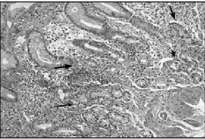
Figure 1 – Oxyntic gastric mucosa of patient with advanced atrophic body showing: diffuse mononuclear cell infiltration of the entire thickness of the lamina propria; few remained acidopeptic glands (short arrow), and pseudopyloric metaplasia (long arrow) (HE staining, 100×)
HE: hematoxylin and eosin.
Figure 2 – Immunoreactive cells to chromogranin in diffuse (long arrow) and nodular (short arrow) endocrine cell hyperplasia of patient with ABG (IHQ staining, 400×)
ABG: atrophic body gastritis; IHQ: immunohistochemical.
by chromogranin staining. Fifty-one patients (94.4%) had multiple hyperplasic endocrine nodules in the lamina propria in addition to diffuse endocrine hyperplasia, and three (5.6%) presented diffuse endocrine cell hyperplasia without the presence of hyperplasic nodules. Some of the hyperplasic nodules were reasonably apparent in the HE sections, but most of them were detected after chromogranin staining (Figure 2). The immunohistochemical expression of ghrelin and preproghrelin was variably present in hyperplasic endocrine cells from all patients studied (Figure 3).
Most of the hyperplasic nodules also presented variable number of immunoreactive cells to chromogranin, ghrelin and preproghrelin, but only those nodules with more than 50% of immunoreactive cells were considered for comparative analysis between the different antibodies
Table 1
Immunohistochemical expression of
ghrelin and preproghrelin in glands
with intestinal metaplasia and in glands with
pseudopyloric metaplasia in patients
with ABG
Type of
metaplasia
n
Ghrelin
n (%)
Preproghrelin
n (%)
Intestinal 42 4 (9.5%) 9 (21.4%)
Psudopyloric 51 37 (72.5%) 26 (50.9%)
ABG: atrophic body gastritis.
Table 2
Number of endocrine hyperplasic nodules
per patient containing more than 50%
of immunoreactive endocrine cells to
chromogranin A and ghrelin
Immunoreaction
to
Mean
± SD
Paired
t test
p
Chromogranin A
8.6 ± 10.1
3.01 0.004
Ghrelin 5.1 ±
6.4 SD: standard deviation.
Table 3
Number of endocrine hyperplasic nodules
per patient containing more than 50%
of immunoreactive endocrine cells to
chromogranin A and preproghrelin
Immunoreaction
to
Mean
± SD
Paired
t test
p
Chromogranin A
8.6 ± 10.1
2.42 0.0189
Preproghrelin 5.6 ±
6.7 SD: standard deviation.
used. The mean number of hyperplasic nodules per patient demonstrated by chromogranin staining was 8.6. Most of these hyperplasic nodules presented ghrelin- and preproghrelin-immunoreactive cells, 5.1 (p = 0.004) and 5.6 (p = 0.018), respectively (Tables 2 and 3).
Discussion
The atrophy of specialized glands of the gastric mucosa associated with chronic inflammation is a common pathological condition in humans. In general, the more severe cases of atrophy of the gastric mucosa do not involve diagnostic difficulties by the two methods commonly used, i.e., endoscopic and histopathological examinations. The current importance of the recognition of gastric mucosal atrophy is the frequency at which this condition is associated with the development of precancerous lesions, i.e., intestinal metaplasia and dysplasia of the gastric epithelium, which may develop into gastric cancer in the sequence known as Correa cascade(4). The two pathological conditions most frequently associated with the development of gastric mucosa atrophy are H. pylori -associated multifocal chronic gastritis and ABG, which in most cases has autoimmune etiology. Being selective for the oxyntic mucosa, autoimmune gastritis leads to progressive loss of parietal cells and chief cells, resulting in the majority of clinical manifestations presented by patients in the later stages of the disease, due to the occurrence of achlorhydria, hypergastrinemia, anemia and their consequences.
It is known that the development of autoimmune gastritis, with or without pernicious anemia, is more common among members of one family in successive generations. Although this is not clearly explained, it is assumed that there is a propensity to develop anti-parietal cell antibodies in genetically predisposed individuals. The existence of genetic predisposition contributing to the induction of atrophy of the oxyntic glands is
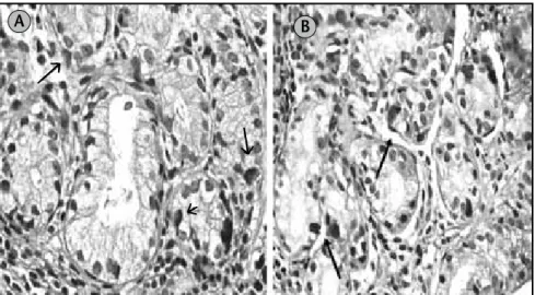
Figure 3 – Diffuse hyperplasic immunoreactive cells (arrows) to ghrelin (A) and to preproghrelin (B) in pseudopyloric metaplasia of gastric mucosa of patient with ABG (IHQ staining, 400×)
ABG: atrophic body gastritis; IHQ: immunohistochemical. B A
suspected since ABG is often associated with other diseases with an immunological background. Moreover, racial preferences seem to exist since this disease appears to be significantly more conspicuous in Caucasian than in Asian patients(1, 7, 16, 21). Despite all of this evidence, the mechanisms of inheritance and the importance of environmental factors related to ABG are not known. Among the putative environmental factors, the bacterium H. pylori has been cited as the most important(17). All patients in this study were negative for H. pylori infection which is expected to occur in cases of severe atrophy of the gastric mucosa, regardless its etiology. Among the patients studied here, women prevailed over men and age was similar in both groups, 58 ± 19 and 53 ± 14 years, respectively. These data are similar to most of those reported in the western literature(3, 11).
aspects of these hyperplasic lesions may be useful in improving our understanding of the pathogenesis of neuroendocrine tumors of the stomach. In a previous study we observed that both ghrelin peptide and its precursor molecule preproghrelin are present in a large number of neoplasic endocrine cells in gastric neuroendocrine tumors associated with ABG(14). Other studies have shown that ghrelin is present not only in neuroendocrine tumors of the stomach but also in neuroendocrine tumors in other locations(6, 13).
It is known that ghrelin-producing cells are present in different tissues, including the intestinal mucosa, however sparingly and inconstantly. In contrast, in the body gastric mucosa these cells reach their highest density and are considered to be the main source of plasma ghrelin(8, 9). In the present study ghrelin- and preproghrelin-immunoreactive cells showed similar frequencies both in areas of intestinal metaplasia (9.5% and 21.4%, respectively) and in areas of pseudopyloric metaplasia (72.5% and 50.9%, respectively). These results show that ghrelin-producing cells was uncommon in areas of intestinal metaplasia and, conversely, was frequent in areas of pseudopyloric metaplasia, the latter showing gastric differentiation. Therefore, regarding the differentiation of endocrine elements, one may assume that the areas of intestinal metaplasia and pseudopyloric metaplasia, present in the gastric mucosa of patients with
ABG, and consequently with autoimmune gastritis, should still retain many of the genetic characteristics of the original tissues they represent.
We conclude that the ghrelin- and preproghrelin-immunoreactive cells are frequently found in the different types of endocrine hyperplasia of the gastric mucosa in advanced stages of ABG. However, these peptides should not be used as immunohistochemical markers for the endocrine hyperplasia that occurs in ABG, since they are significantly less sensitive than chromogranin. Ghrelin- and preproghrelin-immunoreactive cells occur in a relatively frequent manner in glands with pseudopyloric metaplasia and are uncommon in glands with intestinal metaplasia, two types of histological changes frequently seen in ABG. These cells are frequently present in nodular endocrine hyperplasia of patients with ABG. However, the extent to which the proliferation of hyperplasic nodules and their progression to cancer are under the influence of these peptides remains a matter for further research.
Acknowledgements
The authors thank Dr. Eugênio M. A. Goulart for the helpful technical assistance in statistics.
References
1. ALONSO, M. et al. Plasma ghrelin concentrations in type 1 diabetic patients with autoimmune atrophic gastritis.
Eur J Gastroenterol, v. 157, p. 763-9, 2007.
2. BALDELLI, R. et al. Ghrelin: a new hormone with endocrine and non-endocrine activities. Pediatr Endocrinol Rev, v. 2, n. 1, p. 8-14, 2004.
3. CHLUMSKA, A. et al. Autoimmune gastritis. A clinicopathologic study of 25 cases. Cesk Patol, v. 41, p. 137-42, 2005.
4. CORREA, P. Chronic gastritis as a cancer precursor. Scand J Gastroenterol Suppl, v. 104, p. 131-6, 1984. 5. DATE, Y. et al. Ghrelin, a novel growth
hormone-releasing acylated peptide, is synthesised in a distinct endocrine cell type in the gastrointestinal tracts of rats and humans. Endocrinology, v. 141, p. 4255-61, 2000.
6. IWAKURA, H. et al. Ghrelin expression in islet cell tumors: augmented expression of ghrelin in a case of glucagonoma with multiple endocrine neoplasm type I.
J Clin Endocrinol Metab, v. 87, p. 4885-8, 2002.
7. KEKKI, M. et al. Classification, principles and genetics of chronic gastritis. Scan J Gastroenterol, v. 22, Suppl. 141, p. 1-28, 1987.
8. KOJIMA, M. et al. Ghrelin is a growth-hormone-releasing acylated peptide from stomach. Nature, v. 402, p. 656-60, 1999.
9. KOJIMA, M.; HOSODA, H.; KANGAWA, K. Purification and distribution of ghrelin: the natural endogenous ligand for the GH (growth hormone) secretagogue receptor.
Horm Res, v. 56, Suppl., p. 93-7, 2001.
10. KORBONITS, M. et al. The expression of the GHS receptor ligand ghrelin in normal and abnormal human pituitary and other NE tumors. J Clin Endocrinol Metab, v. 86, n. 2, p. 881-7, 2001.
11. LAHNER, E. et al. Occurrence and risk factors for autoimmune thyroid disease in patients with atrophic body gastritis. Am J Med, v. 121, p. 136-41, 2008. 12. LELY, A. J. et al. Biological, phisiological, pathophysiological
and pharmacological aspects of ghrelin. Endocr, v. 25,
13. LEONITIOU, C. A. et al. Ghrelin in neuroendocrine organs and tumours. Pituitary, v. 10, n. 3, p. 213-25, 2007. 14. MOREIRA, L. F.; CARVALHO, M. R. N.; BARBOSA, A. J. A.
Ghrelin-, and pré-proghrelin immunoreactive cells in gastric neuroendocrine tumors associated with atrophic body gastritis. J Bras Patol Med Lab, v. 46, p. 329-34, 2010.
15. PAPOTTI, M. et al. Grelin-producing endocrine tumors of the stomach and intestine. J Clin Endocrinol Metab, v. 86, n. 10, p. 5052-9, 2001.
16. PARK, J. Y. et al. Gastric lesions in patients with autoimmune metaplastic atrophic gastritis (AMAG) in a tertiary care setting. Am J Surg Pathol, v. 34, p. 1591-8, 2010.
17. PRESOTTO, F. et al. Helicobacter pylori infection and gastric autoimmune diseases: is there a link?
Helicobacter, v. 8, p. 578-84, 2003.
Mailing address
Alfredo J. A. Barbosa Faculdade de Medicina da UFMG Laboratório de Patologia Digestiva e Neuroendócrina
Av. Alfredo Balena, 190
CEP: 30130-100 – Belo Horizonte-MG
18. RINDI, G. et al. Gastric carcinoids and neuroendocrine carcinomas: pathogenesis, pathology and behavior – clinicopathologic analysis of 205 cases. World J Surg, v. 20, p. 168-72, 1996.
19. RINDI, G. et al. Ghrelin expression in gut endocrine growths.
Histochem Cell Biol, v. 117, n. 6, p. 521-5, 2002. 20. SOLCIA, E. et al. Histopathological classification of
nonantralgastric endocrine growths in man. Digestion, v. 41, p. 185-200, 1988.
21. TOZZOLI, R. Recent advances in diagnostic technologies and their impact in autoimmune diseases. Autoimmun Rev, v. 10, p. 80-3, 2007.
22. WANG, H. S. et al. Elevated serun ghrelin exerts an orexigenic effect that may maintain body mass index in patients with metastatic neuroendocrine tumors.