Introduction
Sciatic nerve lesions are characterized by the loss or diminution of sensibility and motricity in the innervated areas, which can compromise an individual’s functional activities1,2. Common symptoms of these nervous injuries include dormancy, paresthe-sia, weakness and atrophy of the effector muscles, as well as the impairment of the normal functions and movements of the lower limbs, since this musculature affects the motricity of the joints3, 4
, including the ankle, which is responsible for providing a stable base of support for the body, in addition to effectively propelling it during locomotion5.
Thus, nerve lesions lead to the disuse of the limb and, as a consequence, there is a reduction in the mechanical stimulus necessary to maintain the morphological properties of the joint6-9, which can lead to rigidity of the joint capsule and consequent restriction in movement10, as well as atrophic changes in the ar-ticular cartilage with a reduction in its thickness and the number of chondrocytes11,12. Moreover, according to Hui, McCarty, Masuda, Firestein, Sah13, the composition and function of synovial luid are also altered in cases of joint injury or disease. These changes can result in reduced ability to lubricate the articular cartilage, which can contribute to its deterioration, as already described in the literature12. Free swimming14,15, swimming associated with electrical stimulation16 and resistance exercises17,18,19 have been investigated as therapeutic modalities in peripheral nerve lesions.
As with nerve damage, some studies have demonstrated that physical exercise promotes protection20 or the recovery of joint components21, such as a reduction in joint stiffness,
repair to articular cartilage and improvement in synovial low
and nutrition22,23,24.
Despite the motor disuse promoted by nerve lesions, there are no reports in the literature about the effects of sciatic nerve injury on the morphology of the articular cartilage and synovial membrane of the ankle. Furthermore, despite the potential of physical exercises to help in the recovery of articular constituents, a therapeutic protocol for joint protection or rehabilitation after nerve injury has not been established. Thus, it was necessary to carry out a study to elucidate the implications of peripheral nerve injury on joint morphology, as well as to prove the effect of exercise on joint function after nerve injury. Consequently, this study analyzed the effects of sciatic nerve compression and treatment with resisted stair climbing exercise on the morphol-ogy of the ankle joint of Wistar rats.
Methods
Characterization of samples
Thirty-two Wistar rats were used in the experiments. They were aged 10 ± 1 week, had an average weight of 376±22 grams, and Original article (short paper)
Resistance exercise recovers the structure of cartilage and synovial
membrane of the ankle joint of rats after sciatic compression
Lizyana Vieira Keli Lovison Regina Inês Kunz Juliana Sobral Antunes Gladson Ricardo Flor Bertolini
Rose Meire Costa Brancalhão Lucinéia de Fátima Chasko Ribeiro
Universidade Estadual do Oeste do Paraná, Cascavel, PR, Brazil
Abstract — Aim: to determine the effects of sciatic compression and treatment with resistance exercise on the morphology of the ankle joint of Wistar rats. Methods: 32 male rats, aged 10 ± 1 week, weighing 376±22 grams were divided into the following four groups (n=8/group): CG (control), LG (lesion), EG (exercise) and LEG (lesion and exercise). Three days after sciatic compression, the animals in the EG and LEG were submitted to resistance exercise
by climbing stairs (ive days/week) for three weeks and a load of 100 grams was added. The exercise was carried out
in two sets of ten consecutive ascents of the steps. The ankle joint tissues were analyzed for their morphometry and morphology using light microscopy. Results: Regarding the number of chondrocytes, the LG and EG had more cells in the anterior articular cartilage in the tibia (62 and 43%) and in the talus (57 and 45%) when compared to the CG. In the LEG there was a 25% and 26% reduction of chondrocytes in the anterior cartilage in the tibia and talus when compared
to the LG. Changes were observed in the tibia and talus in the LG, with the presence of locculation, invagination of the
subchondral bone, discontinuity of tidemark and pannus covering the subchondral bone in the talus, as well as changes in the synovial membrane. These alterations were minimized in the articular cartilage and synovial membrane in the LEG. Conclusions: exercise restores the tissue morphology of ankle joint in Wistar rats after sciatic compression.
were obtained from the Central Animal Facility of the State University of the West of Paraná (UNIOESTE), Cascavel, Brazil. The animals were kept in polypropylene cages under controled environmental conditions, a light/dark cycle of 12 hours, tem-perature of 24 ºC ± 1 ºC, with free access to food and water. All the methodological procedures were approved by the Ethics Committee on Animal Use (CEUA) at UNIOESTE Cascavel.
After the period of environmental adaptation, the animals were randomly divided into the following four groups (n = 8/ group): control group (CG); lesion group (LG), composed of animals that were submitted to an experimental model of sciatic nerve compression; exercise group (EG), composed of animals that were submitted to resistance exercise by climbing stairs; and lesion and exercise group (LEG), composed of animals submit-ted to a nerve compression model and treasubmit-ted with resistance exercise by climbing stairs.
Experimental model of sciatic compression
The compression model adopted in this study is classiied as an
axonotmesis-type peripheral nerve injury25 and it was conducted in the animals in the LG and LEG groups.
After weighing the animals, intraperitoneal anesthesia with ketamine (95 mg/Kg) and xylazine (12 mg/Kg) was applied and the state of consciousness of the animals was checked (noted by the absence of motor response to the clamping of the tail and interdigital folds). They were then placed in the prone position with hindlimbs in abduction. Trichotomy of the middle third of the right thigh was performed and then an incision was made
parallel to the ibers of the femoral biceps muscle, exposing
the sciatic nerve. The nerve was pressed for 30 seconds with hemostatic forceps. The clamping pressure was standardized for all animals, using as reference the closure of the forceps second degree, with all pinching performed by the same researcher25.
At the end of the nerve compression procedure, the nerve was anatomically repositioned and the cutaneous plane was
sutured with simple stitches using monoilament nylon yarn.
Poly(vinylpyrrolidone)-iodine (Povidine®) was applied over the incision and the animals were then housed in the same pre-surgical conditions.
Exercise protocol
The resistance exercise protocol was adapted from Hornberger and Farrar18. A vertical ladder made of wood with 67 iron steps was used, spaced 1 cm between each step. The total height was 118 cm and the width was 20.5 cm; the inclination was kept at approximately 60 degrees. At the top of the ladder there was a cage (18.5 cm high and 15 cm wide) which served as a shelter for the rest periods between the sets of exercises.
Before the sciatic nerve compression surgery, all the animals were submitted to a familiarization protocol with the ladder so that they would all present the same conditions after surgery.
The training consisted of three trials per day, ive days a week
for two weeks, totalling ten days of adaptation. A load of 100
grams was added in the second week, which was attached to the proximal portion of the animal’s tail. Because the literature contains differences in relation to the burden on animals17,26 this study used an overload of 35-40% of the total body weight of the animal.
The stair climbing exercise started on the third day after surgery. Animals from the EG and LEG groups were
submit-ted to resistance exercise ive days a week for three weeks.
The exercise was carried out in two sets of ten consecutive ascents of the steps, with an interval of 60 seconds between sets for rest.
The standardization of the treatment beginning on the third day after surgery was based on the study by Gaffuri et al.27 and the period of 21 days of treatment was based on Gorio, Carmignotto, Finesso, Polato, Nunzi28.
Euthanasia of animals and histological analysis
Twenty-four hours after the end of treatment the animals were weighed, anesthetized with ketamine (95 mg/Kg) and xylazine (12 mg/Kg), and euthanized. The right hindlimb was dissected and the ankle joints were collected. These were subsequently
ixed in Metacarn, washed in distilled water, and decalciied
in 5% trichloroacetic acid (TCA) for about 15 days. Then the
protocol for ixing in parafin and microtome was performed,
with 7 µm cuts using an Olympus® CUT 4055 microtome. The slides of the sagittal section of the joint were stained with hematoxylin and eosin29 for general morphology and with Fast Green30 for the articular cartilage.
The morphological and morphometric analyses of the articular tissues were performed using a light microscope (Olympus®) and the visual ields of interest were photomicro -graphed using 20 and 40 lenses. The protocol for the measure-ment of the articular cartilage thickness and the number of chondrocytes was the same as used by Kunz, Coradini, Silva, Bertolini, Brancalhão, Ribeiro12, which deined the areas of interest as the regions of the anterior joint (P1), middle joint (P2) and posterior joint (P3). Apart from the morphometry, the morphological characteristics of the articular cartilage were also observed, such as the appearance of the articular
surface and the presence of cracks, pannus, locculation and
cell clones, as well as the subchondral bone and the syno-vial membrane.
Statistical analysis
Results
Histomorphometric analysis of the thickness of the articular cartilage
In the analysis of the thickness of the articular cartilage there
were no signiicant differences between the measurements of
the joint regions (P1, P2 and P3) of both the talus and the tibia in both groups (Table 1).
Histomorphometric analysis of the number of
chondrocytes in the articular cartilage of the tibia and the talus
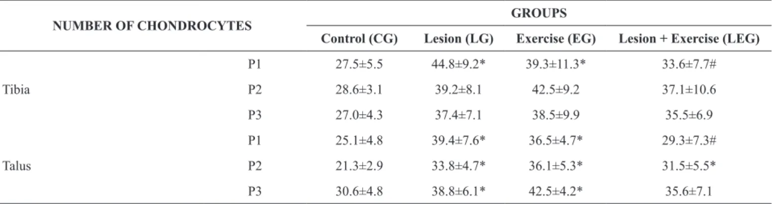
Regarding the number of chondrocytes in the tibia, there were
only signiicant differences between groups in relation to P1.
As regards this articular region, the LG and EG groups had
62% and 43% more cells in the cartilage respectively, when compared to the CG group. The animals in the LEG had 25% less chondrocytes in the anterior articular region compared with the LG. In the other articular regions of the tibia (P2 and P3) no
statistically signiicant differences were observed in relation to
the number of chondrocytes (Table 2).
Regarding the talus, there was an increase of 57% and 45% in the number of chondrocytes in the anterior articular cartilage (P1) in the animals in the EG and LG, respectively, when com-pared to the CG. In the LEG there was a 26% reduction in the number of chondrocytes in relation to the LG. Regarding the middle articular region (P2), all the groups showed higher cell density compared to the CG: 59% in the LG, 69% in the EG and 48% higher in the LEG. In terms of the posterior articular region (P3), there was a 27% and 39% respective increase in the LG and EG, respectively in the number of chondrocytes compared to the CG.
Table 1. Thickness of articular cartilage (μm) of the tibia and talus.
THICKNESS OF CARTILAGE GROUPS
Control (CG) Lesion (LG) Exercíse (EG) Lesion + Exercise (LEG)
Tibia
P1 142.3±36.4 175.34±24.7 185.6±29.5 189.1±29.8
P2 177.7±29.4 149.5±9.0 175.8±26.1 186.3±42.8
P3 151.6±42.2 134.4±38.3 117.9±36.9 153.9±48.3
Talus
P1 135.6±19.2 143.6±28.3 152.3±23.6 165.4±15.9
P2 142.9±18.7 154.8±13.8 140.7±23.7 142.0±33.7
P3 144.3±25.2 161.9±62.4 149.8±42.8 170.8±44.0
P1 – anterior articular region; P2 – middle articular region; P3 – posterior articular region. The results are expressed as mean ± standard deviation. There was no signiicant difference.
Table 2. Number of chondrocytes in the articular cartilage of the tibia and talus.
NUMBER OF CHONDROCYTES GROUPS
Control (CG) Lesion (LG) Exercise (EG) Lesion + Exercise (LEG)
Tibia
P1 27.5±5.5 44.8±9.2* 39.3±11.3* 33.6±7.7#
P2 28.6±3.1 39.2±8.1 42.5±9.2 37.1±10.6
P3 27.0±4.3 37.4±7.1 38.5±9.9 35.5±6.9
Talus
P1 25.1±4.8 39.4±7.6* 36.5±4.7* 29.3±7.3#
P2 21.3±2.9 33.8±4.7* 36.1±5.3* 31.5±5.5*
P3 30.6±4.8 38.8±6.1* 42.5±4.2* 35.6±7.1
P1 – anterior articular region; P2 – middle articular region; P3 – posterior articular region. The results are expressed as mean ± standard deviation; *p<0.05 when compared to control group; #p<0.05 when compared to lesion group).
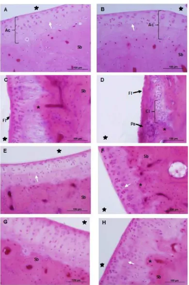
Morphological analysis of ankle joint
The ankle joints of the CG showed characteristic morphology in the tibia and talus, with smooth articular surface and regular
organization of the cellular characteristics (Fig. 1A and 1B). Higher cell density was visible in the surface area, with the
chon-drocytes arranged in horizontal clusters with a lattened aspect.
isolated layout and isogenous groups. The chondrocytes were arranged in gaps that corresponded to the deep zone, separated
from the calciied zone by a line, the tidemark.
However, in the LG, morphological changes were observed in
the cartilage of the tibia such as locculation, invagination of the
subchondral bone, and discontinuity of the tidemark (Fig. 1C). With regard to the talus, the presence of cell clones was noted,
as well as locculation, pannus covering the subchondral bone
(Fig. 1D), invagination of the subchondral bone, an increase in the number of chondrocytes, and the tidemark was discontinuous.
In the EG there were no changes in the morphology of the articular cartilage of the tibia (Fig. 1E). In the articular cartilage of the talus of animals subjected to exercise, a slight discontinuity of the tidemark was observed, mainly in the anterior articular region (P1), where an increase in the number of chondrocytes and invagination of the subchondral bone was also observed (Fig. 1F).
In the LEG a slight invagination of the subchondral bone in the articular cartilage of the tibia and the talus was present in the region corresponding to P1, as well as a slight increase in the number of chondrocytes and discreet disorganization of the tidemark (Fig. 1G and 1H).
The synovial membrane in the CG presented normal char-acteristics, i.e. two to three layers of type A and type B syn-oviocytes in the synovial intima, and connective tissue with a predominance of adipose cells in the subintima (Fig 2A). In the LG the synovial membrane appeared slightly thicker, with the intima disorganized in relation to the distribution of synovio-cytes, and in the subintima there was discrete substitution of
the type of connective tissue, from adipose to ibrous (Fig 2B).
The synovial membrane of the animals in the EG showed no change from normal morphology (Fig 2C). Recovery of tissue organization was observed in the LEG (Fig. 2D).
Fig. 1: Photomicrographs of the ankle joint of Wistar rats, showing the articular cartilage. Sagittal section, hematoxylin and eosin staining. A, C, E and G - cartilage of the tibia; B, D, F and H - cartilage of the talus. Control group (A and B), Lesion group (C and D), Exercise group (E and F), and Lesion and Exercise group (G and H). In A and B, showing the articular cartilage (Ac) and the presence of tidemark (white arrow) with normal
appearance. In C, the presence of locculation (Fl) on the surface of the cartilage and invagination of the subchondral bone (asterisk). In D, forma
-tion of pannus (Pa) and the presence of cell clones (Cl) and loccula-tion (Fl) is more evident. In E and F, slight discontinuity of tidemark (white
Discussion
The present study is a precursor in describing the morphological changes in the ankle joint resulting from a nerve injury, such
as locculation, subchondral bone invagination and tidemark discontinuity in the tibia, and presence of cell clones, loccula -tions, invagination and panus in the subchondral bone of the talus, in addition to the increase in the number of chondrocytes. Since the nerve lesions interrupt neuromuscular communica-tion, which can lead to atrophy of the effector muscles8,31,32, the biomechanics of the ankle are compromised, causing a reduction in the range of motion of the joint33 and a decrease in weight loss in the affected limb. As a consequence, changes in joint structures, such as those cited in the present study, occur
because of the inluence that load transfer plays on the joint
tissues homeostasis34,35.
In both tibia and talus, the thickness of the articular car-tilage was not changed by the sciatic nerve lesion (LG), stair climbing exercise (EG) or exercise after lesion (LEG). Trudel,
Jabi, Uhthoff36 also found no change in thickness, although the articular cartilage did become irregular in the analyzed locations. Hagiwara, Ando, Chimoto, Saijo, Ohmori-Matsuda, Itoi37 noted that the thickness of the articular cartilage of the knee increased in the transition region, which may have been related to the lack of use of the limb and decreased lubrication of the joint. Roos and Dahlberg22 recorded an increase in the thickness of the knee cartilage after cardio and strengthening exercises. Corroborating the results of the present study, Kunz, Coradini, Silva, Bertolini, Brancalhão, Ribeiro12 also found no change in the thickness of articular cartilage, suggesting that the ankle joint is more resistant to changes in cartilage thickness.
The increase in the number of chondrocytes that was observed in the animals in the LG and LEG was also observed in studies by Renner, Carvalho, Soares, Mattiello-Rosa11 and Del Carlo et al.23. Other authors found a reduction in cellulardensity24,36,37. According to Whiting and Zernicke2, prolonged physical exercise in animals can produce hypertrophy of the chondrocytes and an increase in their number. The articular kinematics may promote Fig 2: Photomicrographs of the synovial membrane of the ankle joint of Wistar rats. Sagittal section, hematoxylin and eosin staining. Control group (A), Lesion group (B), Exercise group (C) and Exercise and Lesion group (D). In A, membrane with thin synovial intima (black arrow) containing synoviocytes and subintima (S), with a predominance of adipose cells (Ad). In B, thickening of the synovial membrane, which is
discretely ibrous (asterisk) with less adipocytes (Ad). In C and D, synovial membrane with the intima showing normal appearance (black arrow),
the reduction or increase in the number of chondrocytes38, with a consequent alteration in the composition and organization of the cartilage matrix39. Thus, both the decrease in weight bearing caused by the nervous injury and the increase generated by the exercise, mechanically change the cartilage, resulting in changes in the density of chondrocytes.
Furthermore, chondrocytes are mechano-sensitive cells that can respond to a variety of stimuli, including growth factors, mechanical loads, piezoelectric forces and hydrostatic pres-sures. These cells synthesize glycosaminoglycans (GAGs) and type II collagen, which is rich in extracellular matrix that is essential for the maintenance, enhancement and regeneration of healthy cartilage. In situations of stress, such as physical
exercise, the upregulation of pro-inlammatory genes can lead
to cell proliferation and also compromise the synthesis of extracellular matrix, which leads to loss of cartilage integrity and early signs of lesions40,41.
In addition to changing the number of chondrocytes, nerve injury caused the appearance of locculation and invagination of the subchondral bone in the tibia in the LG. In the talus, these changes were more marked, with the presence of cell clones and pannus covering the subchondral bone. According to Melo, Nunes, Rezende, Gomes, Malm, Gheller42, cell clones represent the hyperactivity of chondrocytes in the mid and deep zones in response to abnormal stress caused by an imbalance in the distribution of forces on the articular surface. Nagai et al43 analyzed the surface of knee cartilage after immobiliza-tion and observed a progressive degeneraimmobiliza-tion of the cartilage,
with hypertrophy, cell degeneration, locculation and changes
in the density of coloring. These changes may not be equally distributed throughout the length of the cartilage because of the angle at which the joint was immobilized as well as the duration of stress applied.
Changes in the synovial membrane of animals submitted to nerve damage, such as thickening, disruption of the intima in relation to the distribution of synoviocytes, as well as discrete
replacement of connective adipose tissue from fatty to ibrous
in the sub-intima, have also been described by Melo, Nunes, Rezende, Gomes, Malm, Gheller42, Ando et al.44 and Trudel, Jabi, Uhthoff36. Articular rigidity and a decreased range of motion, due to lower weight bearing caused by nerve injury, may be responsible for thickening of the synovial membrane23, leading to a lower level of proteoglycans45, which interferes with the
production of synovial luid and consequently reduces the sup -ply of nutrients to the cartilage23,35. According to Kojima, Hoso, Watanabe, Matsuzaki, Hibino, Sasaki46, these changes may be
related to the presence of locculation, corrosion and cracks in
the articular cartilage. Taking into consideration the indings of studies by Hadler-Olsen, Fadnes, Sylte, Uhlin-Hansen, Winberg47 and Takaishi, Kimura, Dalah, Okada, D’Armiento48
, thechanges that were observed in the present study in the LG and EG may have been the result of an increase in the rate of synthesis and the secretion of matrix-degrading enzymes by chondrocytes, as well as the production of metalloproteases that are capable of unfolding collagen and proteoglycans, which release their
fragments in the articular luid, thereby weakening the cartilage matrix. Synovial luid and the ibrous articular capsule respond
to these fragments and other biochemical mediators, such as cytokines and leukotrienes, which leads to changes in the other components of the joint. In relation to the animals in the LEG group, in general terms, tissue reorganization of the articular cartilage and the synovial membrane were observed, indicat-ing that the resistance exercise accelerated the recovery of the articular constituents. According to Ando, Hagiwara, Chimoto, Hatori, Onoda, Itoi30, the restoration of movement reduces
ar-ticular stiffness and improves the low of synovial luid in the
articular cavity, which promotes the nutrition of the cartilage and its consequent regeneration. Physical exercise has been seen as an important tool in maintaining the integrity of the cartilage, preventing degeneration and maintaining its biomechanical properties49. Compressive force, or shear force at the joint, can be produced by exercise, which leads to cartilage regeneration37 and exerts a chondroprotective effect50.
Hornberger and Farrar18 subjected animals to resistance exercise by climbing stairs once every three days for eight weeks, with increasing load, and described an animal model that simulates the training parameters and physiological adaptations observed in humans. Cassilhas, Reis, Venâncio, Fernandes, Tuik, Mello26 used the same exercise model with increasing loads of 50% to 100% of the total body weight of the animal and found hypertrophy in the examined muscles.
Despite the fact that stair climbing exercise is established in the literature as resistance, it is not widely applied as a thera-peutic modality after injury. Following a protocol similar to the one used in the present study, Vasilceac, Souza and Mattiello51 startedtreatment using stair climbing two weeks after inducing osteoarthritis to the knee. This was effective in remodeling
ar-ticular collagen; it produced beneits for the cartilage as well as modiications in the content of chondrocytes and proteoglycans. Thus, exercise has proved to be beneicial for articular morphol -ogy when initiated during the acute phase, as seen in the present study, and also in the chronic phase after injury.
According to Martins et al.52 and Marimoto et al.53, physical exercise reduces pain and promotes the functional recovery of joints after sciatic nerve injury, as well as reducing the articular limitation that is present after periods of immobilization. Jang and Lee54 found that treadmill exercise after experimental sci-atic nerve injury improved ankle biomechanics, as well as knee and hip joints during walking and they concluded that exercise produces analgesic effects and restores motor function when started at an early stage55.
Despite the innovative results of the present study regarding the effect of sciatic nerve compression and the practice of stair climbing exercise on the morphology of the articular cartilage and synovial membrane of the ankle, we believe that the absence of a functional test to assess the range of articular movement,
which could establish the function of the ankle, can be identiied
as a limitation of this study.
Conclusions
the anterior articular region of the tibia, minimized the invagina-tion of the subchondral bone, as well as the presence of panus,
cellular clones and locculation in the articular cartilage. In the
synovial membrane, the epithelioid arrangement of the intima and adipose of the subintima was re-established. Thus, it was
veriied that the exercise that was applied restored the tissue
morphology of the ankle joint of Wistar rats after sciatic injury.
References
1. Araujo RT. Efeitos do treinamento físico na água aquecida so-bre a recuperação funcional e a regeneração nervosa periférica após a lesão do nervo ciático em ratos machos adultos. Porto Alegre. Dissertação [Pós-Graduação em Ciências Biológicas: Neurociências] - Universidade Federal do Rio Grande do Sul; 2008.
2. Whiting, WC, Zernicke, RF. Biomecânica da lesão músculo es-quelética. Rio de Janeiro: Guanabara-Koogan; 2001.
3. Hsueh YY, Chang YJ, Huang TC, Fan SC, Wang DH, Chen JJJ, Wu CC, Lin SC. Functional recoveries of sciatic nerve regeneration by combining chitosan-coated conduit and neurosphere cells induce from adipose-derived stem cells. Biomaterials. 2014;35:2234-44. 4. Mohammadi R, Tirgan MA, Amini K. Dexamethasone topically
accelerates peripheral nerve repair and target organ reinnervation: A transected sciatic nerve model in rat. Injury. 2013;44:565-69. 5. Teixeira LF, Olney SJ. Anatomia funcional e biomecânica das
articulações do tornozelo, subtalar e médio-társica. Rev Fisioter Univ São Paulo. 1997;4:50-65.
6. Carvalho DCL, Caravalho MM, Cliquet Junior A. Osteoporose por Desuso: Aplicação na Reabilitação do Lesado Medular. Acta Ortop Bras. 2001;9:34-43.
7. Galvão MR, Del Carlo RJ, Natali AJ, Viloria MIV, Monteiro BS, Del Carlo KN. Imobilização articular prolongada e remobilização. Fisioter Bras. 2008;9:157-63.
8. Tanaka S, Tsubaki A, Tachino K. Effect of exercise training after partial denervation in rat soleus muscles. J Phys Ther Sci. 2005;17:97-101.
9. Yuen EC, So YT. Sciatic Neuropathy. Neurol Clin. 1999;17:617-31. 10. Ando A, Suda H, Hagiwara Y, Onoda, Y, Chimoto E, Itoi E.
Remobilization does not restore immobilization-induced adhesion of capsule and restricted joint motion in rat knee joints. Tohoku J Exp Med. 2012; 227:13-22.
11. Renner AF, Carvalho E, Soares E, Mattiello-Rosa S. The effect of a passive muscle stretching protocol on the articular cartilage. Osteoarthritis Cartilage. 2006;14:196-202.
12. Kunz RI, Coradini JG, Silva LI, Bertolini GRF, Brancalhão RMC, Ribeiro LFC. Effects of immobilization and remobilization on the ankle joint in Wistar rats. Braz J Med Biol Res. 2014;47:842-9. 13. Hui AY, McCarty WJ, Masuda K, Firestein GS, Sah RL. A systems
biology approach to synovial joint lubrication in health, injury, and disease. WIREs Syst Biol Med. 2012;4:15-37.
14. Possamai F, Siepko CM, André ES. Investigação dos efeitos do exercício terapêutico sobre a regeneração nervosa periférica. Acta Fisiátr. 2010;17:142-7.
15. Sobral LL, Oliveira LS, Takeda SYM, Somazz MC, Montebelo MIL, Teodori RM. Immediate versus late exercise for rat sciatic
nerve regeneration after axonotmesis: histomorphometric and functional analysis. Braz. J. Phys. Ther. 2008;12:311-6.
16. Oliveira LS, Sobral LL, Takeda SY, Betini J, Guirro RR, Somazz MC, Teodori RM. Electrical stimulation and swimming in the
acute phase of axonotmesis: their inluence on nerve regeneration
and functional recovery. Rev Neurol. 2008;47:11-5.
17. Ilha J, Araujo RT, Malysz T, Hermel EE, Rigon P, Xavier LL, Achaval M. Endurance and resistance exercise training
pro-grams elicit speciic effects on sciatic nerve regeneration after
experimental traumatic lesion in rats. Neurorehabil Neural Repair. 2008;22:355–66.
18. Hornberger TAJ, Farrar RP. Physiological hypertrophy of the FHL muscle following 8 weeks of progressive resistance exercise in the rat. Can J Appl Physiol. 2004; 29:16-31.
19. Van Meeteren NLU, Brakkee JH, Hamers FPT, Helders PJM, Gispen WH. Exercise training improves functional recovery and motor nerve conduction velocity after sciatic nerve crush lesion in the rat. Arch Phys Med Rehabil. 1997;78:70–7.
20. Newton PM, Mow VC, Gardner TR, Buckwalter JA, Albright JP. The effect of life long exercise on canine articular cartilage. Am J Sport Med. 1997;25:282–7.
21. Dias CN, Renner AF, Santos AA, Vasilceac FA, Mattiello SM. Progression of articular cartilage degeneration after application of muscle stretch. Connect Tissue Res. 2012;53:39-47.
22. Roos EM, Dahlberg L. Positive effects of moderate exercise on glycosaminoglycan content in knee cartilage: a four-month, randomized, controlled trial in patients at risk of osteoarthritis. Arthritis Rheum. 2005;52:3507-14.
23. Del Carlo RJ, Galvão MR, Viloria MIV, Natali AJ, Barbosa ALT, Monteiro BS, et al. Experimental immobilization and remobiliza-tion rat knee joints: clinical and microscopic study. Arq Bras Med Vet Zootec. 2007;59:363-70.
24. Ando A, Suda H, Hagiwara Y, Onoda Y, Chimoto E, Saijo Y, et al. Reversibility of immobilization-induced articular cartilage degeneration after remobilization in rat knee joints. Tohoku J Exp Med. 2011;224:77-85.
25. Possamai F, Pacheco DR, Santos TS, André ES. Repercussões morfológicas e funcionais do exercício sobre a regeneração ner-vosa periférica. Fisioter Mov. 2012; 25:617-27.
26. Cassilhas RC, Reis IT, Venâncio D, Fernandes J, Tuik S, Mello MT. Animal model for progressive resistance exercise: a detailed description of model and its implications for basic research in exercise. Motriz. 2013;19:178-84.
27. Gaffuri J, Meireles A, Rocha BP, Rosa CT, Artifon EL, Silva LI, et al. Physical exercise assessment as na analgesia factor in a sci-atica experimental model. Rev Bras Med Esporte. 2011;17:115-8. 28. Gorio A, Carmignotto G, Finesso M, Polato P, Nunzi MG. Muscle
reinnervation – II. Sprouting, synapse formation and repression. Neuroscience. 1983;8:403-16.
29. Junqueira LC, Junqueira LMMS. Técnicas básicas de citologia e histologia. São Paulo: Santos; 1983.
31. Lieber RL. Skeletal muscle structure, function & plasticity: the physiological basis of rehabilitation. Philadelphia: Lippincott Williams & Wilkins; 2002.
32. Cavalcante EVV, Silva LGM, Montenegro EJN, Filho NTP. Efeito da eletroestimulação no músculo desnervado de animais: revisão sistemática. Fisioter. Mov. 2012;25:669-78.
33. Moore KL, Dalley AF. Anatomia orientada para a clínica. 4ed. Rio de Janeiro: Guanabara Koogan, 2001.
34. Aigner T, Soeder S, Haag J. IL-1B and BMPS-interactive play-ers of cartilage matrix degradation and regeneration. Eur Cells Mater. 2006;12:49-56.
35. Vanwanseele B, Lucchinetti E, Stussi E. The effects of immobili-zation on the characteristics of articular cartilage: current concepts and future directions. Osteoarthritis Cartilage. 2002;10:408-19. 36. Trudel G, Jabi M, Uhthoff HK. Localized and adaptive
synovio-cyte proliferation characteristics in rat knee joint contractures sec-ondary to immobility. Arch Phys Med Rehabil. 2003;84:1350-6. 37. Hagiwara Y, Ando A, Chimoto E, Saijo Y, Ohmori-Matsuda K,
Itoi E. Changes of articular cartilage after immobilization in a rat knee contracture model. J Orthop Res. 2009;27:236-42. 38. Andriacchi TP, Mundermann A, Smith RL. A framework for
understanding the in vivo pathomechanics of osteoarthritis at the knee. Ann Biomed Eng. 2004;32:457-77.
39. Jortikka MO, Inkinen RI, Tammi MI, Parkkinen J, Haapala J, Kiviranta I, et al. Immobilization causes long-lasting matrix changes both in the immobilized and contralateral joint cartilage. Ann Rheum Dis. 1997; 56:255-61.
40. Sophia Fox AJ, Bedi A, Rodeo SA. The basic science of articular cartilage: structure, composition, and function. Sports Health. 2009;1:461-8.
41. Blazek AD, Nam J, Gupta R, Pradhan M, Perera P, Weisleder NL,
Hewett TE, Chaudhari AM, Lee BS, Leblebicioglu B, Butterield
TA, Agarwal S, Exercise-Driven Metabolic Pathways in Healthy Cartilage, Osteoarthritis Cartilage. 2016.
42. Melo EG, Nunes VA, Rezende CMF, Gomes MG, Malm C, Gheller VA. Chondroitin sulfate and sodium hyaluronate in the treatment of the degenerative joint disease in dogs. Histological features of articular cartilage and synovium. Arq Bras Med Vet Zootec. 2008;60:83-92.
43. Nagai M, Aoyama T, Ito A, Tajino J, Iijima H, Yamaguchi S, et al.
Alteration of cartilage surface collagen ibers differs locally after
immobilization of knee joints in rats. J Anat. 2015;226:447-7. 44. Ando A, Hagiwara Y, Onoda Y, Hatori K, Suda H, Chimoto E, et
al. Distribution of type A and type B synoviocytes in the adhesive and shortened synovial membrane during immobilization of the knee joint in rats. Tohoku J Exp Med. 2010;221:161-8.
45. Otterners IG, Eskra JD, Bliven ML, Shay AK, Pelletier J-P, Milici AJ. Exercise protects against articular cartilage degeneration in the hamster. Arthritis Rheum. 1998;41:2068-76.
46. Kojima S, Hoso M, Watanabe M, Matsuzaki T, Hibino I, Sasaki K. Experimental Joint Immobilization and Remobilization in the Rats. J Phys Ther Sci. 2014;26:865–71.
47. Hadler-Olsen E, Fadnes B, Sylte I, Uhlin-Hansen L, Winberg JO. Regulation of matrix metalloproteinase activity in health and disease. FEBS J. 2011;278: 28-45.
48. Takaishi H, Kimura T, Dalah S, Okada Y, D’Armiento J. Joint disease and matrix metalloproteinases: a role for MMP-13. Curr Pharmacol Biotechnol. 2008;9:47-54.
49. Maldonado DC, Silva MCP, El-Rasi Neto S, Souza MR, Souza, RR. The effects of joint immobilization on articular cartilage of the knee in previously exercised rats. J Anat. 2013;222:518-25. 50. Brismar, BH, Lei W, Hjerpe A, Svensson O. The effect of body
mass and physical activity on the development of guinea pig osteoarthrosis. Acta Orthop Scand. 2003; 74:442–8.
51. Vasilceac FA, Souza MC, Mattiello SM. Efeito do exercício resistido na cartilagem articular de modelo animal de osteo-artrite. São Carlos. Dissertação [Programa de Pós-Graduação em Fisioterapia da UFSCar], Universidade Federal de São Carlos; 2012.
52. Martins DF, Mazzaro-Martins L, Gadotti VM, Nascimento FP, Lima DAN, Speckhann B, et al. Ankle joint mobilization reduces axonotmesis-induced neuropathic pain and glial activation in the spinal cord and enhances nerve regeneration in rats. Pain. 2011;152:2653-61.
53. Morimoto A, Winaga H, Sakurai H, Ohmichi M, Yoshimoto T, Ohmichi Y, et al. Treadmill running and static stretching improve long-lasting hyperalgesia, joint limitation, and muscle atrophy induced by cast immobilization in rats. Neuroscience Letters. 2013;534:295-300.
54. Jang S-H, Lee J-H. Effects of physical exercise on the functional recovery of rat hindlimbs with impairments of the sciatic nerve as assessed by 2D video analysis.J Phys Ther Sci. 2015;27:935–8. 55. Kim K-H, Hwangbo G, Kim S-G. The effect of weight-bearing
exercise and non-weight-bearing exercise on gait in rats with sciatic nerve crush injury. J Phys Ther Sci. 2015;27:1177-9.
Acknowledgements
The authors wish to thank the Coordination for Higher Education Staff Develop-ment (CAPES) for providing the MA scholarship, UNIOESTE and the MA pro-gram in Biosciences and Health for the opportunity to develop this research, and the
Physical Rehabilitation Center at UNIOESTE for inancial support.
Corresponding author
Lizyana Vieira
Laboratory of Structural and Functional Biology, Master’s in Biosciences and Health, State University of West Paraná, Cascavel, Brazil. Rua Universitária, nº 2069. Cascavel,Paraná, Brazil
Email: [email protected]
Manuscript received on January 24, 2017
Manuscript accepted on May 05, 2017