Bacterial Colonization and Risk Factors for Sludge
Formation
Jochen Schneider1,2, Alexander Hapfelmeier3, Julia Fremd1, Philipp Schenk1, Andreas Obermeier4, Rainer Burgkart4, Stefanie Forkl2, Susanne Feihl2, Nina Wantia2, Bruno Neu1, Monther Bajbouj1, Stefan von Delius1, Roland M. Schmid1, Hana Algu¨l1, Andreas Weber1*
1II. Medizinische Klinik und Poliklinik, Klinikum rechts der Isar, Technische Universita¨t Mu¨nchen, Mu¨nchen, Germany,2Institut fu¨r Medizinische Mikrobiologie und Hygiene, Technische Universita¨t Mu¨nchen, Mu¨nchen, Germany,3Institut fu¨r Medizinische Statistik und Epidemiologie, Technische Universita¨t Mu¨nchen, Mu¨nchen, Germany,4Klinik fu¨r Orthopa¨die und Sportorthopa¨die, Technische Universita¨t Mu¨nchen, Mu¨nchen, Germany
Abstract
Bacterial colonization of biliary stents is one of the driving forces behind sludge formation which may result in stent occlusion. Major focus of the study was to analyze the spectrum and number of microorganisms in relation to the indwelling time of stents and the risk factors for sludge formation. 343 stents were sonicated to optimize the bacterial release from the biofilm and identified by matrix-associated laser desorption/ionization-time of flight mass spectrometer (MALDI-TOF). 2283 bacteria were analyzed in total. The most prevalent microorganisms wereEnterococcus species (spp.)
(504;22%), followed byKlebsiella spp.(218;10%) andCandida spp.(188;8%). Colonization of the stents mainly began with aerobic gram-positive bacteria (43/49;88%) and Candida spp. (25/49;51%), whereas stents with an indwelling time.60 days(d) showed an almost equal colonization rate by aerobic gram-negative (176/184;96%) and aerobic gram-positive bacteria (183/184;99%) and a high proportion of anaerobes (127/184;69%). Compared to stents without sludge, more
Clostridium spp. [(P = 0.02; Odds Ratio (OR): 2.4; 95% confidence interval (95%CI): (1.1–4.9)]) and Staphylococcus spp. [(P = 0.03; OR (95%CI): 4.3 (1.1–16.5)] were cultured from stents with sludge. Multivariate analysis revealed a significant relationship between the number of microorganisms [P,0.01; OR (95%CI): 1.3(1.1–1.5)], the indwelling time [P,0.01; 1– 15 d vs. 20–59 d: OR (95%CI): 5.6(1.4–22), 1–15 d vs. 60–3087 d: OR (95% CI): 9.5(2.5–35.7)], the presence of sideholes [P, 0.01; OR (95%CI): 3.5(1.6–7.9)] and the occurrence of sludge. Stent occlusion was found in 70/343(20%) stents. In 35% of cases, stent occlusion resulted in a cholangitis or cholestasis. In conclusion, microbial colonization of the stents changed with the indwelling time. Sludge was associated with an altered spectrum and an increasing number of microorganisms, a long indwelling time and the presence of sideholes. Interestingly, stent occlusion did not necessarily lead to a symptomatic biliary obstruction.
Citation:Schneider J, Hapfelmeier A, Fremd J, Schenk P, Obermeier A, et al. (2014) Biliary Endoprosthesis: A Prospective Analysis of Bacterial Colonization and Risk Factors for Sludge Formation. PLoS ONE 9(10): e110112. doi:10.1371/journal.pone.0110112
Editor:Raymond Schuch, Rockefeller University, United States of America
ReceivedJune 18, 2014;AcceptedSeptember 8, 2014;PublishedOctober 14, 2014
Copyright:ß2014 Schneider et al. This is an open-access article distributed under the terms of the Creative Commons Attribution License, which permits unrestricted use, distribution, and reproduction in any medium, provided the original author and source are credited.
Data Availability:The authors confirm that all data underlying the findings are fully available without restriction. All relevant data are within the paper. Funding:These authors have no support or funding to report.
Competing Interests:The authors have declared that no competing interests exist. * Email: [email protected]
Introduction
Endoscopic stent therapy is a well-established therapeutic approach in patients with biliary obstructive diseases [1–3]. However, stent occlusion represents a common complication in patients undergoing stent therapy, which can result in cholestasis or cholangitis [4,5]. The risk for stent occlusion increases with the indwelling time of the stent [6]. According to literature, median stent patency ranges between 80 and 126 days [7,8]. Therefore, stent exchanges at an interval of 3 months are routinely performed in most endoscopic centres to avoid stent occlusion [9]. Stent occlusion is mainly caused by sludge [9,10]. Sludge consists of a mixture of microorganisms and other compounds like calcium bilirubinate, calcium palmitate, plant fibres or proteins [11–13]. In vitro-studies suggest that bacteria adhesion is the driving force for sludge development [10,14]. Leung et al. [10] perfused biliary
indwelling time and the presence of sideholes on the stent surface. Furthermore, stent patency over time as well as the rate of symptomatic stent occlusions were assessed. To optimize bacterial release from the biofilm, biliary stents were exposed to low frequency ultrasound.
Patients and Methods
Study population
From November 2012 to December 2013, 130 patients with an elective or emergency stent exchange were consecutively included into the study. 6 patients rejected their participation to the study or could not be cleared up. Stent exchange was conducted at the II. Medizinische Klinik und Poliklinik, Klinikum rechts der Isar, Technische Universita¨t Mu¨nchen.
Ethics Statement
The study was approved by the Ethics Committee, Klinikum rechts der Isar, Technische Universita¨t Mu¨nchen, which operates according to the Declaration of Helsinki. Written consent was obtained from most participants of the study. In a few patients, it was not possible to get a written consent and therefore oral consent was considered as sufficient basis for inclusion into the study provided that patients were cognitively able to give oral consent. All participants were fully informed about the benefits and risks of the study.
Interventional procedure
Initial Endoscopic retrograde cholangiography (ERC): ERC was performed with a standard videoduodenoscope (TJF-160 VR). During the first ERC selective bile duct cannulation was conducted using a papillotome and a Terumo guide. In case of difficult bile duct cannulation precut techniques such as transpan-creatic precut sphincterotomy or needle knife precut sphincterot-omy were used. Thereafter, cholangiograms were performed by injection of contrast fluid into the bile duct. Subsequently, in most patients endoscopic sphincterotomy (EST) was carried out after placement of a stiff guide wire (e.g. Teflon guide wire). After EST, stones (or biliary sludge) were removed by using a basket or stone balloon; and strictures were dilated with a bougie or stricture balloon. In cases of biliary strictures or incomplete stone removal, (a) polyethylene stent(s) was (were) inserted. The caliber of the inserted stents varied between 7F and 11.5F.
Stent exchange and stent removal: First, the position of the indwelling biliary stent was documented with an abdominal x-ray. Thereafter, stent(s) was (were) extracted either through the working channel of the videoduodenoscope or by complete removal of the videoduodenoscope. Subsequently, a 6F ERCP catheter was inserted into the biliary tract and contrast fluid was injected. The morphological situation was (re-)evaluated by comparing previous and current cholangiograms. Depending on that, either a new polyethylene stent was inserted or stent therapy was ended. The caliber of the subsequent stents also varied between 7F and 11.5F.
Stent characteristics and preparation
All extracted stents were made of polyethylene (Peter Pflugbeil, GmbH, Germany; Cook Incorporation, Ireland). Scanning electron microscope analysis showed similar surface conditions. To provide good preanalytic conditions, extracted stents were immediately transported to the institute of microbiology and directly prepared according to a standardized protocol: In order to minimize the risk of contamination, 1.5 centimeter of the proximal and distal end of the stent were removed using sterile scalpel and
the outer surface of the stent was wiped off by a sterile compress soaked with 70% ethanol. Subsequently, the stent was opened longitudinally using a sterile scalpel and the inside of the stent assessed. The presence of sideholes was documented except for the sideholes situated at the truncated terminal ends.
Sonication process
The prepared stent was put into an autoclaved container (Lock&Lock- container, Bandelin, Germany) and completely covered with 60 milliliters of Ringer’s solution. To planktonize the microorganisms in the biofilm on the surface of the stent, the stent was vortexed for 30 seconds and subsequently exposed to low frequency (40 kHz) ultrasound for 60 seconds. The sonication process was performed in a specially for microbiological analysis designed ultrasound bath (BactoSonic, Bandelin, Germany). After the sonication process, the container was vortexed again for 30 seconds.
Microbiological analysis
20 milliliters of the sonication fluid was centrifuged at 3000 G for 10 minutes. The supernatant was discarded, the sediment was cultivated on aerobic and anaerobic agar plates (Columbia sheep blood agar, chocolate agar, McConkey agar, Scha¨dler anaerobic agar, Scha¨dler KV anaerobic agar, and Sabouroud agar) and incubated in aerobic and anaerobic atmosphere at 37uC for 48 hours. Identification was conducted by matrix-associated laser desorption/ionization-time of flight mass spectrometer (MALDI-TOF, Bruker Corporation, Billerica, U.S.A.)
Definitions of stent occlusion and sludge formation Sludge formation was qualitatively assessed. If the sonication fluid turned after the sonication process into a yellow-brownish color, the stent was considered as sludge positive. The sonication process was standardized, using 60 ml Ringer’s solution. Encrust-ed sludge completely narrowing the stent lumen was definEncrust-ed as stent occlusion.
Statistical analysis
Statistical analysis was performed by SPSS (Version 22.0, IBM). Observations taken from several stents of the same patient were assumed to be independent. The distribution of quantitative and qualitative data is presented as median (range) or absolute and relative frequencies, respectively. Pearson’s Chi-squared Test and Fisher’s exact Test were used to investigate the relation of the spectrum of microorganisms to the categorized indwelling time and the occurrence of sludge, depending on the cell counts of corresponding contingency tables. In addition to this univariate analysis of potential risk factors, a multivariate analysis was performed by logistic regression. Risk factors that showed statistical significance in univariate analysis were transferred to multivariate analysis. All tests were performed on a two-sided 5% significance level.
Results
Patient and stent characteristics
Microbiological analysis
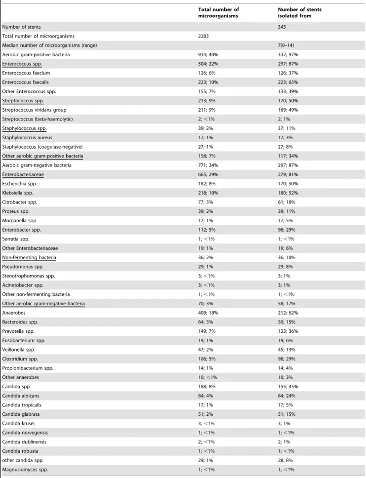
Overall, 2283 microorganisms from 343 stents were isolated (Table 2), including 914(40%) aerobic gram-positive bacteria, 771(34%) aerobic gram-negative bacteria, 409(18%) anaerobic bacteria and 188(8%)Candida spp.. In total, 56 genera with 185 different species were identified.
Enterococcus spp. were the most prevalent microorganisms (504;22%), isolated from 297 of 343 stents; followed byKlebsiella spp. (218;10%), Streptococcus spp. (213;9%), Candida spp.
(188;8%) and Escherichia spp. (182;8%), which were cultivated from 180(52%), 170(50%), 155(45%) and 170(50%) stents, respectively. 409(18%) anaerobic bacteria were found on 212(62%) stents. 149(7%) Prevotella spp. were isolated from 123(36%) stents, representing the most common anaerobic microorganisms. Clostridium spp. (106; 5%) were the second,
Bacteroides spp. (64; 3%) the third most isolated anaerobic bacteria, cultured from 98(29%) and 50(15%) stents, respectively.
Candida spp. grewon 155(45%) biliary stents.Candida albicans
(84; 4%) was the most prevalent Candida spp., followed by
Candida glabrata(51; 2%).
Analysis of spectrum of microorganisms (Table 3) in relation to an indwelling time of 1 d–15 d (group 1), 20 d–59 d (group 2) and 60 d–3087 d (group 3) revealed that stents extracted after a indwelling period of 1 d–15 d were mainly colonized by aerobic gram-positive bacteria (43/49;88%) and Candida spp. (25/ 49;51%). Aerobic gram-negative bacteria and anaerobes had a proportion of 45%(22/49) and 24%(12/49), respectively. In contrast, stents extracted after an indwelling time of 60 d– 3087 d showed an almost equal colonization rate by aerobic gram-negative (176/184;96%) and aerobic gram-positive bacteria (183/184;99%) and a high proportion of anaerobes (127/ 184;69%). Among aerobic gram-positive bacteria [P,0.01; 88% (group 1), 98% (group 2), 99% (group 3)], the incidence of
Enterococcus spp.significantly changed with the indwelling time Table 1.Baseline characteristics.
Number of patients 130
Median age in years (range) 67(22–91)
Male 77
Reason for stent therapy
Malignant genesis: 45
-Cholangiocarcinoma 20
-Pancreas cancer 14
-Hepatocellular carcinoma 1
-Liver metastases with intrahepatic biliary obstruction:
-Breast cancer 1
-Colorectal cancer 6
-Gastric cancer 1
-Malignant melanoma 1
-Lymphoma 1
Benign genesis: 80
-Anastomotic stricture after liver transplantation 7
-Biliary leakage after liver transplantation 4
-Chronic pancreatitis with extrahepatic bile duct obstruction 4
-Biliary stricture after cholecystectomy 8
-Adenoma of the papilla vateri 1
-Insufficiency of the ductus cysticus after cholecystectomy 1
-Radiation induced biliary stricture 2
-Incomplete removal of biliary stones 47
-Primary sclerosing cholangitis 2
-Secondary sclerosing cholangitis 2
-Biloma 2
Idiopathic biliary stricture: 5
Total number of biliary stents 343
Number of biliary stents with sludge 149
Number of biliary stent with occlusion 70
Median duration of indwelling time in days (range) 70 (1–3087)
Total number of treatment episodes with stent extraction 234
Number of treatment episodes with multi-stenting 90
Number of elective stent extractions 197
Table 2.Spectrum of microorganisms isolated from the biliary stents.
Total number of microorganisms
Number of stents isolated from
Number of stents 343
Total number of microorganisms 2283
Median number of microorganisms (range) 7(0–14)
Aerobic gram-positive bacteria 914; 40% 332; 97%
Enterococcus spp. 504; 22% 297; 87%
Enterococcus faecium 126; 6% 126; 37%
Enterococcus faecalis 223; 10% 223; 65%
Other Enterococcus spp. 155; 7% 133; 39%
Streptococcus spp. 213; 9% 170; 50%
Streptococcus viridans group 211; 9% 169; 49%
Streptococcus (beta-haemolytic) 2;,1% 2; 1%
Staphylococcus spp. 39; 2% 37; 11%
Staphylococcus aureus 12; 1% 12; 3%
Staphylococcus (coagulase-negative) 27; 1% 27; 8%
Other aerobic gram-positive bacteria 158; 7% 117; 34%
Aerobic gram-negative bacteria 771; 34% 297; 87%
Enterobacteriaceae 665; 29% 279; 81%
Escherichia spp. 182; 8% 170; 50%
Klebsiella spp. 218; 10% 180; 52%
Citrobacter spp. 77; 3% 61; 18%
Proteus spp. 39; 2% 39; 11%
Morganella spp. 17; 1% 17; 5%
Enterobacter spp. 112; 5% 98; 29%
Serratia spp. 1;,1% 1;,1%
Other Enterobacteriaceae 19; 1% 19; 6%
Non-fermenting bacteria 36; 2% 36; 10%
Pseudomonas spp. 29; 1% 29; 8%
Stenotrophomonas spp. 3;,1% 3; 1%
Acinetobacter spp. 3;,1% 3; 1%
Other non-fermenting bacteria 1;,1% 1;,1%
Other aerobic gram-negative bacteria 70; 3% 58; 17%
Anaerobes 409; 18% 212; 62%
Bacteroides spp. 64; 3% 50; 15%
Prevotella spp. 149; 7% 123; 36%
Fusobacterium spp. 19; 1% 19; 6%
Veillonella spp. 47; 2% 45; 13%
Clostridium spp. 106; 5% 98; 29%
Propionibacterium spp. 14; 1% 14; 4%
Other anaerobes 10;,1% 10; 3%
Candida spp. 188; 8% 155; 45%
Candida albicans 84; 4% 84; 24%
Candida tropicalis 17; 1% 17; 5%
Candida glabrata 51; 2% 51; 15%
Candida krusei 3;,1% 3; 1%
Candida norvegensis 1;,1% 1;,1%
Candida dublinensis 2;,1% 2; 1%
Candida robusta 1;,1% 1;,1%
other candida spp. 29; 1% 28; 8%
Magnusiomyces spp. 1;,1% 1;,1%
[P,0.01; 49% (group 1), 90% (group 2), 95% (group 3)]. In addition, the colonization rate of anaerobes [P,0.01; 24% (group 1), 67% (group 2), 69% (group 3)] and aerobic gram-negative microorganisms [P,0.01; 45% (group 1), 90% (group 2), 96% (group 3)], in particular the family of Enterobacteriaceae [P,0.01; 35% (group 1), 82% (group 2), 93% (group 3)] altered with the indwelling time.
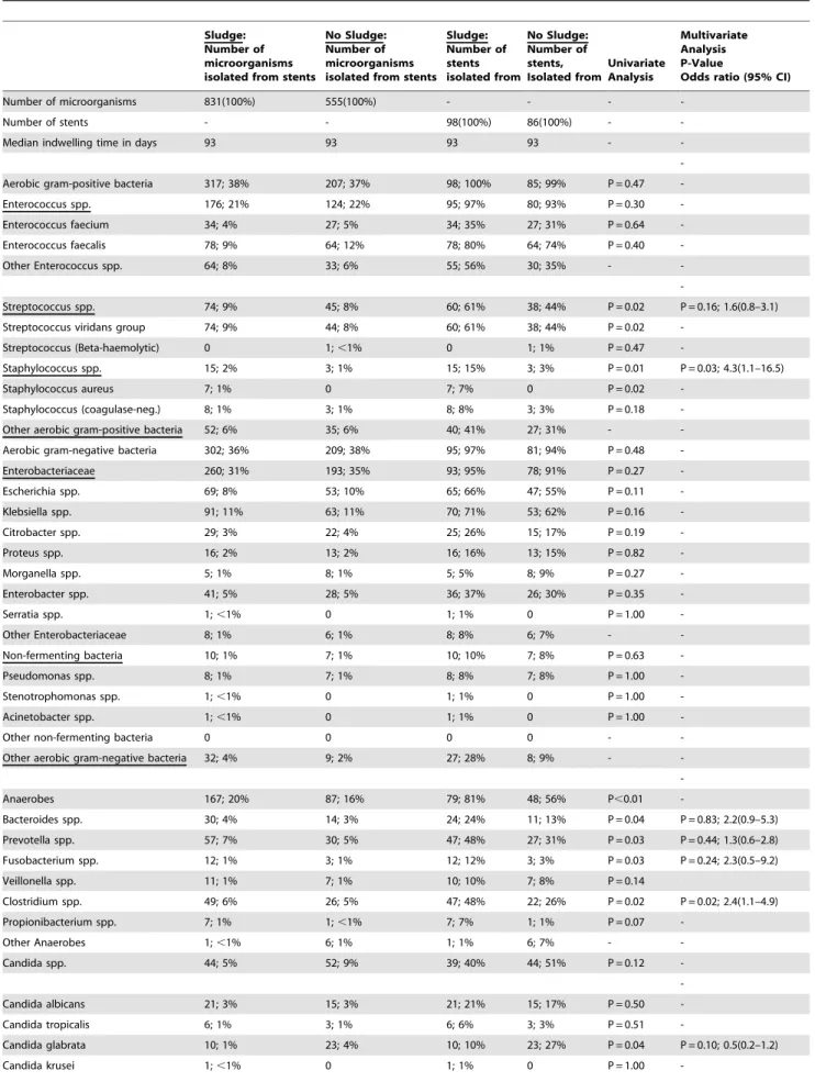
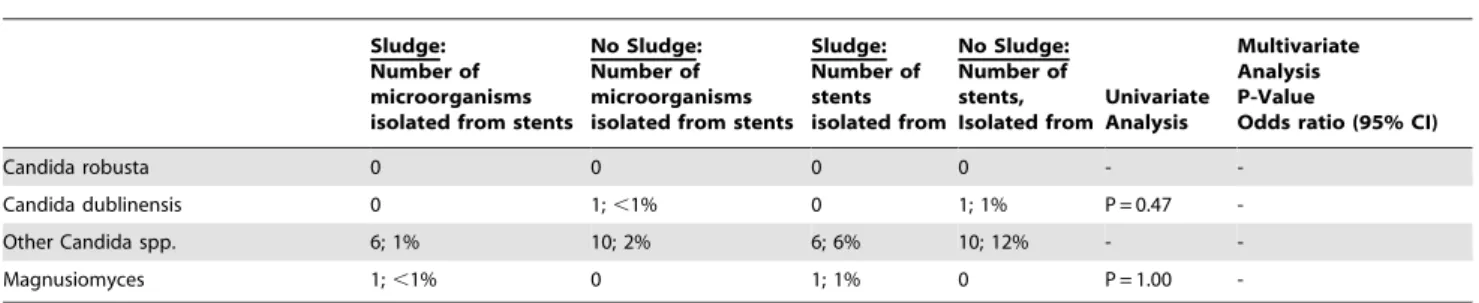
Table 4 illustrates the analysis of the spectrum of microorgan-isms comparing stents with and without sludge. To avoid confounding by the indwelling time, only stents with an indwelling time $60 days (group 3) were included to the stents. In the univariate analysis, significantly more stents with sludge were colonized byStreptococcus spp. [P = 0.02; 60(61%) vs. 38(44%)],
Staphylococcus spp. [P = 0.01; 15(15%) vs. 3(3%)],Bacteroides spp. [P = 0.04; 24(24%) vs. 11(13%)], Prevotella spp. [P = 0.03; 47(48%) vs. 27(31%)], Fusobacterium spp. [P = 0.03; 12(12%) vs. 3(3%)] and Clostridium spp. [P = 0.02; 47(48%) vs. 22(26%)], compared to stents without sludge. All microorganisms with a significant difference in colonization rates between stents with and without sludge in the univariate analysis were analyzed in a multivariate logistic regression model. According to multivariate analysis, there was only a significant relationship between the occurrence of sludge and the isolation of Clostridium spp. [(P = 0.02; OR (95% CI): 2.4(1.1–4.9)] and Staphylococcus spp. [(P = 0.03; OR (95% CI): 4.3(1.1–16.5)].
The relationship between the number of microorganisms, sideholes, the diameter as well as the indwelling time of the stent and the occurrence of sludge was also analyzed by multivariate logistic regression analysis. There was a significant relationship between the number of microorganisms [P,0.01; OR (95% CI): 1.3(1.1–1.5)], the indwelling time [P,0.01; 1–15 d vs. 20–59 d: OR (95% CI): 5.6(1.4–22), 1–15 d vs. 60–3087 d: OR (95% CI): 9.5(2.5–35.7)], the presence of sideholes [P,0.01; OR (95% CI): 3.5(1.6–7.9)] and the occurrence of sludge, whereas no significant association between sludge and the diameter of the stent was found [P = 0.32; OR (95% CI): 1.1(0.8–1.5)].
Stent occlusion occurred in 70/343(20%) stents after a median indwelling time of 70 days. In 35% (19/54) of cases, stent occlusion resulted in cholangitis or cholestasis.
Discussion
Previous analyses of the spectrum of microorganisms isolated from explanted biliary stents were performed with a small series of stents [17,18]. The strength of this study lies in the large sample size and the sonication method, allowing a better release of pathogens from the biofilm on the surface of the stent [19–21]. In the current study, 2283 microorganisms isolated from 343 stents were included into the analysis. The incidence of anaerobes (18%) was high, implying that the preanalytic conditions were correct and preparation of the stents was adequately performed. In literature, the proportion of anaerobes, isolated either from bile or directly from biliary stents ranges between 6% and 40% [16,22– 24] and Bacteroides spp. are reported to be most frequently isolated biliary anaerobes [16,23,25–27], which poses a contrast to the present study: Bacteroides spp. were only the third most frequently isolated anaerobic microorganisms afterPrevotella spp. and Clostridium spp.. The reason for our discrepant findings compared to literature may be that identification of microorgan-isms was performed by matrix-associated laser desorption/ ionization-time of flight mass spectrometer (MALDI-TOF), allowing a more precise differentiation of microorganisms compared to other microbiological testing methods. However, some Bacteroides spp. were re-classified as Prevotella spp. [28].
The incidence of anaerobes was associated with the presence of sludge which can be attributed to ideal anaerobic conditions inside the sludge. Another explanation is that anaerobic bacteria may have a direct impact on the sludge formation. Thus,Clostridium spp.were significantly more frequently detected on stents in the presence of sludge. Bacterial enzymes such as beta-glucuronidase and phosphlipase C play an important role in sludge formation [29]: Bilirubin is deconjungated by beta-glucuronidase and precipates as calcium bilirubinate. Furthermore, phospholipase C can hydrolyse lecithin precipating as calcium palmitate. Both enzymes are produced byClostridium spp.. The high proportion of
Clostridium spp. in occluded stents might also be of clinical relevance, as stent occlusion results in cholangitis. Infections with
Clostridium perfringens manifest through a broad variety of clinical course ranging from mild to severe septic courses and the most common origin of septicaemia withClostridium perfringens
is the hepatobiliary system [30]. Apart from Clostridium spp., Staphylococcus spp. were also significantly more often isolated from stents with sludge, compared to stents without sludge. Similar to our finding, Di Rosa et al. [31] also reported a higher proportion of Staphylococcus spp. in occluded stents compared to non-occluded stents. However, we can only hypothesize that the change in biliary spectrum observed in occluded stents may be of clinical relevance in case of acute cholangitis. Most of the stent extractions were performed electively and therefore blood cultures were collected only in a small number of patients presenting acute cholangitis. Consequently, no analysis could be performed to find out whether those microorganisms isolated from blood cultures were similar to the microorganisms isolated from the biofilm on the occluded stents. Furthermore, we can only assume that the microorganisms isolated from the biofilm on the stents surface are similar to the microorganisms in the bile fluid, because no concomitant bile culture was collected. In literature, the clinical relevance of bile collection is controversially discussed. Park et al. [32] analyzed 258 bacteremic cholangitis episodes: Complete agreement with blood cultures was observed in 80 (31%) bile samples, 129 (50%) bile samples showed a partial agreement with the blood culture findings, whereas 49 (19%) bile samples presented completely different microorganisms compared to the blood culture results. Gram-negative bacteria showed a significant higher coincidence rate than gram-positive bacteria. The degree of coincidence between bile and blood culture forEscherichia coli,
Klebsiella spp., Enterococcus spp. and Streptococcus spp. was 71.2%, 53.1%, 34.5% and 26.9%, respectively. There are studies showing a clear association between an indwelling biliary stent and an altered spectrum of pathogens in the bile fluid [16,33]. In the current series,Enterococcus spp. were by far the most prevalent genera. This high proportion ofEnterococcus spp.is in line with our preceding study [16], revealing that bile collected from patients with stents had a significantly higher incidence of
Enterococcus spp.compared to bile from patients without a stent. Furthermore, the number ofEnterococcus spp. isolated from blood cultures was also higher in cholangitis episodes with than without an indwelling stent.
Number of microorganisms isolated from stents, with an indwelling time (1–15)d
Number of microorganisms isolated from stents, with an indwelling time (20–59)d
Number of microorganisms isolated from stents, with an indwelling time (60–3087)d
Number of stents isolated from, with an indwelling time (1–15)d
Number of stents isolated from, with an indwelling time (20–59)d
Number of stents isolated from, with an indwelling
time (60–3087)d P-Value
Number of stents* 49 100 184
Total number (microorganisms) 178 660 1386
Median number (microorganisms) (range)
3(0–10) 6(2–13) 8(2–14)
Aerobic gram-positive bacteria 92; 52% 277; 42% 524; 38% 43; 88% 98; 98% 183; 99% P,0.01
Enterococcus spp. 28; 16% 162; 25% 300; 22% 24; 49% 90; 90% 175; 95% P,0.01
Enterococcus faecium 16; 9% 46; 7% 61; 4% 16; 33% 46; 46% 61; 33% P = 0.08
Enterococcus faecalis 6; 3% 69; 10% 142; 10% 6; 12% 69; 69% 142; 77% P,0.01
Other Enterococcus spp. 6; 3% 47; 7% 97; 7% 6; 12% 37; 37% 85; 46% P,0.01
Streptococcus spp. 29; 16% 59; 9% 119; 9% 21; 43% 47; 47% 98; 53% P = 0.34
Streptococcus viridans group 29; 16% 58; 9% 118; 9% 21; 43% 46; 46% 98; 53% P = 0.30
Streptococcus (Beta-haemolytic) 0 1;,1% 1;,1% 0 1; 1% 1; 1% P = 1.00
Staphylococcus spp. 12; 7% 8; 1% 18; 1% 10; 20% 8; 8% 18; 10% P = 0.06
Staphylococcus aureus 1; 1% 3;,1% 7; 1% 1; 2% 3; 3% 7; 4% P = 1.00
Staphylococcus coagulase-negative 11; 6% 5; 1% 11; 1% 9; 18% 5; 5% 11; 6% P = 0.02
Other aerobic gram-positive bacteria
23; 13% 48; 7% 87; 6% 18; 37% 32; 32% 67; 36%
-Aerobic gram-negative bacteria 35; 20% 204; 31% 511; 37% 22; 45% 90; 90% 176; 96% P,0.01
Enterobacteriaceae 26; 15% 165; 25% 453; 33% 17; 35% 82; 82% 171; 93% P,0.01
Escherichia spp. 8; 4% 44; 7% 122; 9% 8; 16% 43; 43% 112; 61% P,0.01
Klebsiella spp. 5; 3% 54; 8% 154; 11% 4; 8% 48; 48% 123; 67% P,0.01
Citrobacter spp. 2; 1% 23; 3% 51; 4% 2; 4% 18; 18% 40; 22% P = 0.02
Proteus spp. 0(0) 8; 1% 29; 2% 0 8; 8% 29; 16% P,0.01
Morganella spp. 0(0) 4; 1% 13; 1% 0 4; 4% 13; 7% P = 0.12
Enterobacter spp. 11; 6% 27; 4% 69; 5% 8; 16% 23; 23% 62; 34% P = 0.02
Serratia spp. 0(0) 0(0) 1;,1% 0 0 1; 1% P = 1.00
Other Enterobacteriaceae 0(0) 5; 1% 14; 1% 0 5; 5% 14; 8%
-Non-fermenting bacteria 4; 2% 15; 2% 17; 1% 4: 8% 15; 15% 17; 9% P = 0.27
Pseudomonas spp. 1; 1% 13; 2% 15; 1% 1; 2% 13; 13% 15; 8% P = 0.07
Stenotrophomonas spp. 0(0) 2;,1% 1;,1% 0 2; 2% 1; 1% P = 0.56
Acinetobacter spp. 2; 1% 0(0) 1;,1% 2; 4% 0 1; 1% P = 0.08
Other non-fermenting bacteria 1; 1% 0(0) 0 1; 2% 0 0
-Other aerobic gram-negative bacteria
5; 3% 24; 4% 41; 3% 3; 6% 20; 20% 35; 19%
-Biliary
Endoprosthe
sis:
Sludge
Formation
and
Microbial
Colonizatio
n
ONE
|
www.ploson
e.org
6
October
2014
|
Volume
9
|
Issue
10
|
Number of microorganisms isolated from stents, with an indwelling time (1–15)d
Number of microorganisms isolated from stents, with an indwelling time (20–59)d
Number of microorganisms isolated from stents, with an indwelling time (60–3087)d
Number of stents isolated from, with an indwelling time (1–15)d
Number of stents isolated from, with an indwelling time (20–59)d
Number of stents isolated from, with an indwelling
time (60–3087)d P-Value
Anaerobes 18; 10% 125; 19% 254; 18% 12; 24% 67; 67% 127; 69% P,0.01
Bacteroides spp. 2; 1% 18; 3% 44; 3% 2; 4% 13; 13% 35; 19% P = 0.03
Prevotella spp. 10; 6% 48; 7% 87; 6% 8; 16% 39; 39% 73; 40% P = 0.01
Fusobacterium spp. 0(0) 2;,1% 15; 1% 0 2; 2% 15; 8% P = 0.02
Veillonella spp. 5; 3% 21; 3% 18; 1% 5; 10% 21; 21% 17; 9% P = 0.08
Clostridium spp. 1; 1% 28; 4% 75; 5% 1; 2% 26; 26% 69; 38% P,0.01
Propionibacterium spp. 0(0) 5; 1% 8; 1% 0 5; 5% 8; 4% P = 0.30
Other anaerobes 0(0) 3;,1% 7; 1% 0 3; 3% 7; 4%
-Candida spp. 33; 19% 54; 8% 96; 7% 25; 51% 42; 42% 83; 45% P = 0.58
Candida albicans 17; 10% 27; 4% 36; 3% 17; 35% 27; 27% 36; 20% P = 0.62
Candida tropicalis 1; 1% 7; 1% 9; 1% 1; 2% 7; 7% 9; 5% P = 0.48
Candida glabrata 5; 3% 12; 2% 33; 2% 5; 10% 12; 12% 33; 18% P = 0.24
Candida krusei 0 2;,1% 1;,1% 0 2; 2% 1; 1% P = 0.56
Candida norvegensis 1; 1% 0 0 1; 2% 0 0 P = 0.15
Candida dublinensis 0 1;,1% 1;,1% 0 1; 1% 1; 1% P = 1.00
Candida robusta 0 1;,1% 0 0 1; 1% 0 P = 0.45
Other Candida spp. 9; 5% 4; 1% 16; 1% 8; 16% 4; 4% 16; 9%
-Magnusiomyces 0 0 1;,1% 0 0 1; 1% P = 1.00
*8 stents could not be included to this analysis due to an unknown indwelling time. doi:10.1371/journal.pone.0110112.t003
Biliary
Endoprosthe
sis:
Sludge
Formation
and
Microbial
Colonizatio
n
ONE
|
www.ploson
e.org
7
October
2014
|
Volume
9
|
Issue
10
|
Table 4.Comparison of the spectrum of microorganisms between stents with and without sludge.
Sludge: Number of microorganisms isolated from stents
No Sludge: Number of microorganisms isolated from stents
Sludge: Number of stents isolated from
No Sludge: Number of stents, Isolated from
Univariate Analysis
Multivariate Analysis P-Value
Odds ratio (95% CI)
Number of microorganisms 831(100%) 555(100%) - - -
-Number of stents - - 98(100%) 86(100%) -
-Median indwelling time in days 93 93 93 93 -
-Aerobic gram-positive bacteria 317; 38% 207; 37% 98; 100% 85; 99% P = 0.47
-Enterococcus spp. 176; 21% 124; 22% 95; 97% 80; 93% P = 0.30
-Enterococcus faecium 34; 4% 27; 5% 34; 35% 27; 31% P = 0.64
-Enterococcus faecalis 78; 9% 64; 12% 78; 80% 64; 74% P = 0.40
-Other Enterococcus spp. 64; 8% 33; 6% 55; 56% 30; 35% -
-Streptococcus spp. 74; 9% 45; 8% 60; 61% 38; 44% P = 0.02 P = 0.16; 1.6(0.8–3.1)
Streptococcus viridans group 74; 9% 44; 8% 60; 61% 38; 44% P = 0.02
-Streptococcus (Beta-haemolytic) 0 1;,1% 0 1; 1% P = 0.47
-Staphylococcus spp. 15; 2% 3; 1% 15; 15% 3; 3% P = 0.01 P = 0.03; 4.3(1.1–16.5)
Staphylococcus aureus 7; 1% 0 7; 7% 0 P = 0.02
-Staphylococcus (coagulase-neg.) 8; 1% 3; 1% 8; 8% 3; 3% P = 0.18
-Other aerobic gram-positive bacteria 52; 6% 35; 6% 40; 41% 27; 31% -
-Aerobic gram-negative bacteria 302; 36% 209; 38% 95; 97% 81; 94% P = 0.48
-Enterobacteriaceae 260; 31% 193; 35% 93; 95% 78; 91% P = 0.27
-Escherichia spp. 69; 8% 53; 10% 65; 66% 47; 55% P = 0.11
-Klebsiella spp. 91; 11% 63; 11% 70; 71% 53; 62% P = 0.16
-Citrobacter spp. 29; 3% 22; 4% 25; 26% 15; 17% P = 0.19
-Proteus spp. 16; 2% 13; 2% 16; 16% 13; 15% P = 0.82
-Morganella spp. 5; 1% 8; 1% 5; 5% 8; 9% P = 0.27
-Enterobacter spp. 41; 5% 28; 5% 36; 37% 26; 30% P = 0.35
-Serratia spp. 1;,1% 0 1; 1% 0 P = 1.00
-Other Enterobacteriaceae 8; 1% 6; 1% 8; 8% 6; 7% -
-Non-fermenting bacteria 10; 1% 7; 1% 10; 10% 7; 8% P = 0.63
-Pseudomonas spp. 8; 1% 7; 1% 8; 8% 7; 8% P = 1.00
-Stenotrophomonas spp. 1;,1% 0 1; 1% 0 P = 1.00
-Acinetobacter spp. 1;,1% 0 1; 1% 0 P = 1.00
-Other non-fermenting bacteria 0 0 0 0 -
-Other aerobic gram-negative bacteria 32; 4% 9; 2% 27; 28% 8; 9% -
-Anaerobes 167; 20% 87; 16% 79; 81% 48; 56% P,0.01
-Bacteroides spp. 30; 4% 14; 3% 24; 24% 11; 13% P = 0.04 P = 0.83; 2.2(0.9–5.3)
Prevotella spp. 57; 7% 30; 5% 47; 48% 27; 31% P = 0.03 P = 0.44; 1.3(0.6–2.8)
Fusobacterium spp. 12; 1% 3; 1% 12; 12% 3; 3% P = 0.03 P = 0.24; 2.3(0.5–9.2)
Veillonella spp. 11; 1% 7; 1% 10; 10% 7; 8% P = 0.14
Clostridium spp. 49; 6% 26; 5% 47; 48% 22; 26% P = 0.02 P = 0.02; 2.4(1.1–4.9)
Propionibacterium spp. 7; 1% 1;,1% 7; 7% 1; 1% P = 0.07
-Other Anaerobes 1;,1% 6; 1% 1; 1% 6; 7% -
-Candida spp. 44; 5% 52; 9% 39; 40% 44; 51% P = 0.12
-Candida albicans 21; 3% 15; 3% 21; 21% 15; 17% P = 0.50
-Candida tropicalis 6; 1% 3; 1% 6; 6% 3; 3% P = 0.51
-Candida glabrata 10; 1% 23; 4% 10; 10% 23; 27% P = 0.04 P = 0.10; 0.5(0.2–1.2)
-through the tube is switched to a micro-disturbed fluid stream resulting in an increased resistance of the stent to the bile flow. In theory, sideholes should guarantee the biliary drainage in case of the proximal and distal main orifice being occluded by plugs or blocked by the wall of the bile duct [34]. However, clinical significance of this hypothesis is still missing [34,36]. Sung et al. [36] compared the clinical efficacy of stents with and without sideholes in a prospectively randomised control trial. In both stents with and without sideholes, the median time before stent occlusion was similar (7.8 vs. 7.9 weeks). Several studies showed that antimicrobial coatings on medical devices are effective against microbial biofilm formation. According to Agostinho et al. [37] antimicrobially coated materials are able to significantly inhibit bacterial biofilm formation through microorganisms like Staphy-lococcus spp. over a certain time. A recent study [38] reported similar findings, analyzing novel high efficiency coatings on anti-microbial surgical sutures using chlorhexidine in fatty acid slow-release carrier systems. Furthermore, the use of new coatings like diamond like carbons (DLC) seems to be a promising approach to reduce biofilm formation on medical devices. Due to its low friction coefficient, high biocompability, chemical inertness, and both high hardness as well excellent smoothness [39–41], diamond like carbon is virtually predestined for use as a coating for biliary stents. Laube et al. [42] investigated the ability of diamond-like carbon to decrease formation of crystalline bacterial biofilm as well as stent related side effects and reported an excellent handling, a less painful replacement procedure and high tolerance of
application. Furthermore, no crystalline biofilm formation could be detected on the stent surface in vivo and the number and severity of symptomatic urinary tract infections was decreased.
In the current series, stents occluded after a median indwelling time of 70 days. However, stent occlusion resulted in a cholangitis or cholestasis in only 35% of the cases. This suggests that stent occlusion is not always associated with clinical symptoms.
The study displays the following limitations: Choice and indwelling time of the polyethylene stents inserted in the bile duct were not influenced. Therefore, time point of stent extraction and use of stents were not standardized.
In conclusion, the spectrum of microorganisms colonizing the inner surface of the stent changed with the indwelling time. The occurrence of sludge was associated with a higher isolation rate of
Staphylococcus spp. and Clostridium spp., the presence of sideholes and with an increasing indwelling time. Although these factors may support stent occlusion, a great proportion of stent occlusions proceeded clinically asymptomatic.
Author Contributions
Conceived and designed the experiments: JS AW RMS HA BN. Performed the experiments: JS AO JF PS NW S. Forkl S. Feihl HA RMS BN SVD MB RB AW. Analyzed the data: AH JS S. Forkl AW HA JF PS. Contributed reagents/materials/analysis tools: JS AO JF PS NW S. Forkl S. Feihl HA RB BN SVD MB. Wrote the paper: JS AH AO BN MB S. Feihl RMS.
References
1. van Milligen de Wit AW, van Bracht J, Rauws EA, Jones EA, Tytgat GN, et al. (1996) Endoscopic stent therapy for dominant extrahepatic bile duct strictures in primary sclerosing cholangitis. Gastrointest Endosc 44: 293–299.
2. Aljiffry M, Renfrew PD, Walsh MJ, Laryea M, Molinari M (2011) Analytical review of diagnosis and treatment strategies for dominant bile duct strictures in patients with primary sclerosing cholangitis. HPB: the official journal of the International Hepato Pancreato Biliary Association 13: 79–90.
3. Baron TH, Sr., Davee T (2013) Endoscopic management of benign bile duct strictures. Gastrointest Endosc Clin N Am 23: 295–311.
4. Boulay BR, Gardner TB, Gordon SR (2010) Occlusion rate and complications of plastic biliary stent placement in patients undergoing neoadjuvant chemoradiotherapy for pancreatic cancer with malignant biliary obstruction. J Clin Gastroenterol 44: 452–455.
5. Lawrence C, Romagnuolo J, Payne KM, Hawes RH, Cotton PB (2010) Low symptomatic premature stent occlusion of multiple plastic stents for benign biliary strictures: comparing standard and prolonged stent change intervals. Gastrointest Endosc 72: 558–563.
6. Meyenberger C, Fantin AC (2003) [Biliary stents]. Ther Umsch 60: 225–232. 7. van Berkel AM, Boland C, Redekop WK, Bergman JJ, Groen AK, et al. (1998)
A prospective randomized trial of Teflon versus polyethylene stents for distal malignant biliary obstruction. Endoscopy 30: 681–686.
8. Davids PH, Groen AK, Rauws EA, Tytgat GN, Huibregtse K (1992) Randomised trial of self-expanding metal stents versus polyethylene stents for distal malignant biliary obstruction. Lancet 340: 1488–1492.
9. Dumonceau JM, Tringali A, Blero D, Deviere J, Laugiers R, et al. (2012) Biliary stenting: indications, choice of stents and results: European Society of Gastrointestinal Endoscopy (ESGE) clinical guideline. Endoscopy 44: 277–298. 10. Leung JW, Ling TK, Kung JL, Vallance-Owen J (1988) The role of bacteria in
the blockage of biliary stents. Gastrointest Endosc 34: 19–22.
11. Speer HF, Costerton JW, Cotton PB (1986) The role of bacterial biofilm in clogging of biliary stents. Gastrointest Endosc 32: 156.
12. Groen AK, Out T, Huibregtse K, Delzenne B, Hoek FJ, et al. (1987) Characterization of the content of occluded biliary endoprostheses. Endoscopy 19: 57–59.
13. van Berkel AM, van Marle J, Groen AK, Bruno MJ (2005) Mechanisms of biliary stent clogging: confocal laser scanning and scanning electron microscopy. Endoscopy 37: 729–734.
14. Dowidar N, Kolmos HJ, Matzen P (1992) Experimental clogging of biliary endoprostheses. Role of bacteria, endoprosthesis material, and design. Scand J Gastroenterol 27: 77–80.
15. Naber KG, Schito G, Botto H, Palou J, Mazzei T (2008) Surveillance study in Europe and Brazil on clinical aspects and Antimicrobial Resistance Epidemi-ology in Females with Cystitis (ARESC): implications for empiric therapy. Eur Urol 54: 1164–1175.
16. Weber A, Schneider J, Wagenpfeil S, Winkle P, Riedel J, et al. (2013) Spectrum of pathogens in acute cholangitis in patients with and without biliary endoprosthesis. J Infect 67: 111–121.
17. Guaglianone E, Mastrantonio P, Di Rosa R, Penni A, Puggioni G, et al. (2008) Role of multispecies microbial biofilms in the occlusion of biliary stents. Microb Ecology in Health and Disease 20: 207–209.
Table 4.Cont.
Sludge: Number of microorganisms isolated from stents
No Sludge: Number of microorganisms isolated from stents
Sludge: Number of stents isolated from No Sludge: Number of stents, Isolated from Univariate Analysis Multivariate Analysis P-Value
Odds ratio (95% CI)
Candida robusta 0 0 0 0 -
-Candida dublinensis 0 1;,1% 0 1; 1% P = 0.47
-Other Candida spp. 6; 1% 10; 2% 6; 6% 10; 12% -
-Magnusiomyces 1;,1% 0 1; 1% 0 P = 1.00
18. Zhang H, Tsang TK, Jack CA (2003) Bile glycoprotein mucin in sludge occluding biliary stent. J Lab Clin Med 142: 58–65.
19. Rieger UM, Pierer G, Luscher NJ, Trampuz A (2009) Sonication of removed breast implants for improved detection of subclinical infection. Aesthetic Plast Surg 33: 404–408.
20. Trampuz A, Piper KE, Jacobson MJ, Hanssen AD, Unni KK, et al. (2007) Sonication of removed hip and knee prostheses for diagnosis of infection. N Engl J Med 357: 654–663.
21. Piper KE, Jacobson MJ, Cofield RH, Sperling JW, Sanchez-Sotelo J, et al. (2009) Microbiologic diagnosis of prosthetic shoulder infection by use of implant sonication. J Clin Microbiol 47: 1878–1884.
22. Sheen-Chen S, Chen W, Eng H, Sheen C, Chou F, et al. (2000) Bacteriology and antimicrobial choice in hepatolithiasis. Am J Infect Control 28: 298–301. 23. Brook I (1989) Aerobic and anaerobic microbiology of biliary tract disease.
J Clin Microbiol 27: 2373–2375.
24. Finegold SM (1979) Anaerobes in biliary tract infection. Arch Intern Med 139: 1338–1339.
25. Fu HQ (1989) [Anaerobes in biliary tract infection]. Zhonghua Wai Ke Za Zhi 27: 454–456, 507.
26. Tabata M, Nakayama F (1984) Bacteriology of hepatolithiasis. Prog Clin Biol Res 152: 163–174.
27. England DM, Rosenblatt JE (1977) Anaerobes in human biliary tracts. J Clin Microbiol 6: 494–498.
28. Shah HN, Collins DM (1990) Prevotella, a new genus to include Bacteroides melaninogenicus and related species formerly classified in the genus Bacteroides. Int J Syst Bacteriol 40: 205–208.
29. Donelli G, Guaglianone E, Di Rosa R, Fiocca F, Basoli A (2007) Plastic biliary stent occlusion: factors involved and possible preventive approaches. Clin Med Res 5: 53–60.
30. van Bunderen CC, Bomers MK, Wesdorp E, Peerbooms P, Veenstra J (2010) Clostridium perfringens septicaemia with massive intravascular haemolysis: a case report and review of the literature. Neth J Med 68: 343–346.
31. Di Rosa R, Donelli G, Penni A, Savatori FM, Fiocca F, et al. (1999) A microbiological and morphological study of blocked biliary stents Microbial Ecology in Health and Disease 11: 84–88.
32. Park JW, Lee JK, Lee KT, Lee KH, Sung YK, et al. (2014) How to interpret the bile culture results of patients with biliary tract infections. Clin Res Hepatol Gastroenterol 38: 300–309.
33. Rerknimitr R, Fogel EL, Kalayci C, Esber E, Lehman GA, et al. (2002) Microbiology of bile in patients with cholangitis or cholestasis with and without plastic biliary endoprosthesis. Gastrointest Endosc 56: 885–889.
34. Coene PP, Groen AK, Cheng J, Out MM, Tytgat GN, et al. (1990) Clogging of biliary endoprostheses: a new perspective. Gut 31: 913–917.
35. Rey JF, Maupetit P, Greff M (1985) Experimental study of biliary endoprosthesis efficiency. Endoscopy 17: 145–148.
36. Sung JJ, Chung SC, Tsui CP, Co AL, Li AK (1994) Omitting side-holes in biliary stents does not improve drainage of the obstructed biliary system: a prospective randomized trial. Gastrointest Endosc 40: 321–325.
37. Agostinho A, James G, Wazni O, Citron M, Wilkoff BD (2009) Inhibition of Staphylococcus aureus biofilms by a novel antibacterial envelope for use with implantable cardiac devices. Clin Transl Sci 2: 193–198.
38. Obermeier A, Schneider J, Wehner S, Matl FD, Schieker M, et al. (2014) Novel high efficient coatings for anti-microbial surgical sutures using chlorhexidine in Fatty Acid slow-release carrier systems. PLoS One 9: e101426.
39. Lappalainen R, Anttila A, Heinonen H (1998) Diamond coated total hip replacements. Clin Orthop Relat Res: 118–127.
40. Roy RK, Lee KR (2007) Biomedical applications of diamond-like carbon coatings: a review. J Biomed Mater Res B Appl Biomater 83: 72–84. 41. Castellino M, Stolojan V, Virga A, Rovere M, Cabiale K, et al. (2013)
Chemico-physical characterisation and in vivo biocompatibility assessment of DLC-coated coronary stents. Anal Bioanal Chem 405: 321–329.