APOE Effects on Default Mode Network in
Chinese Cognitive Normal Elderly:
Relationship with Clinical Cognitive
Performance
Haiqing Song1, Haixia Long2, Xiumei Zuo1, Chunshui Yu3, Bing Liu2, Zhiqun Wang3, Qi Wang1, Fen Wang1, Ying Han1, Jianping Jia1*
1Department of Neurology, Xuanwu Hospital Capital Medical University, Beijing, China,2Institute of Automation, Chinese Academy of Sciences, Beijing, China,3Department of Radiology, Xuanwu Hospital Capital Medical University, Beijing, China
Abstract
Background
Functional connectivity in default mode network (DMN) may be changed in Alzheimer’s dis-ease (AD) patients and related risk populations, such as amnestic mild cognitive impairment (aMCI) patients and APOEε4 carriers. Exploring DMN changes and related behavioral per-formance of APOEε4 population might provide valuable evidence for better understanding the development of AD.
Methods
Subjects were enrolled from a population-based cohort established in a multi-center study in China. Forty-nine cognitive normal individuals were enrolled after standardized cognitive evaluations, MRI examination and APOE genotyping. Regions of interest (ROI)-based func-tional connectivity analyses were performed, and voxel-based analyses were used to vali-date these findings. The correlation between DMN functional connectivity and behavioral performance was further evaluated between APOEε4ε3 andε3ε3 carriers.
Results
Comparing toε3ε3 carriers, functional connectivity between left parahippocampal gyrus and right superior frontal cortex (LPHC-R.Sup.F), left parahippocampal gyrus and medial prefrontal cortex (ventral) (LPHC-vMPFC) were significantly increased inε4ε3 carriers, while connectivity between cerebellar tonsils and retrosplenial was decreased. LPHC-R. Sup.F connectivity was positively correlated with the changes of delay recall from baseline to follow-up (r = 0.768, p = 0.009), while LPHC-vMPFC connectivity had a positive correla-tion with MMSE at baseline (r = 0.356, p = 0.018), and a negative correlacorrela-tion with long-delayed recognition at follow-up (r = -0.677, p = 0.031). Significantly increased functional OPEN ACCESS
Citation:Song H, Long H, Zuo X, Yu C, Liu B, Wang Z, et al. (2015) APOE Effects on Default Mode Network in Chinese Cognitive Normal Elderly: Relationship with Clinical Cognitive Performance. PLoS ONE 10(7): e0133179. doi:10.1371/journal. pone.0133179
Editor:Kewei Chen, Banner Alzheimer's Institute, UNITED STATES
Received:March 7, 2015
Accepted:June 23, 2015
Published:July 15, 2015
Copyright:© 2015 Song et al. This is an open access article distributed under the terms of the Creative Commons Attribution License, which permits unrestricted use, distribution, and reproduction in any medium, provided the original author and source are credited.
Data Availability Statement:Ethical restrictions prevent public sharing of data. Data requests may be sent to the corresponding author.
connectivity in vMPFC was confirmed in voxel-based analyses by taking LPHC as seed region.
Conclusion
APOEε4 carriers present both increased and decreased functional connectivity in DMN, which is correlated with clinical cognitive performances. DMN changes might be an early sign for cognitive decline.
Introduction
Alzheimer’s disease (AD) is the most common neurodegenerative disease in elderly, but effec-tive treatments are limited. Therefore, researches are trying to understand the development of AD, and look for early sign of cognitive decline which might be treatable at early stage.
Neuroimaging studies is an useful tool to evaluate structural, functional, and metabolic changes of the brain, and widely used in investigation of AD and associated diseases. Default mode network (DMN) is one of the highlighted network, which consists of a collection of brain structures including the medical prefrontal cortex (mPFC), posterior cingulate cortex (PCC), precuneus, anterior cingulate cortex (ACC), parietal cortex, and hippocampus[1]. It presents increased connectivity during rest and decreased connectivity during specific goal directed behaviors[2].
DMN is considered to be associated with AD, since the regions involved are core structures for memory system, and vulnerable to deposition of amyloid protein[3,4]. Moreover, there are numerous evidence of disrupted functional connectivity (FC) of DMN in AD patients[5,6] and related risk populations, such as amnestic mild cognitive impairment (aMCI) patients[7,8]and APOEε4 carriers[9–12]. Besides, some studies showed increased FC in AD and related dis-eases, which has been explained by compensatory-recruitment hypothesis[13].
APOEε4 allele has been associated with an increased risk for AD[14], and therefore the APOEε4 carriers are considered to be good candidates for learning the development of AD. Similar imaging studies on DMN have been carried out in APOE4 carriers. Both decreased and increased functional connectivity were observed within different regions in the DMN in older asymptomatic APOE4 carriers[9–12]; while increased functional connectivity was also found in younger APOE4 carriers[15,16], which suggested a compensatory mechanism to maintain normal cognitive performances decades before clinical manifestation.
However, the clinical meanings of these functional connectivity have not been fully investi-gated, and relevant literatures are limited. Westlye and colleagues found significant negative correlation between memory performance and resting hippocampal connectivity synchroniza-tion in older cognitive normal individuals using the Norwegian translasynchroniza-tion of the California Verbal Learning Test, second version (CVLT-II), suggesting a potential relationship between functional connectivity and clinical cognition[10]. Therefore, It is important to confirm these findings, which would be very useful for better understanding the associations between imag-ing findimag-ings and clinical manifestations.
To validate the significant relationships between DMN functional connectivity changes and clinical cognitive performances, we presented here an imaging study in cognitive normal elderly from a population with two visits of cognitive measurements in Beijing, China, to inves-tigate the APOEε4 effects on DMN connectivity in Chinese, and further evaluate the associa-tions between the changed functional connectivity and cognitive performances.
Technology R&D Program in the Eleventh Five-year Plan Period (2006BAI02B01) and the key project (30830045) and general projects (31371007, 30970823) of the National Natural Science Foundation of China. The funders had no role in study design, data collection and analysis, decision to publish, or preparation of the manuscript.
Materials and Methods
Subjects
Cognitive normal elderly were enrolled from a population-based cohort established by a multi-center study in China. This cohort was set up in 2009, aiming to screen MCI and AD patients in a well-defined population. Totally 10,276 individuals aged over 65 years were enrolled in this cohort. Cognitive measurements were evaluated at baseline, and blood samples were col-lected. Follow-ups were scheduled every year. Individuals from Beijing with: (a) Clinical Dementia Rating (CDR) score of 0; (b) no neurological or psychiatric disorders; (c) normal activities described in a daily living scale were invited and voluntary to participant in this study at baseline, of which 161 participants took part in the MRI examination. All scans were reviewed by neuroimaging experts. Individual with brain tumors, recent infarctions, or global abnormal signals were excluded. 99 individuals were defined as normal after cognitive evalua-tions at baseline, of which 64 subjects has APOE genotype information. 14 (21.9%) subjects were excluded because they wereε2 carriers, which is suggested to be a protective factor of cog-nition (ε3ε2 n = 11;ε4ε2 n = 3). One more subjects (ε3ε3) was excluded due to incompletion of fMRI data. Therefore, our final study population comprised of 49 cognitive normal individu-als, including 14ε4ε3 carriers and 35ε3ε3 carriers.
Ethics
The study was approved by the Ethics Committee of Xuanwu Hospital Capital Medical Univer-sity. All subjects were informed the purpose of this study, and informed consent was signed by each subject or their legal guardian.
Cognitive measurements
Standardized general and neurologic examination were performed on each participant. Neuro-psychological tests were performed by neurologists, including mini-mental state examination (MMSE), Montreal cognitive assessment (MoCA), World Health Organization-University of California-Los Angeles auditory verbal learning test (WHO-UCLA AVLT), center for epidemi-ologic studies-depression (CESD), and Hachinski ischemic index (HIS). The clinical dementia rating scale (CDR) was also administered and detailed cognitive impairment history was inquired. Subjects with CDR scored as”0”and without any complaint of cognitive decline were considered to be normal.
Genotyping
Peripheral blood samples were collected at baseline of the cohort. Genomic DNA was extracted via a modified phenol/chloroform extraction procedure. APOE genotype was determined by one-stage polymerase chain reaction (PCR) as suggested by Wenham et al [17]. Distributions of APOE genotypes were in Hardy-Weinberg Equilibrium in all subjects, and the frequency of ε4 carriers was in accordance with the known frequency of this allele in Chinese population. Observed genotypic frequencies were as follows:ε3ε2 17.2%,ε4ε2 4.7%,ε4ε3 21.9%,ε3ε3 54.7%.
MRI data acquisition and preprocessing
thickness = 3.0 mm (with 1mm gap), FOV = 220×220 mm, matrix = 64×64, flip angle (FA) = 90°. For each subject, the fMRI scan during the resting-state provided 270 volumes. 3D T1-weighted magnetization-prepared rapid gradient echo (MPRAGE) sagittal images were col-lected by using the following parameters: TR = 1900 ms, TE = 2.2 ms, inversion time (TI) = 900 ms, FA = 9°, resolution = 256 × 256 matrix, slices = 176, thickness = 1.0 mm.
We preprocessed the fMRI data with the following steps using SPM5 (http://www.fil.ion. ucl.ac.uk/spm/software/spm5/) and in-house software: (1) slice timing; (2) realigning the vol-umes to the first volume; (3) spatially normalizing to a standard EPI template and making a resample to 2 mm2 mm2 mm; (4) spatially smoothing; (5) performing linear regression to
remove the influence of head motion, whole brain signals and linear trends; (6) temporal band-pass filtration (0.01–0.08Hz). The parameters obtained from movement correction sug-gested that the maximum displacement in each cardinal direction (x, y, z) was less than 2 mm, and the maximum spin (x, y, z) was less than 2° for each participant.
ROI-based individual DMN functional connectivity
We used the same regions of interest (ROI) to define the default network and performed the similar default network functional connectivity analyses here as reported in our previous stud-ies[18,19]. The detailed coordinates of these 13 ROIs are shown inTable 1. All ROIs were defined as a spherical region with a radius of 6 mm at the center of the obtained coordinates for a specific ROI. We extracted the averaged BOLD time series separately for 13 ROIs in each subject, and then calculated the Pearson’s correlation coefficient between the any two averaged time series. We then transformed the resulting correlation into Z score by r-to-z transforma-tion. Finally, we got a 1313 DMN functional connectivity graphs for each subject.
Parahippocampal functional connectivity analyses
To validate the ROI-based DMN findings, we extracted left parahippocampal gyrus, which was suggested to be involved in the changed functional connectivity in preliminary analysis, as a seed region, and then performed voxel-based whole brain functional connectivity analyses. We computed the whole brain functional connectivity by calculating the Pearson correlation coeffi-cients between the averaged time series of left parahippocampal gyrus and each voxel in the whole brain for each individual.
Statistical analysis
Demographic information and cognitive measurements were analyzed using two-samplet-test
andChi-squaretest. We used a two-samplet-test to examine differences in the strength of
con-nectivity between the two APOE genotype groups, meanwhile, a permutation-based correction method[20] was used to correct for multiple comparisons. For each seed region in whole brain analyses, we performed a two-samplet-test to examine the significance of the differences in the voxel-based default network between individuals with two different genotypes. The Pearson correlation was calculated to evaluate the correlation between DMN functional connectivity and behavioral performance.
Results
Demographic information and cognitive measurements
and 2ε4ε3 carriers) at follow-up with completed cognitive measurements were included in fur-ther analysis for associations between functional connectivity and behavior performance. One more subject (ε3ε3 carrier) was excluded from the analysis involving baseline MMSE due to the far deviation from the average score (>3SD). Average scores of Mini-Mental State
Exami-nation (MMSE), Delay recall and Long-delayed recognition were not significantly different betweenε3ε3 andε4ε3 carriers both at baseline and follow-up (Table 2).
APOE
ε
4 and DMN Functional connectivity
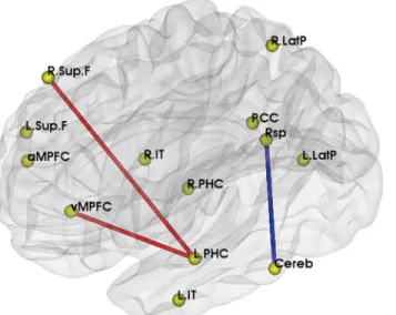
We first compared ROIs functional connectivity betweenε3ε3 andε4ε3 carriers. Comparing toε3ε3 carriers, functional connectivity between left parahippocampal gyrus and right supe-rior frontal cortex (LPHC-R.Sup.F, praw= 0.028, padjusted= 0.026), left parahippocampal gyrus and medial prefrontal cortex (ventral) (LPHC-vMPFC, praw= 0.004, padjusted= 0.010) were sig-nificantly increased inε4ε3 carriers, while connectivity between cerebellar tonsils and retro-splenial (Cereb-Rsp, praw= 0.030, padjusted= 0.016) was decreased, after controlling for the effects of age and gender (Fig 1).
Table 1. Thirteen Regions of Interest in DMN.
Brain region Abbreviations MNI Coordinates
Medial prefrontal cortex (anterior) aMPFC (-3,54,18)
Left superior frontal cortex L.Sup.F (-15,54,42)
Right superior frontal cortex R.Sup.F (18,42,48)
Medial prefrontal cortex (ventral) vMPFC (-6,36,-9)
Left inferior temporal cortex L.IT (-60,-9,-24)
Right inferior temporal cortex R.IT (57,0,-27)
Left parahippocampal gyrus L.PHC (-24,-18,-27)
Right parahippocampal gyrus R.PHC (27,-18,-27)
Posterior cingulated cortex PCC (-3,-48,30)
Retrosplenial Rsp (9,-54,12)
Left lateral parietal cortex L.LatP (-48,-69,39)
Right lateral parietal cortex R.LatP (48,-66,36)
Cerebellar tonsils Cereb (-6,-54,-48)
doi:10.1371/journal.pone.0133179.t001
Table 2. Comparison of demographic information and cognitive measurements between APOEε3ε3 andε4ε3 carriers.
Variable APOEε3ε3 APOEε4ε3 P
Demographic,N 35 14
Age 72.11±4.09 75.93±5.28 0.009
Gender, Male, %(N) 37.1 (13) 42.9 (6) 0.711
Cognitive measurements(baseline),N 34 13
MMSE 26.56±3.33 28.23±1.96 0.097
Delay recall 9.79±2.20 9.85±2.70 0.946
Long-delayed recognition 12.15±3.32 13.38±1.76 0.210
Cognitive measurements(follow-up),N 11 2
MMSE 25.73±3.41 24.50±3.54 0.650
Delay recall 8.55±2.58 5.50±0.71 0.137
Long-delayed recognition 10.64±2.34 11.50±0.71 0.625
Functional connectivity and cognitive measurements
To evaluate the effects on clinical manifestations from these changed functional connectivity, we further analyzed the association between each connection and neuropsychological tests (Fig 2). After controlling for age and gender, we found that the LPHC-R.Sup.F functional con-nectivity was positively correlated with changes of delay recall from baseline to follow-up (r = 0.768, p = 0.009). For LPHC-vMPFC functional connectivity, we found that it had a posi-tive correlation with MMSE at baseline (r = 0.356, p = 0.018), and a negaposi-tive correlation with long-delayed recognition at follow-up (r = -0.677, p = 0.031).
Voxel-based LPHC functional connectivity analyses
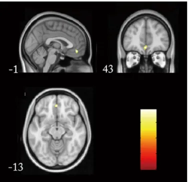
To verify our ROI-based findings, we also performed the voxel-based whole brain functional connectivity analyses by taking LPHC as a seed region. When the individuals’functional con-nectivity was calculated and compared between APOEε4 carriers andε3ε3 genotypes, we found significantly increased functional connectivity in medial prefrontal cortex (cluster
Fig 1. Significantly different DMN functional connectivity between APOEε4ε3 andε3ε3.Brain model for
the changed functional connectivity: the red line represented the increased functional connectivity inε4ε3 carriers, and blue line represented the decreased one.
doi:10.1371/journal.pone.0133179.g001
Fig 2. Changed functional connectivity was associated with cognitive performances.A. The increased LPHC-R.Sup.F functional connectivity was positively correlated with the changes of delay recall from baseline to follow-up. B. The increased LPHC-vMPFC functional connectivity had a positive correlation with MMSE at baseline, and C. a negative correlation with long-delayed recognition at follow-up.
size = 20, p = 0.005,Fig 3), which partially supported the ROI-based functional connectivity findings.
Discussion
Our study demonstrated APOEε4 related changes in DMN in cognitive normal elderly, with decreased connectivity between cerebellar tonsils and retrosplenial, and increased LPHC-R. Sup.F and LPHC-vMPFC connectivity inε4ε3 carriers, compared toε3ε3 carriers. Whole brain analysis confirmed increased functional connectivity between MPFC and LPHC, which suggested specific effects of APOE on DMN. Moreover, we found the significant correlations between functional connectivity and clinical cognitive performances, indicating potential func-tional role of DMN.
Neuroimaging studies have identified the effects of APOEε4 allele on both brain structure and metabolism[21–24]. APOEε4 carriers may present gray matter reductions, decreased rest-ing glucose metabolism in brain regions with potential AD pathology, includrest-ing the posterior cingulate, parietal, temporal, and prefrontal cortices, and also increased task-related activation in relative regions. Recently, altered DMN connectivity has been reported in APOEε4 carriers. Westlye and colleagues reported increased functional connectivity between hippocampus and the posterior DMN in APOE4 carriers [10], and their whole-brain analysis revealed similar effects in the PCC, parietal, and parahippocampal regions. These findings remained significant even hippocampal volumes and gray matter maps were considered as covariates. Another case-control study used seed-based voxel-wise connectivity analysis, and found increased connectiv-ity in the cingulate gyrus, medial prefrontal cortex, bilateral insular cortex, striatum, and thala-mus inε4 carriers when ACC was considered as a seed region[12]. They also found decreased connectivity between PCC and the posterior DMN in APOE4 carriers. Fleisher et al reported increased DMN connectivity in the medial and dorsolateral prefrontal cortex and temporal lobe structures in cognitively normal APOE4 carriers, with posterior cingulate/retrospenial region (pC/rsp) as a seed region[9]. Sheline et al presented a study in 2010 using bilateral pre-cuneus as a seed region, and suggested that most of DMN regions, particularly bilateral hippo-campus and left parahippohippo-campus, had decreased connectivity with precuneus in carriers[25]. Not only in the old population, similar findings were presented in young APOE4 carriers. Filip-pini et al found increased DMN coactivation (including retrosplenial, medial temporal, and medial-prefrontal cortical areas) in APOE4 young carriers (20–35 years)[15]. Later, Dennis and colleagues replicated this findings[16]. This suggested a long-term effect of APOE4 on DMN connectivity, even decades before the onset of AD. Our results were consistent with these studies though there was some differences in regions involved.
The decreased functional connectivity in DMN has been considered to be attributed to the early pathology of AD in APOE4 carriers, which is in line with the disruption of white matter tracts and beta amyloid deposition in DMN reported recently[26,27]. Amyloid-βplaques are an important early event in the pathogenesis of AD. Since the APOEε4 is the risk allele for AD, we speculate that the group APOEε3ε4 allele might have more amyloid-βdeposition, which result in the myelin breakdown, and in turn disrupt functional connectivity. On the other hand, the increased functional connectivity has been interpreted as a compensation for normal cognitive performances. When the functional connectivity between some regions showed decrease in the group APOEε3ε4, the balance was disrupted, the subjects commonly recruit more other regions and strengthen the connectivity to counteract the neurobiological changes due to APOEε4.
z-scores in the precuneus in APOE4 carriers[9], while anther study reported a negative correla-tion between DMN synchronizacorrela-tion and memory performance[10]. we found a positive corre-lation between increased LPHC-vMPFC functional connectivity and baseline MMSE scores in this study, which proved the impact of changed functional connectivity on clinical cognitive manifestations. Moreover, the negative correlation between increased functional connectivity and cognitive performance at follow-up supported the hypothesis that these increased func-tional connectivity is a compensative mechanism to maintain normal cognition, but with changes over time, this compensation might not be enough for perseveration of cognitive per-formance, and decline of cognition would be presented.
Currently, changes of DMN are identified not only in AD, but also in AD risk population, such as APOE4 carriers and MCI patients. This suggests its potential value for early prediction and diagnosis of AD. Sheline and colleagues performed a fMRI study in individuals without preclinical fibrillar amyloid deposition (Pittsburgh Compound B, PIB−)[25] and found that most regions associated with decreased connectivity in whole brain analysis were located in DMN, suggesting early damage of DMN even before pathological manifestation. In addition, Koch et al. investigated diagnostic power of DMN in the detection of AD using both ROI-based signal time course evaluations and independent component analyses (ICA)[28]. Multivariate model combining both the activity of various parts in DMN and also the interconnectivity between these regions was proved to enhance the diagnostic power. Moreover, a model devel-oped for AD identified an AD typical pattern in 11 of 17 MCI patients, with a similar percent-age of MCI subjects presenting an AD typical pattern in a recent PET study (79% in subjects with impairment in multiple cognitive domains and 31% in aMCI patients)[29]. These findings suggested that DMN might be a good candidate to be biomarkers of AD prediction and early diagnosis. Changes of DMN connectivity were found to be associated with progress of clinical
Fig 3. Whole brain functional connectivity by taking LPHC as a seed region.Significantly increased functional connectivity presented in medial prefrontal cortex (cluster size = 20, p = 0.005) in APOEε4 carriers were presented from different directions.
cognitive performance in our study, which also supported its potential predicting role in cogni-tive decline.
Different analytical approaches could influence the results for DMN connectivity. Here we used ROI-based analysis with the ROI that has been proved in our previous studies to define DMN[18,19], which might minimize the impact of inaccurate ROIs. Furthermore, we used seed-based correlation analysis to verify the findings from ROI-based analysis. Though the age of two groups were not perfectly matched, our subjects were enrolled from a population-based cohort, which might be more representative. Moreover, we used statistical methods to control the effects of age and gender. It is a pity that there were several missing data in clinical cognitive measurements, therefore the sample size for related analysis was limited, especially in follow-ups. Further studies should be carried out in larger population with completed clinical infor-mation to verify the findings between changed functional connectivity and neurocognitive performance.
Another analytic issue is multiple comparison for ROI-based analyses in our study. Here we used a permutation-based correction to assess the significance of the P (t-test) value for any given connectivity. The permutation test is similar to the Bonferroni correction in that it con-trols the probability of finding one connectivity by chance in the hypotheses tested; however, a stringent Bonferroni correction is known to be extremely conservative and can thus lead to unacceptable levels of Type II (i.e. false negative) errors in multiple testing; especially when the test statistics are highly interdependent. Meanwhile, a permutation-based correction is data-dependent and has been widely accepted and recommended in studies that involved multiple statistical testing[20,30,31]. Under the threshold at raw P = 0.05 or adjusted P = 0.05, we can get the same 3 significantly different connectivity within the default network.
Conclusion
Our study suggested significant effects of APOE4 on functional connectivity in DMN in older Chinese population, which might have important correlations with clinical cognition. Clini-cians might need to consider the genetic influence on DMN and cognitive performance in clin-ical practice.
Author Contributions
Conceived and designed the experiments: HQS BL JPJ. Performed the experiments: HQS XMZ CSY ZQW QW FW YH. Analyzed the data: HQS HXL BL. Wrote the paper: HQS BL YH JPJ.
References
1. Buckner RL, Andrews-Hanna JR, Schacter DL. The brain's default network: anatomy, function, and rel-evance to disease. Ann N Y Acad Sci. 2008 Mar; 1124:1–38. doi:10.1196/annals.1440.011PMID: 18400922
2. Raichle ME, MacLeod AM, Snyder AZ, Powers WJ, Gusnard DA, Shulman GL. A default mode of brain function. Proc Natl Acad Sci U S A. 2001 Jan 16; 98(2):676–82. PMID:11209064
3. Minoshima S, Giordani B, Berent S, Frey KA, Foster NL, Kuhl DE. Metabolic reduction in the posterior cingulate cortex in very early Alzheimer's disease. Ann Neurol. 1997 Jul; 42(1):85–94. PMID:9225689
4. Buckner RL, Snyder AZ, Shannon BJ, LaRossa G, Sachs R, Fotenos AF, et al. Molecular, structural, and functional characterization of Alzheimer's disease: evidence for a relationship between default activity, amyloid, and memory. J Neurosci. 2005 Aug 24; 25(34):7709–17. PMID:16120771
5. Zhou J, Greicius MD, Gennatas ED, Growdon ME, Jang JY, Rabinovici GD, et al. Divergent network connectivity changes in behavioural variant frontotemporal dementia and Alzheimer's disease. Brain. 2010 May; 133(Pt 5):1352–67.
7. Sorg C, Riedl V, Muhlau M, Calhoun VD, Eichele T, Laer L, et al. Selective changes of resting-state net-works in individuals at risk for Alzheimer's disease. Proc Natl Acad Sci U S A. 2007 Nov 20; 104 (47):18760–5. PMID:18003904
8. Gili T, Cercignani M, Serra L, Perri R, Giove F, Maraviglia B, et al. Regional brain atrophy and functional disconnection across Alzheimer's disease evolution. J Neurol Neurosurg Psychiatry. 2011 Jan; 82 (1):58–66. doi:10.1136/jnnp.2009.199935PMID:20639384
9. Fleisher AS, Sherzai A, Taylor C, Langbaum JB, Chen K, Buxton RB. Resting-state BOLD networks versus task-associated functional MRI for distinguishing Alzheimer's disease risk groups. Neuroimage. 2009 Oct 1; 47(4):1678–90. doi:10.1016/j.neuroimage.2009.06.021PMID:19539034
10. Westlye ET, Lundervold A, Rootwelt H, Lundervold AJ, Westlye LT. Increased hippocampal default mode synchronization during rest in middle-aged and elderly APOE epsilon4 carriers: relationships with memory performance. J Neurosci. 2011 May 25; 31(21):7775–83. doi:10.1523/JNEUROSCI. 1230-11.2011PMID:21613490
11. Sheline YI, Price JL, Yan Z, Mintun MA. Resting-state functional MRI in depression unmasks increased connectivity between networks via the dorsal nexus. Proc Natl Acad Sci U S A. 2010 Jun 15; 107 (24):11020–5. doi:10.1073/pnas.1000446107PMID:20534464
12. Machulda MM, Jones DT, Vemuri P, McDade E, Avula R, Przybelski S, et al. Effect of APOE epsilon4 status on intrinsic network connectivity in cognitively normal elderly subjects. Arch Neurol. 2011 Sep; 68(9):1131–6. doi:10.1001/archneurol.2011.108PMID:21555604
13. Mevel K, Chetelat G, Eustache F, Desgranges B. The default mode network in healthy aging and Alz-heimer's disease. Int J Alzheimers Dis. 2011; 2011:535816. doi:10.4061/2011/535816PMID: 21760988
14. Strittmatter WJ, Saunders AM, Schmechel D, Pericak-Vance M, Enghild J, Salvesen GS, et al. Apolipo-protein E: high-avidity binding to beta-amyloid and increased frequency of type 4 allele in late-onset familial Alzheimer disease. Proc Natl Acad Sci U S A. 1993 Mar 1; 90(5):1977–81. PMID:8446617
15. Filippini N, MacIntosh BJ, Hough MG, Goodwin GM, Frisoni GB, Smith SM, et al. Distinct patterns of brain activity in young carriers of the APOE-epsilon4 allele. Proc Natl Acad Sci U S A. 2009 Apr 28; 106 (17):7209–14. doi:10.1073/pnas.0811879106PMID:19357304
16. Dennis NA, Browndyke JN, Stokes J, Need A, Burke JR, Welsh-Bohmer KA, et al. Temporal lobe func-tional activity and connectivity in young adult APOE varepsilon4 carriers. Alzheimers Dement. 2010 Jul; 6(4):303–11. doi:10.1016/j.jalz.2009.07.003PMID:19744893
17. Wenham PR, Price WH, Blandell G. Apolipoprotein E genotyping by one-stage PCR. Lancet. 1991 May 11; 337(8750):1158–9.
18. Song M, Liu Y, Zhou Y, Wang K, Yu C, Jiang T. Default network and intelligence difference. Conf Proc IEEE Eng Med Biol Soc. 2009; 2009:2212–5. doi:10.1109/IEMBS.2009.5334874PMID:19964951
19. Liu B, Song M, Li J, Liu Y, Li K, Yu C, et al. Prefrontal-related functional connectivities within the default network are modulated by COMT val158met in healthy young adults. J Neurosci. 2010 Jan 6; 30 (1):64–9. doi:10.1523/JNEUROSCI.3941-09.2010PMID:20053888
20. Camargo A, Azuaje F, Wang H, Zheng H. Permutation—based statistical tests for multiple hypotheses. Source Code Biol Med. 2008 Oct 21; 3:15. doi:10.1186/1751-0473-3-15PMID:18939983
21. Wishart HA, Saykin AJ, McAllister TW, Rabin LA, McDonald BC, Flashman LA, et al. Regional brain atrophy in cognitively intact adults with a single APOE epsilon4 allele. Neurology. 2006 Oct 10; 67 (7):1221–4. PMID:17030756
22. Reiman EM, Caselli RJ, Yun LS, Chen K, Bandy D, Minoshima S, et al. Preclinical evidence of Alzhei-mer's disease in persons homozygous for the epsilon 4 allele for apolipoprotein E. N Engl J Med. 1996 Mar 21; 334(12):752–8. PMID:8592548
23. Small GW, Ercoli LM, Silverman DH, Huang SC, Komo S, Bookheimer SY, et al. Cerebral metabolic and cognitive decline in persons at genetic risk for Alzheimer's disease. Proc Natl Acad Sci U S A. 2000 May 23; 97(11):6037–42. PMID:10811879
24. Reiman EM, Chen K, Alexander GE, Caselli RJ, Bandy D, Osborne D, et al. Functional brain abnormali-ties in young adults at genetic risk for late-onset Alzheimer's dementia. Proc Natl Acad Sci U S A. 2004 Jan 6; 101(1):284–9. PMID:14688411
25. Sheline YI, Morris JC, Snyder AZ, Price JL, Yan Z, D'Angelo G, et al. APOE4 allele disrupts resting state fMRI connectivity in the absence of amyloid plaques or decreased CSF Abeta42. J Neurosci. 2010 Dec 15; 30(50):17035–40. doi:10.1523/JNEUROSCI.3987-10.2010PMID:21159973
27. Heise V, Filippini N, Ebmeier KP, Mackay CE. The APOE varepsilon4 allele modulates brain white mat-ter integrity in healthy adults. Mol Psychiatry. 2011 Sep; 16(9):908–16. doi:10.1038/mp.2010.90PMID: 20820167
28. Koch W, Teipel S, Mueller S, Benninghoff J, Wagner M, Bokde AL, et al. Diagnostic power of default mode network resting state fMRI in the detection of Alzheimer's disease. Neurobiol Aging. 2012 Mar; 33(3):466–78. doi:10.1016/j.neurobiolaging.2010.04.013PMID:20541837
29. Mosconi L, Tsui WH, Herholz K, Pupi A, Drzezga A, Lucignani G, et al. Multicenter standardized 18F-FDG PET diagnosis of mild cognitive impairment, Alzheimer's disease, and other dementias. J Nucl Med. 2008 Mar; 49(3):390–8. doi:10.2967/jnumed.107.045385PMID:18287270
30. Conneely KN, Boehnke M. So many correlated tests, so little time! Rapid adjustment of P values for multiple correlated tests. Am J Hum Genet. 2007 Dec; 81(6):1158–68. PMID:17966093