Culture-Independent Identification of
Nontuberculous Mycobacteria in Cystic
Fibrosis Respiratory Samples
Lindsay J. Caverly1*, Lisa A. Carmody1, Sarah-Jane Haig2, Nadine Kotlarz2, Linda M. Kalikin1, Lutgarde Raskin2, John J. LiPuma1
1Department of Pediatrics and Communicable Diseases, University of Michigan Medical School, Ann Arbor, Michigan, United States of America,2Department of Civil & Environmental Engineering, University of Michigan, Ann Arbor, Michigan, United States of America
*caverlyl@med.umich.edu
Abstract
Respiratory tract infections with nontuberculous mycobacteria (NTM) are increasing in prev-alence and are a significant cause of lung function decline in individuals with cystic fibrosis (CF). NTM have been detected in culture-independent analyses of CF airway microbiota at lower rates than would be expected based on published prevalence data, likely due to poor lysing of the NTM cell wall during DNA extraction. We compared a standard bacterial lysis protocol with a modified method by measuring NTM DNA extraction by qPCR and NTM detection with bacterial 16S rRNA gene sequencing. The modified method improved NTM DNA recovery from spiked CF sputum samples by a mean of 0.53 log10copies/mL forM. abscessuscomplex and by a mean of 0.43 log10copies/mL forM.aviumcomplex as mea-sured by qPCR targeting theatpEgene. The modified method also improved DNA sequence based NTM detection in NTM culture-positive CF sputum and bronchoalveolar lavage samples; however, both qPCR and 16S rRNA gene sequencing remained less sen-sitive than culture for NTM detection. We highlight the limitations of culture-independent identification of NTM from CF respiratory samples, and illustrate how alterations in the bac-terial lysis and DNA extraction process can be employed to improve NTM detection with both qPCR and 16S rRNA gene sequencing.
Introduction
Nontuberculous mycobacterial (NTM) infections affect approximately 13% of individuals with cystic fibrosis (CF) and are increasing in prevalence[1,2]. While NTM infections can result in significant lung function decline and death, the clinical course after NTM acquisition is highly variable[3,4]. Predictors of NTM acquisition and subsequent disease course are largely unknown.
Understanding the relationship(s) between NTM infection and other members of the microbial communities inhabiting the CF airways offers potential insight into NTM disease
a11111
OPEN ACCESS
Citation:Caverly LJ, Carmody LA, Haig S-J, Kotlarz N, Kalikin LM, Raskin L, et al. (2016) Culture-Independent Identification of Nontuberculous Mycobacteria in Cystic Fibrosis Respiratory Samples. PLoS ONE 11(4): e0153876. doi:10.1371/journal. pone.0153876
Editor:Amit Gaggar, University of Alabama-Birmingham, UNITED STATES
Received:January 28, 2016
Accepted:April 5, 2016
Published:April 19, 2016
Copyright:© 2016 Caverly et al. This is an open access article distributed under the terms of the
Creative Commons Attribution License, which permits unrestricted use, distribution, and reproduction in any medium, provided the original author and source are credited.
Data Availability Statement:All relevant data are within the paper.
Funding:This work was supported by National Institute of Child Heath and Human Development,
https://www.nichd.nih.gov/Pages/index.aspx, grant number: HD 028820, to LJC; Cystic Fibrosis Foundation,https://www.cff.org/, grant number: CAVERL15I0 to LJC; University of Michigan Department of Pediatrics and Communicable Diseases,https://medicine.umich.edu/dept/pediatrics/
pathogenesis. Although prior investigations have not consistently identified co-infection with other bacterial pathogens as risk factors for NTM acquisition or disease course[1,5,6], these studies have been limited by the use of culture-based bacterial detection, which focuses on identifying known CF pathogens[7]. Studies employing DNA-sequencing based approaches to characterize CF airway bacterial communities offer an opportunity to assess the impact of a broader range of species on NTM acquisition and/or disease. Culture-independent analyses have, for example, identified differences in the airway microbiomes of non-CF patients with pulmonaryMycobacterium tuberculosis(MTb) infection compared to healthy controls[8,9]. Differences in airway microbiota in individuals with pulmonary MTb are also associated with treatment response[10].
We previously sequenced the bacterial 16S rRNA genes in more than a thousand sputum samples from individuals with CF[7,11–14]. Review of these data showed an unexpected lack of DNA sequences classified asMycobacterium, particularly in 16 samples from eight individu-als that were culture-positive for NTM. Suspecting that the failure to detect NTM DNA resulted from inadequate lysing of NTM cells during DNA extraction from sputum, we assessed the effect of various alterations to our standard bacterial cell lysis protocol on enhanc-ing NTM cell lysis and developed a modified bacterial cell lysis protocol.
We compared the performance of this modified lysis protocol to our standard protocol with respect to NTM DNA recovery from both NTM-spiked sputum samples and NTM culture-positive respiratory samples. We also assessed the impact of the lysis protocol modifications on 16S rRNA gene sequence based measures of bacterial community structure in the NTM cul-ture-positive respiratory samples.
Materials and Methods
Development of modified lysis protocol
Prior efforts by other investigators to enhance NTM cell lysis for DNA extraction have demon-strated that a combination of bead beating and enzymatic extraction results in better NTM cell lysis as compared to either enzymatic or chemical lysis alone[15–18]. In preliminary experi-ments, we confirmed these findings on CF respiratory samples, as well as confirmed findings that NTM DNA yield did not improve with prolonged duration of enzymatic lysis[19], alter-nate methods of physical lysis (e.g. freeze-thaw)[18,20], or with extending bead beating times beyond two minutes[19,21]. We observed that the greatest increase in NTM DNA yield from CF respiratory samples was obtained by altering the bead beating conditions of our standard protocol. Specifically, we modified our standard lysis protocol by changing from glass beads to the denser zirconium beads[21] and by decreasing the sample volume during bead-beating.
Respiratory samples and NTM strains
The sputum and bronchoalveolar lavage (BAL) samples included in this study are part of a large collection of respiratory samples obtained during the course of routine care of individuals with CF followed at the University of Michigan Health System (UMHS) CF care centers from 2006–2015. Sample collection and medical record review were approved by the University of Michigan Institutional Review Board (HUM00048991 and HUM00080378). Waiver of subject informed consent was granted as this study was limited to a retrospective analysis of existing unique-identifier-encoded samples and clinical data. The Electronic Medical Record Search Engine (EMERSE) was used to extract relevant clinical data from the medical record[22]. Spu-tum and BAL samples were aliquoted and stored neat at -80°C. Prior to DNA extraction, spu-tum and BAL sample aliquots were thawed on ice then incubated with an equal volume of 10% Sputolysin1at 37°C for 30 min with pulse vortexing every 5 min to achieve homogenization.
design, data collection and analysis, decision to publish, or preparation of the manuscript.
All NTM strains used in spiking experiments were stored in Mueller-Hinton liquid media with glycerol at -80°C prior to being grown on Middlebrook 7H11 solid media at 37°C. Bacteria were suspended in sterile phosphate-buffered saline and density was quantified by optical den-sity and confirmed by plating serial dilutions.
NTM- spiked sputum samples
Sputum samples were identified from an individual with CF who had no history of NTM infec-tion based on annual negative NTM sputum cultures. Sputum samples were pooled and homogenized, then spiked with 107cfu/ml of one of four NTM strains:Mycobacterium absces-suscomplex (MABSC) ATCC 19977,Mycobacterium aviumcomplex (MAC) ATCC 25291, and one clinical isolate each of MABSC and MAC that were obtained from the UMHS clinical microbiology laboratory. Replicate 350μL aliquots of spiked sputum were processed by the
standard or the modified lysis protocols prior to DNA extraction and qPCR as described below. Six paired, spiked sputum samples were tested for each ATCC strain, and four paired, spiked sputum samples were tested for each clinical strain.
NTM culture-positive samples
Twelve individuals with CF and a history of NTM infection were identified with EMERSE. From these 12 individuals, 15 samples (11 sputum and four BAL) were identified in our collec-tion that were NTM culture-positive (seven MABSC and eight MAC). NTM culture had been performed by the UMHS clinical microbiology laboratory. Smear positivity was defined as the presence of at least one organism per high powered field. NTM species identification was con-firmed in each case by the Michigan Department of Community Health. Sample appearance was assessed with respect to consistency and presence/absence of gross blood at the time of DNA extraction. After thawing and homogenization, duplicate aliquots of the NTM culture-positive samples were processed by the standard or the modified lysis protocol prior to DNA extraction, qPCR, and 16S rRNA gene sequencing as described below.
Bacterial lysis and DNA extraction
Standard lysis protocol. A 350μL aliquot of the homogenized sample was mixed with 0.9
volume of MagNA Pure Bacterial Lysis Buffer (Roche Applied Science, Indianapolis, IN), lyso-zyme (final concentration, 2.9 mg/mL; Sigma-Aldrich Corp., St. Louis, MO), and lysostaphin (final concentration, 0.14 mg/mL; Sigma-Aldrich), then incubated for 30 min at 37°C[23]. Samples were transferred to a tube containing 0.1 mm glass beads (MoBio Laboratories, Carls-bad, CA) and agitated in a Mini-Beadbeater-9 (Biospec Products Inc., Bartlesville, OK) for 1 min at the maximum setting. Samples were digested with Proteinase K (final concentration, 1 mg/mL) and incubated for 10 min at 65°C, agitated for an additional 1 min in the Mini-Bead-beater-9, and then incubated for an additional 10 min at 95°C. DNA purification was per-formed using an automated nucleic acid purification platform (MagNa Pure Compact System, Roche) using the manufacturer’s DNA Bacteria v3.1 protocol.
Modified lysis protocol. A 350μL aliquot of the homogenized sample was placed into a
Quantitative PCR
NTM DNA extraction was quantified with qPCR targeting theatpEgene[24] under the follow-ing conditions: each 20 uL reaction contained 1X SYBR Green Master Mix, 500 nM of each primer, 0.75 mg/mL BSA, and 2 uL of undiluted sputum DNA. Cycling conditions were as fol-lows: 95°C for 10 min x 1, then (95°C for 15 sec, 60°C x 1 min) for 40 cycles. Each run con-tained non-template control wells and a 10-fold dilution series of MABSC (ATCC 19977) genomic DNA. All samples were assayed in triplicate. The limit of detection for the qPCR reac-tion was 14 copies/reacreac-tion.
Differences in yield of total bacterial DNA between the lysis protocols were quantified with qPCR using primers targeting conserved regions of the bacterial 16S rRNA gene as described previously[11,25]. qPCR data were log-transformed and analyzed with paired t-tests. Data were analyzed with GraphPad Prism 6 (GraphPad Software, Inc., La Jolla, CA).
DNA sequencing and sequence analyses
For the NTM culture-positive sputum and BAL samples, the V4 region of the 16S rRNA gene was amplified with barcoded primers and sequenced using the MiSeq Reagent Kit V2 (Illu-mina, San Diego, CA) and the MiSeq Illumina platform[14]. Raw sequencing data were pro-cessed with the mothur software package (version 1.35.0, downloaded on 5/21/15) as described in the MiSeq standard operating procedure [26]. OTU assignment, determination of presence or absence of NTM sequence reads, and relative abundance calculations were performed on sequence reads prior to subsampling. For calculations of alpha and beta diversity measures and Metastats[27] analysis, sequence reads were subsampled to 3,729, the smallest number of sequences obtained among the 30 samples. Sequencing data were analyzed with mothur and R [28].
Results
NTM detection by qPCR and 16S rRNA gene sequencing
Based on qPCR of the mycobacterialatpEgene, the use of the modified lysis protocol signifi-cantly increased the recovery of NTM DNA from the NTM-spiked sputum samples for each of the four NTM strains (Fig 1). The mean (SD) increases in log10atpEgene copies/mL using the
modified protocol compared to the standard protocol were 0.56 (0.19) for MABSC ATCC 19977 (p = 0.001, paired t-test), 0.51 (0.03) for the MABSC clinical isolate (p<0.001, paired
t-test), 0.45 (0.23) for MAC ATCC 25291 (p = 0.005, paired t-t-test), and 0.4 (0.1) for the MAC clinical isolate (p = 0.004, paired t-test).
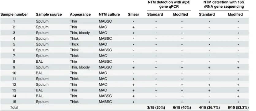
While NTM DNA was detected by qPCR targeting theatpEgene in three (20%) of the 15 culture-positive samples processed with the standard bacterial lysis protocol, six (40%) of the 15 samples were PCR-positive when processed with the modified lysis protocol (Table 1). All of the samples in which NTM DNA was detected by qPCR with either protocol were NTM smear-positive. NTM DNA was detected by qPCR in three (37.5%) of the eight smear-positive samples processed with the standard lysis protocol and in six (75%) of these samples processed with the modified lysis protocol. The qPCR-positive samples included both sputum and BAL samples, and samples that were culture-positive for either MABSC or MAC (Table 1).
gene sequences were detected in four (50%) of the eight smear-positive samples after process-ing with the standard lysis protocol, and in seven (87.5%) of these smear-positive samples after processing with modified lysis protocol. The samples that were positive for NTM 16S rRNA gene sequences included both sputum and BAL samples, samples with both thick and thin con-sistency, and samples that were culture-positive for either MABSC or MAC (Table 1).
The relative abundances of NTM sequence reads in the eight samples in which NTM were detected by 16S rRNA gene sequencing after processing with the modified lysis protocol were compared to the relative abundances of NTM sequence reads detected in the same samples after processing with the standard lysis protocol. The mean relative abundance of NTM sequence reads increased from 0.098% (range 0.0%–0.33%) with the standard lysis protocol to 1.21% (range 0.003% - 3.95%) with the modified lysis protocol; however, this difference was not statistically significant (p = 0.08, paired t-test) (Fig 2A). Based on qPCR targeting the bacte-rial 16S rRNA gene, total bactebacte-rial DNA did not differ between paired NTM culture-positive samples processed with the standard lysis protocol compared to the modified protocol (Fi 2B).
Impact on community structure
The influence of the lysis protocols on bacterial community structure of the NTM culture-posi-tive samples was assessed with a Bray-Curtis-based nonmetric multidimensional scaling (NMDS) plot (Fig 3). The majority of the sample pairs clustered closely together despite differ-ences in lysis protocol. However, in five of the 15 sample pairs (solid symbols inFig 3) some separation between samples was noted on the ordination plot. These 5 sample pairs tended to have higher levels of alpha diversity (higher Shannon diversity and evenness; blue and red
Fig 1. Improvement in NTM DNA extraction from spiked sputum samples with the modified as compared to the standard lysis protocol.Log10atpE
gene copies/mL in DNA extracted from sputum spiked with either (A) MABSC or (B) MAC using the standard (blue circles) or the modified (red squares) lysis protocols. Error bars indicate mean and SD.
doi:10.1371/journal.pone.0153876.g001
Table 1. Characteristics of NTM culture-positive samples.
NTM detection withatpE gene qPCR
NTM detection with 16S rRNA gene sequencing Sample number Sample source Appearance NTM culture Smear Standard Modified Standard Modified
1 Sputum Thin MABSC - - - -
-2 Sputum Thin MAC + - - -
-3 Sputum Thin, bloody MAC + - + - +
4 Sputum Thick MABSC - - - -
-5 Sputum Thick MAC - - - -
-6 Sputum Thick MABSC - - - -
-7 Sputum Thick MAC - - - -
-8 BAL Thin MABSC - - - - +
9 Sputum Thin, bloody MABSC + + + + +
10 BAL Thin MAC - - - -
-11 Sputum Thick MAC + + + - +
12 Sputum Thin MAC + - + + +
13 BAL Thin MAC + + + + +
14 BAL Thin MABSC + - - - +
15 Sputum Thick MABSC + - + + +
Total 3/15 (20%) 6/15 (40%) 4/15 (26.7%) 8/15 (53.3%)
symbols inFig 4) than the 10 sample pairs that remained more tightly clustered despite differ-ences in lysis protocol. Overall, the samples processed with the modified protocol had higher measures of richness and Shannon diversity than the samples processed with the standard pro-tocol (Fig 4).
The potential for differential abundance of bacterial genera between the NTM culture-posi-tive samples processed with the standard as compared to the modified lysis protocol was assessed. The ten most abundant OTUs in the sample set representedStaphylococcus, Stenotro-phomonas,Pseudomonas,Streptococcus,Prevotella,Veillonella,Haemophilus, Enterobacteria-ceae,Firmicutes, andActinomyces; these did not significantly differ between the lysis protocols (Metastats[27] with Bonferroni correction for multiple comparisons).
Discussion
With this study we illustrate the limitations of culture-independent detection of NTM from CF respiratory samples and demonstrate how alterations in the bacterial cell lysis method can be
Fig 2. (A) Relative abundances of NTM OTUs and (B) total bacterial load in NTM culture-positive samples. (A) The mean relative abundance of NTM OTUs in the samples processed with the standard protocol was not significantly different from that observed when these samples were processed with the modified protocol (mean 0.098% and 1.21%, respectively; p = 0.08, paired t-test). (B) Total bacterial load in NTM culture-positive samples as measured by 16S rRNA gene qPCR did not significantly differ between lysis protocols. (p = 0.91, paired t-test). Error bars indicate mean and SD.
doi:10.1371/journal.pone.0153876.g002
Fig 3. Impact of lysis method on community structure.Bray-Curtis-based nonmetric multidimensional scaling (NMDS) plot showing pairwise comparison of samples processed with the standard (blue symbols) or modified (red symbols) lysis protocols. Solid symbols represent paired samples with greater separation.
employed to improve NTM sequence-based detection. Despite the significant improvements seen in NTM DNA extraction with the modified lysis protocol, NTM sequences were detected by 16S rRNA gene sequencing in only about half (53%) of the NTM culture-positive samples (87% of smear-positive and 14% of smear-negative samples) included in our study. This decreased sensitivity of 16S rRNA gene sequencing for NTM as compared to culture are consis-tent with findings of MTb pulmonary infection in a non-CF population, in which Mycobacte-riumwas identified by 16S rRNA gene sequencing in only 39% (29/75) of sputum samples from MTb-infected individuals[10]. Consistent with a recent study in which qPCR was less sensitive than 16S rRNA gene sequencing for detection ofStaphylococcus aureusfrom CF respiratory samples[29], qPCR of theatpEgene in our study was less sensitive than 16S rRNA gene sequencing in detecting NTM in CF respiratory samples.
The improvement in NTM DNA sequence detection with use of the modified lysis protocol supports inadequate bacterial lysis as a contributing factor to the underrepresentation of NTM in culture-independent studies of the CF microbiome. NTM are notoriously difficult to lyse due to the layer of glycopeptidolipids and mycolic acid that make up the NTM cell wall[30]. Previous work by others has shown that NTM lysis and DNA yields can be improved by enhancing the physical disruption of mycobacterial cell walls[15,18,19,21]. We similarly observed that the greatest increase in NTM DNA yield from CF respiratory samples was obtained by altering bead beating conditions. More specifically, the changes from glass beads to the denser zirconium beads and decreasing the sample volume as described in our modified protocol provided the greatest increase in NTM yield, with a minimal impact on the 16S rRNA gene sequence based measures of bacterial community structure in the majority of the NTM culture-positive respiratory samples.
These and other efforts to optimize NTM DNA extraction have focused on cultured NTM isolates[15,16,18,21], or on respiratory samples from individuals with MTb infection[31]. We are not aware of previous studies to enhance NTM DNA extraction from CF respiratory sam-ples. CF sputum is a complex biologic matrix dominated by neutrophil-derived DNA polymers and F-actin that differs from the respiratory secretions found in other chronic pulmonary pro-cesses such as asthma or bronchitis[32]. We reiterate that while our modified cell lysis protocol increased NTM yield from CF respiratory samples, other approaches may be more appropriate for other biological or environmental sample types.
In addition to the difficulty of lysing NTM bacterial cells, other factors likely also contribute to the poor performance of culture-independent approaches in identifying NTM in CF respira-tory samples. MABSC and MAC, the primary NTM species found in CF infections, have genomes that harbor single 16S rRNA gene operons[33,34]. This is in contrast to other bacte-rial genera, which typically have genomes that include multiple copies of this operon[35]. The genome ofPseudomonas aeruginosa, for example, includes four copies of the 16S rRNA gene operon[35]. NTM infection also often may involve lower levels of bacterial burden than infec-tion with other known CF pathogens. For instance, NTM smear positivity is observed with a bacterial density of103cfu/mL[36]. Smear positivity is typically thought to indicate a high burden of infection[37], but occurs in the minority of individuals with CF and NTM pulmo-nary infection[1]. This is in contrast to infection withP.aeruginosa, where bacterial density in the CF airways can often exceed ~107cfu/mL[38]. The difficulty in lysing NTM bacterial cells,
Fig 4. Differences in alpha diversity between lysis protocols.Samples processed with the modified protocol had higher levels of (A) richness (p = 0.04, paired t-test), and higher (C) Shannon diversity (p = 0.004, paired t-test) than samples processed with the standard protocol. (B) Evenness did not differ between the lysis protocols (p = 0.06, paired t-test). Blue and red symbols represent paired samples with greater separation on NMDS. Error bars indicate mean and SD.
the single 16S rRNA gene operon, and the low density of NTM all likely contributed to the lack of culture-independent NTM detection in the smear-negative samples despite the lysis protocol modifications.
We identified NTM sequences by 16S rRNA gene sequencing in 27% of the NTM culture-positive samples processed with the standard protocol. This is in contrast to the absence of NTM sequences found in the 16 culture-positive sputum samples that were analyzed by next-generation sequencing of the 16S rRNA gene in our prior studies[7,11–14]. We note, however, that in these previous studies, DNA sequencing was performed using the Roche 454 sequencing platform. We suspect that the greater sequencing depth of the MiSeq Illumina sequencing plat-form used in the current study may account for the detection of NTM sequences in the small number of samples processed with the standard protocol. We recognize that multiple variables, or combinations of variables, may influence DNA sequence-based detection of NTM. In addi-tion to bacterial lysis, DNA extracaddi-tion, and sequencing platform, these variables also include the variable region of the 16S rRNA gene targeted and the analytic pipeline used for 16S rRNA sequence analysis[39].
In this study, we limited our testing of the lysis protocols to sputum and BAL samples con-taining MABSC and MAC, as these are the NTM species most commonly isolated from the CF airways[1,2]. The cell wall mycolic acid composition varies between NTM species in ways that influence susceptibility to lysis methods[16,21]. Although our findings were consistent across the four NTM strains tested, and across multiple CF clinical samples, we cannot confidently extrapolate our results to all other NTM species.
Understanding the limitations of DNA sequence-based detection of NTM from CF respira-tory samples is a necessary first step for future culture-independent studies of the microbial ecology of NTM infection in the CF. Identifying CF airway microbial community structures (e.g., members of the communities and their distribution) that relate to NTM acquisition, NTM pulmonary disease, and/or treatment response, will be critical steps in identifying clini-cally useful biomarkers of disease and in understanding the pathophysiology of NTM disease in CF.
Acknowledgments
We gratefully acknowledge the many individuals who provided the sputum samples used in this study. We thank the UMHS clinical microbiology laboratory for the generous assistance in obtaining sputum samples for this study. We also thank Amanda Forde and Michelle Azar for their assistance with this effort.
Author Contributions
Conceived and designed the experiments: LJC LAC SJH NK LMK LR JJL. Performed the exper-iments: LJC. Analyzed the data: LJC LAC SJH NK JJL. Contributed reagents/materials/analysis tools: LJC LAC SJH NK LMK JJL. Wrote the paper: LJC LAC SJH NK LMK LR JJL.
References
1. Olivier KN, Weber DJ, Wallace RJ Jr, Faiz AR, Lee JH, et al. (2003) Nontuberculous mycobacteria. I: multicenter prevalence study in cystic fibrosis. Am J Respir Crit Care Med 167: 828–834. PMID: 12433668
2. Salsgiver EL, Fink AK, Knapp EA, LiPuma JJ, Olivier KN, et al. (2015) Changing Epidemiology of the Respiratory Bacteriology of Patients with Cystic Fibrosis. Chest 149: 390–400.
4. Martiniano SL, Sontag MK, Daley CL, Nick JA, Sagel SD (2014) Clinical significance of a first positive nontuberculous mycobacteria culture in cystic fibrosis. Ann Am Thorac Soc 11: 36–44. doi:10.1513/ AnnalsATS.201309-310OCPMID:24251858
5. Esther CR Jr, Henry MM, Molina PL, Leigh MW (2005) Nontuberculous mycobacterial infection in young children with cystic fibrosis. Pediatr Pulmonol 40: 39–44. PMID:15858802
6. Levy I, Grisaru-Soen G, Lerner-Geva L, Kerem E, Blau H, et al. (2008) Multicenter cross-sectional study of nontuberculous mycobacterial infections among cystic fibrosis patients, Israel. Emerg Infect Dis 14: 378–384. doi:10.3201/eid1403.061405PMID:18325250
7. Mahboubi MA, Carmody LA, Foster BK, Kalikin LM, VanDevanter DR, et al. (2015) Culture-based and culture-independent bacteriologic analysis of cystic fibrosis respiratory specimens. J Clin Microbiol 54: 613–619. doi:10.1128/JCM.02299-15PMID:26699705
8. Cheung MK, Lam WY, Fung WY, Law PT, Au CH, et al. (2013) Sputum microbiota in tuberculosis as revealed by 16S rRNA pyrosequencing. PLoS One 8: e54574. doi:10.1371/journal.pone.0054574 PMID:23365674
9. Cui Z, Zhou Y, Li H, Zhang Y, Zhang S, et al. (2012) Complex sputum microbial composition in patients with pulmonary tuberculosis. BMC Microbiol 12: 276. doi:10.1186/1471-2180-12-276PMID: 23176186
10. Wu J, Liu W, He L, Huang F, Chen J, et al. (2013) Sputum microbiota associated with new, recurrent and treatment failure tuberculosis. PLoS One 8: e83445. doi:10.1371/journal.pone.0083445PMID: 24349510
11. Zhao J, Schloss PD, Kalikin LM, Carmody LA, Foster BK, et al. (2012) Decade-long bacterial commu-nity dynamics in cystic fibrosis airways. Proc Natl Acad Sci U S A 109: 5809–5814. doi:10.1073/pnas. 1120577109PMID:22451929
12. Carmody LA, Zhao J, Schloss PD, Petrosino JF, Murray S, et al. (2013) Changes in cystic fibrosis air-way microbiota at pulmonary exacerbation. Ann Am Thorac Soc 10: 179–187. doi:10.1513/ AnnalsATS.201211-107OCPMID:23802813
13. Carmody LA, Zhao J, Kalikin LM, LeBar W, Simon RH, et al. (2015) The daily dynamics of cystic fibrosis airway microbiota during clinical stability and at exacerbation. Microbiome 3: 12. doi: 10.1186/s40168-015-0074-9PMID:25834733
14. Zhao J, Evans CR, Carmody LA, LiPuma JJ (2015) Impact of storage conditions on metabolite profiles of sputum samples from persons with cystic fibrosis. J Cyst Fibros 14: 468–473. doi:10.1016/j.jcf. 2015.02.004PMID:25725986
15. Plain KM, Waldron AM, Begg DJ, de Silva K, Purdie AC, et al. (2015) Efficient, validated method for detection of mycobacterial growth in liquid culture media by use of bead beating, magnetic-particle-based nucleic acid isolation, and quantitative PCR. J Clin Microbiol 53: 1121–1128. doi:10.1128/JCM. 03521-14PMID:25609725
16. Amaro A, Duarte E, Amado A, Ferronha H, Botelho A (2008) Comparison of three DNA extraction meth-ods forMycobacterium bovis,Mycobacterium tuberculosisandMycobacterium aviumsubsp.avium. Lett Appl Microbiol 47: 8–11. doi:10.1111/j.1472-765X.2008.02372.xPMID:18498320
17. Kaser M, Ruf MT, Hauser J, Marsollier L, Pluschke G (2009) Optimized method for preparation of DNA from pathogenic and environmental mycobacteria. Appl Environ Microbiol 75: 414–418. doi:10.1128/ AEM.01358-08PMID:19047396
18. Tell LA, Foley J, Needham ML, Walker RL (2003) Comparison of four rapid DNA extraction techniques for conventional polymerase chain reaction testing of threeMycobacteriumspp. that affect birds. Avian Dis 47: 1486–1490. PMID:14709001
19. Radomski N, Kreitmann L, McIntosh F, Behr MA (2013) The critical role of DNA extraction for detection of mycobacteria in tissues. PLoS One 8: e78749. doi:10.1371/journal.pone.0078749PMID:24194951
20. Odumeru J, Gao A, Chen S, Raymond M, Mutharia L (2001) Use of the bead beater for preparation of
Mycobacterium paratuberculosistemplate DNA in milk. Can J Vet Res 65: 201–205. PMID:11768125
21. Madiraju MV, Qin MH, Rajagopalan M (2000) Development of simple and efficient protocol for isolation of plasmids from mycobacteria using zirconia beads. Lett Appl Microbiol 30: 38–41. PMID:10728558
22. Hanauer DA, Mei Q, Law J, Khanna R, Zheng K (2015) Supporting information retrieval from electronic health records: A report of University of Michigan's nine-year experience in developing and using the Electronic Medical Record Search Engine (EMERSE). J Biomed Inform 55: 290–300. doi:10.1016/j. jbi.2015.05.003PMID:25979153
23. Zhao J, Carmody LA, Kalikin LM, Li J, Petrosino JF, et al. (2012) Impact of enhancedStaphylococcus
24. Radomski N, Roguet A, Lucas FS, Veyrier FJ, Cambau E, et al. (2013)atpEgene as a new useful spe-cific molecular target to quantify Mycobacterium in environmental samples. BMC Microbiol 13: 277. doi:10.1186/1471-2180-13-277PMID:24299240
25. Nadkarni MA, Martin FE, Jacques NA, Hunter N (2002) Determination of bacterial load by real-time PCR using a broad-range (universal) probe and primers set. Microbiology 148: 257–266. PMID: 11782518
26. Kozich JJ, Westcott SL, Baxter NT, Highlander SK, Schloss PD (2013) Development of a dual-index sequencing strategy and curation pipeline for analyzing amplicon sequence data on the MiSeq Illumina sequencing platform. Appl Environ Microbiol 79: 5112–5120. doi:10.1128/AEM.01043-13PMID: 23793624
27. White JR, Nagarajan N, Pop M (2009) Statistical methods for detecting differentially abundant features in clinical metagenomic samples. PLoS Comput Biol 5: e1000352. doi:10.1371/journal.pcbi.1000352 PMID:19360128
28. R: A Language and Environment for Statistical Computing. (2015) Vienna, Austria: R Foundation for Statistical Computing.
29. Johnson EJ, Zemanick ET, Accurso FJ, Wagner BD, Robertson CE, et al. (2016) Molecular Identifica-tion of Staphylococcus aureus in Airway Samples from Children with Cystic Fibrosis. PLoS One 11: e0147643. doi:10.1371/journal.pone.0147643PMID:26808658
30. McNeil MR, Brennan PJ (1991) Structure, function and biogenesis of the cell envelope of mycobacteria in relation to bacterial physiology, pathogenesis and drug resistance; some thoughts and possibilities arising from recent structural information. Res Microbiol 142: 451–463. PMID:1871433
31. Chakravorty S, Tyagi JS (2001) Novel use of guanidinium isothiocyanate in the isolation of Mycobacte-rium tuberculosis DNA from clinical material. FEMS Microbiol Lett 205: 113–117. PMID:11728724
32. Rubin BK (2009) Mucus, phlegm, and sputum in cystic fibrosis. Respir Care 54: 726–732; discussion 732. PMID:19467160
33. Bercovier H, Kafri O, Sela S (1986) Mycobacteria possess a surprisingly small number of ribosomal RNA genes in relation to the size of their genome. Biochem Biophys Res Commun 136: 1136–1141. PMID:3013168
34. Nessar R, Cambau E, Reyrat JM, Murray A, Gicquel B (2012) Mycobacterium abscessus: a new antibi-otic nightmare. J Antimicrob Chemother 67: 810–818. doi:10.1093/jac/dkr578PMID:22290346
35. Coenye T, Vandamme P (2003) Intragenomic heterogeneity between multiple 16S ribosomal RNA operons in sequenced bacterial genomes. FEMS Microbiol Lett 228: 45–49. PMID:14612235
36. Pollock HM, Wieman EJ (1977) Smear results in the diagnosis of mycobacterioses using blue light fluo-rescence microscopy. J Clin Microbiol 5: 329–331. PMID:404313
37. Griffith DE, Aksamit T, Brown-Elliott BA, Catanzaro A, Daley C, et al. (2007) An official ATS/IDSA state-ment: diagnosis, treatment, and prevention of nontuberculous mycobacterial diseases. Am J Respir Crit Care Med 175: 367–416. PMID:17277290
38. Stressmann FA, Rogers GB, Marsh P, Lilley AK, Daniels TW, et al. (2011) Does bacterial density in cystic fibrosis sputum increase prior to pulmonary exacerbation? J Cyst Fibros 10: 357–365. doi:10. 1016/j.jcf.2011.05.002PMID:21664196