Available online at www.ijpsdr.com
489
International Journal of Pharmaceutical Sciences and Drug Research
2015; 7(6): 489-492
Research Article
ISSN: 0975-248X
CODEN (USA): IJPSPP
Effect of Temperature and Light on Phytochemical Profiling and
Antimicrobial Activity of
Andrographis paniculata
B. Jaikumar, R. Sharmila
*PG-Research Department of Biotechnology and Bioinformatics, Bishop Heber College Trichy-17, Tamil Nadu, India
ABSTRACT
Andrographis paniculata (Acanthaceae) is an annual herb. It is found in Sri Lanka, and throughout the plains of
India especially Tamilnadu, Maharashtra, Karnataka, and Orissa. Various medicinal properties like cholevetic, antidiarrheal, immunostimulant and anti-inflammatory have been attributed to this plant in the traditional system of Indian medicine. Further reported activities are hepatoprotective, antimalarial, anticancer, antihypertensive, antipyretic, antithrombotic and antidote for snake bites. The present study aimed to evaluate the anti-microbial activity for the isopropanol extract of A. paniculata against different bacterial strains such as E.
coli, K. pneumoniae, P. vulgaris, S. pneumoniae, P. aeruginosa, S. aureus by determining inhibitory concentration and
zone of inhibition. Minimum inhibitory concentration and zone of inhibition values and the high bioactive metabolites production was observed in different extracts of A. paniculata under different physical stress (Light and temperatures). The results revealed that, the isopropanol extract of A. paniculata is potent for inhibiting bacterial growth and various secondary metabolites production in dark condition at 37°C than other tested parameters.
Keywords: Antimicrobial activity, Phytochemical analysis, Minimum Inhibitory Concentration, Andrographis paniculata.
INTRODUCTION
In recent times, there have been increases in antibiotic resistant strains of clinically important pathogens, which have led to the emergence of new bacterial strains that are multi-resistant. [1] The non-availability and high cost of new generation antibiotics with limited effective span have resulted in increase in morbidity and mortality. [2] Therefore, there is a need to look for substances from other sources with proven antimicrobial activity. Consequently, this has led to the
*Corresponding author: Mrs. R. Sharmila,
PG-Research Department of Biotechnology and Bioinformatics, Bishop Heber College Trichy-17, Tamil Nadu, India;
E-mail: [email protected]
Received: 04 September, 2015; Accepted: 17 November, 2015
search for more effective antimicrobial agents among materials of plant origin, with the aim of discovering potentially useful active ingredients that can serve as source and template for the synthesis of new antimicrobial drugs. [3]
Jaikumar et al. / Effect of Temperature and Light on Phytochemical Profiling and Antimicrobial Activity…..……
Int. J. Pharm. Sci. Drug Res. November-December, 2015, Vol 7, Issue 6 (489-492)
490
cosmetics and pesticides. Many new and interesting bioactive metabolites such as antibiotics, antiviral, anticancer and antioxidant compounds having pharmaceutical value produced by plants. [6]
A.paniculata is well known plant in Southeast Asia and
itis used in traditional Siddha and Ayurvedic systems of medicine as well as in tribal medicine in India and some other countries for multiple clinical applications.
[7] A. paniculata commonly known as “king of bitter”.
Andrographolide, the chief constituent extracted from the leaves of this plant, is bitter water-soluble lactones exhibiting various pharmacological activities. [8] Chemical profile of plants and accumulation level of a special metabolite in plant tissues can be influenced by several environmental factors such as temperature light quality and light intensity In this sense, determination of optimum temperatures and light intensities for chemical accumulation as well as plant growth and development is an important topic in obtaining the increased concentration of phytochemical. [9] There are few reports are available on effect of physical stress (temperature and light) on secondary metabolite production in A. paniculata. On the basis of this background, in-vitro antimicrobial activities and phytochemical profile of the extracts A. paniculata as affected by light and temperature were tested against clinically important pathogens.
MATERIAL AND METHODS Sample Collection
The plant of A. paniculata was collected around Tiruchirappalli District, Tamilnadu. The plant material were cleaned with distilled water and shade dried at room temperature. The shade dried plant material was powdered by using electric blunder.
Preparation of Plant extracts
The plant leaf powder (500 g) of A.paniculata was extracted separately to exhaustion in a soxhelet apparatus using isopropanol. The extracts were filtered through a cotton plug followed by whatman no: 1 filter paper and stored in different physiological condition.
Test organisms
Bacterial isolates used in this study were (E. coli, K. pneumoniae, P. vulgaris, S. pneumoniae, P. aeruginosa, S.
aureus.) collected from the Government Hospital,
Tiruchirapalli, Tamilnadu and they were maintained on Mueller-Hinton Agar medium. Twenty-four hour old pure cultures were prepared for use each time.
Culture media and Inoculums preparation
Nutrient broth (NA) (HIMEDIA, 1993) was used as media for culturing of bacterial strains. A loop full of microbial cultures was inoculated in the nutrient broth stored in room temperature for 24 hours.
Phytochemical Analysis
Photochemical test were done to find the presence of the bioactive chemicals constituents such as alkaloids, flavonoids, terpenoids, carbohydrate, cardiac glycosides, phenols, phlobatannis, saponins, sterols, tennis, quinines, oxalate, reducing sugar, amino acids,
anthraquinones, triterpenoids, leucoantholyanin, coumurins, fatty acids, diterpenes, physterols, protein, lactones, anthrocyanins, xanthoprotein, carboxylicacid, rasin, vitamin-c, starch, anthracenoxids, and catechin compounds and by the following procedure. [10-13]
Antimicrobial Activity Disc Diffusion Method
In vitro antimicrobial was carried out by disc diffusion
technique in whatman no;1 filter paper disc with 4mm diameter were impregnated with known amount test sample of the disc were loaded each with 10µl of the extract by the first applying 5µl with the pipette allowed to evaporate than applying another 5µl than drying again. The positive control contained a standard antibiotic disc sterile disc use as negative control. The impregnated disc along with control (streptomycin) was kept at the center of agar plates, seeded with test bacterial cultures. The discs were then placed individually using a sterile forceps in appropriate grids which were marked on the under surface of the plates Petri plates and kept for incubation at room temperature for 24 hours. After incubation plates were observed for zones of inhibition and recorded in millimeters.
Minimum Inhibitory Concentration and the Minimum Bactericidal Concentration
Minimum inhibitory concentration (MIC) was determined by the micro dilution method. A twofold serial dilution of the extract/fractions was prepared in Mueller Hinton Broth (MHB) and 100μl (approximately 1.5 × 10⁸CFU/ml) of bacteria suspension was added. The samples were incubated for 24 h at 37°C. Resazurin solution (0.01%) was used as an indicator by color change visualization: any color changes from purple to pink were recorded as bacterial growth. The lowest concentration at which no color change occurred was taken as the MIC. Afterwards, cultures were seeded in MHA medium and incubated for 24h at 37°C to determine the minimum bactericidal concentration (MBC) which corresponds to the minimum concentration of extract/fractions that eliminated the bacteria. [14]
RESULTS AND DISCUSSION
Temperature and light are the major environmental factors affecting plant physiology, especially the photosynthesis and development. The physiological changes in plants in response to different stress factors may stimulate the secondary metabolite production for the restoration of the defensive systems. [15] In this study Antimicrobial activity of A. paniculata leaves extracts were assessed by using disc diffusion method against some bacterial strains are showed considerable effect (Table 1-3). Among the different physical parameters tested, the dark condition found to be a good result than light condition for its antimicrobial activity at 37°C. However the various tested organisms (E. coli, K. pneumoniae, P. vulgaris, S. pneumoniae, P.
Jaikumar et al. / Effect of Temperature and Light on Phytochemical Profiling and Antimicrobial Activity…..……
Int. J. Pharm. Sci. Drug Res. November-December, 2015, Vol 7, Issue 6 (489-492)
491
inhibited by the isopropanol extracts of A. paniculata
under dark condition at 37°C when comparing with standard drug [streptomycin]. The increase in secondary metabolite concentrations of plants observed in the present study under moderate temperatures and dark conditions may be attributed to those possible physiological changes. It is also possible that biosynthesis of the secondary metabolites under stress conditions. [16] The extract was seen to be active against many opportunistic as well as pathogenic microorganisms like E. coli (Crohn’s disease and
ulcerative colitis), B. subtilis (food poisoning), P.
aeruginosa (nosocomial infections) and K. peumoniae
(urinary tract infections and pulmonary infections). Among the microorganisms used, S. aureus are potentially pathogenic. S. aureus in particular can cause a range of infections from minor skin infections to life threatening meningitis, toxic shock syndrome, endocarditis and septicemia. Some of the main components were seen to be present in both the extracts, but differed in their relative amounts, indicating their role in antimicrobial activity (Table 5). Hence the crude extract of A. paniculata in isopropanol can be used for further purification and preparation of new antimicrobials for the more resistant type of microorganisms. Bacterial infection is one of the most serious global health issues in 21st century.
Table 1: Zones of inhibition by Isopropanol extracts of A.
paniculata in light condition at different temperature by Disc
diffusion method
S. No Bacterial Starins
Extract Con. µL
Temperature in °C 4°C 37°C 70°C
1 E. coli 20 1.0 3.7 0.3
2 S. aureus 20 0.5 3.5 0.1
3 K. pneumoniae 20 1.1 2 0.9
4 P. vulgaris 20 1.7 3 0.6
5 S. pneumoniae 20 1.5 2.6 1.0
6 P. aeurginosa 20 1.1 2.1 1.1
Table 2: Zones of inhibition by Isopropanol extracts of A.
paniculata in dark condition at different temperature by Disc
diffusion method
S. No Bacterial Starins
Extract Con.μl
Temperature in °C 4°C 37°C 70°C
1 E. coli 20 3 5 1.3
2 S. aureus 20 2 5.2 0.5
3 K. pneumoniae 20 3.7 5.9 1.7
4 P. vulgaris 20 2.5 6.2 1.1
5 S. pneumoniae 20 2.6 6.8 2.1
6 P. aeurginosa 20 2.6 7.8 1.0
Table 3: Zones of inhibition by Isopropanol extracts of A. paniculata in light & dark condition by Disc diffusion method
S. No
Bacterial Strains
Extract Con.
mL
Light conditi on at 37°C
Dark condit ion at 37°C
Streptomycin
1 E. coli 10 3.7 5 0.6
2 S. aureus 10 3.5 5.2 0.9
3 K. pneumoniae 10 2 5.9 0.8
4 P. vulgaris 10 3 6.2 0.1
5 S. pneumoniae 10 2.6 6.8 0.9
6 P. aeurginosa 10 2.1 7.8 0.7
Fig. 1: Zones of inhibition (mm) by Isopropanol extract of A. paniculata in light condition at different temperature
Fig. 2: Zones of inhibition (mm) by Isopropanol extract of A. paniculata in dark condition at different temperature
Fig. 3: Zones of inhibition (mm) by Isopropanol extract of A. paniculata in light & dark condition compare with Streptomycin.
Fig. 4: Phytochemical screening of Isopropanol extract of A. paniculata
Jaikumar et al. / Effect of Temperature and Light on Phytochemical Profiling and Antimicrobial Activity…..……
Int. J. Pharm. Sci. Drug Res. November-December, 2015, Vol 7, Issue 6 (489-492)
492
contributions to human health and wellbeing. The use of plant extracts with known antimicrobial properties can be of great significance of therapeutic treatments. The present study explicitly exhibited the antimicrobial effect of isopropanol extracts of A. paniculata against various bacterial strains.
Table 4: Minimal inhibitory concentration of Isopropanol extract of A. paniculata (in µg)
S. No Bacterial Starins Light condition at 37°C
Dark condition at 37°C
1 E. coli 500 250
2 S. aureus 500 125
3 P. vulgaris 500 500
4 K. pneumoniae 500 250
5 S. pneumonia 500 250
6 P. aeurginosa 250 16.125
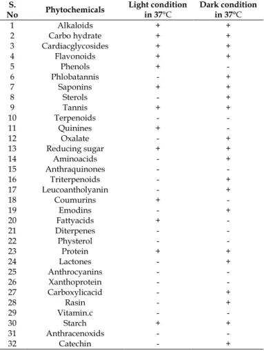
Table 5: Phytochemical screening of Isopropanol extract of A. paniculata
S.
No Phytochemicals
Light condition in 37°C
Dark condition in 37°C
1 Alkaloids + +
2 Carbo hydrate + +
3 Cardiacglycosides + +
4 Flavonoids + +
5 Phenols + -
6 Phlobatannis - +
7 Saponins + +
8 Sterols - +
9 Tannis + +
10 Terpenoids - -
11 Quinines + -
12 Oxalate - +
13 Reducing sugar + +
14 Aminoacids - +
15 Anthraquinones - -
16 Triterpenoids - +
17 Leucoantholyanin - +
18 Coumurins + -
19 Emodins - +
20 Fattyacids + -
21 Diterpenes - -
22 Physterol - -
23 Protein + +
24 Lactones - +
25 Anthrocyanins - -
26 Xanthoprotein - -
27 Carboxylicacid - +
28 Rasin - +
29 Vitamin.c - -
30 Starch + +
31 Anthracenoxids - -
32 Catechin - +
The antimicrobial result were also comparable to that of the antibiotic (streptomycin) used as a standard reference. The result also indicated that isopropanol was a suitable organic solvent for extraction of active principles responsible for antimicrobial activity of A. paniculata. The inhibitory effect of the extracts justified the medicinal use of A. paniculata in the treatment of various diseases by medical practitioners and our results also suggested that temperature and light are important environmental factors to optimize the phytochemical production in extracts of A. paniculata
under dark condition. These factors can significantly increase the phytochemical profile of it. Further study
is mandatory to find out the active compounds of medicinal value.
ACKNOWLEDGMENT
Financial support to Dr. R.Sharmila, Principal Investigator, Minor Research project (MRP-11/14 [SERO/ UGC], University Grant Commission (UGC), New Delhi, India is acknowledged. We are also grateful to the Management of Bishop Heber College for extending their extension services in helping with the instrument analysis.
REFERENCES
1. Aibinu I, Adenipekun E, Odugbemi T. Emergence of
Quinolone Resistance amongst Escherichia coli strains isolated
from clinical infections in some Lagos State Hospitals in
Nigeria. Nigerian Health and Biomed. Sci. 2004;3(2):73-78.
2. Williams R. Antimicrobial resistance a global threat. Esse.
Drug. Moni. 2000; 13: 28-29.
3. Pretorius JC, Magama S, Zietsman PC. Growth inhibition of
plant pathogenic bacteria and fungi by extracts from selected South African plant species. South African J. Bot. 2003; 20: 188-192.
4. Leelavasamee A. Undetectable antibacterial activity of A.
paniculata (Burn) wall ex Nees. J. med. Assoc. of Thailand. 1990; 73:299-304.
5. Balandrin MF, Kjocke AJ, Wurtele E. Natural plant chemicals:
sources of industrial and mechanical materials. Sci. 1985; 228: 1154-1160.
6. Li W, Xu X, Zhang H. Secondary metabolites from A.
paniculata. Chem. Pharm. Bull. (Tokyo) 2007; 55:455-458.
7. Krishnaraju AV, Rao TVN, Sundararaju D. Assessment of
bioactivity of Indian medicinal plants using Brine shrimp (Artemia salina) lethality assay. Int. J. Appl. Sci. Eng. 2005; 2: 125-134.
8. Cacer DD, Hancke JL, Burgos RLA, Wickman JK. Prevention
of common cold with A. paniculata dried extract. A pilot
double blind trial. Phytomed.1997; 4: 101-104.
9. Upadhyaya MK, Furness NH. Influence of light intensity and
water stress on leaf surface characteristics of Cynoglossum
offıcinale, Centaurea spp. and Tragopogon spp., Can. J. Bot.
1994; 72:1379–1386.
10. Mishra S, Tiwari SK, Kakker A, Pandey AK. Chemoprofiling
of A. paniculata (Kelmegh) for its andrographolide content in Madhya Pradesh, India. Int. J. Pharm. Biol. Sci, 2010; 1 (2): 1-5.
11. Trivedi NP, Rawal UM. Hepatoprotetive and antioxidant
property of A. paniculata in BHC-induced liver damage in
mice. Indian J. Exp.Biol. 2001; 39:41-46.
12. Zhao J, Davis LC, Verpoorte R. Elicitor signal transduction
leading to production of plant secondary metabolites.
Biotechnol. Adv.2005; 23: 283-333.
13. Bauer AW, Kirby WMM, Sherris JC. Antibiotic susceptibility
testing by a standardized single disk method. Am. J. Clin Pathol. 1966; 45: 493-496.
14. Gupta S. Antidiarrheal activity of diterpenes of A. paniculata
(kalamegh) against E. coli enterotoxin in in vivo models. Int. J.
Crude. Drug. Res.1990; 28: 273-283.
15. Bourgaud F, Gravot A, Milesi S, Gontier E. Production of
plant secondary metabolites: a historical perspective. Plant Sci. 2001; 161: 839–851.
16. Odabas MS, Uzun S, Gulumser A. The Quantitative Effects of
Temperature and Light on Growth, Development and Yield
of Faba Bean (Vicia faba L.): I. Growth. Int. J. Agric. Res. 2007;
2(9):765-775.