Streptococcus thermophilus
Formation: A Remnant Trait of Ancestral
Commensal Life?
Benoit Couvigny1,2☯, Claire Thérial1,2☯¤, Céline Gautier1,2, Pierre Renault1,2, Romain Briandet1,2, Eric Guédon1,2
*
1INRA, UMR 1319 Micalis, Domaine de Vilvert, F-78352 Jouy-en-Josas, France,2AgroParisTech, UMR MICALIS, Jouy-en-Josas, France
☯These authors contributed equally to this work.
¤ Current address: Laboratoire Eau Environnement et Systèmes Urbains, Faculté des sciences et technologie, Créteil, France
*eric.guedon@jouy.inra.fr
Abstract
Microorganisms have a long history of use in food production and preservation. Their adap-tation to food environments has profoundly modified their features, mainly through genomic flux.Streptococcus thermophilus, one of the most frequent starter culture organisms con-sumed daily by humans emerged recently from a commensal ancestor. As such, it is a use-ful model for genomic studies of bacterial domestication processes. Many streptococcal species form biofilms, a key feature of the major lifestyle of these bacteria in nature. Howev-er, few descriptions ofS.thermophilusbiofilms have been reported. An analysis of the ability of a representative collection of natural isolates to form biofilms revealed thatS. ther-mophiluswas a poor biofilm producer and that this characteristic was associated with an in-ability to attach firmly to surfaces. The identification of three biofilm-associated genes in the strain producing the most biofilms shed light on the reasons for the rarity of this trait in this species. These genes encode proteins involved in crucial stages of biofilm formation and are heterogeneously distributed between strains. One of the biofilm genes appears to have been acquired by horizontal transfer. The other two are located in loci presenting features of reductive evolution, and are absent from most of the strains analyzed. Their orthologs in commensal bacteria are involved in adhesion to host cells, suggesting that they are rem-nants of ancestral functions. The biofilm phenotype appears to be a commensal trait that has been lost during the genetic domestication ofS.thermophilus, consistent with its adap-tation to the milk environment and the selection of starter strains for dairy fermenadap-tations.
Introduction
Adaptation to their external environment is crucial for the survival and replication of bacteria. The modulation of gene expression is one of the major adaptive strategies by which bacteria
a11111
OPEN ACCESS
Citation:Couvigny B, Thérial C, Gautier C, Renault P, Briandet R, Guédon E (2015)Streptococcus thermophilusBiofilm Formation: A Remnant Trait of Ancestral Commensal Life? PLoS ONE 10(6): e0128099. doi:10.1371/journal.pone.0128099
Academic Editor:Ali Al-Ahmad, University Hospital of the Albert-Ludwigs-University Freiburg, GERMANY
Received:June 10, 2014
Accepted:April 23, 2015
Published:June 2, 2015
Copyright:© 2015 Couvigny et al. This is an open access article distributed under the terms of the
Creative Commons Attribution License, which permits unrestricted use, distribution, and reproduction in any medium, provided the original author and source are credited.
Data Availability Statement:All relevant data are within the paper and its Supporting Information files.
Funding:These authors have no support or funding to report.
deal with changing environmental conditions. However, long-term adaptation, during the col-onization of a new habitat for example, requires more profound genomic modifications. These changes include both the acquisition of new gene sets and the discarding of genes that are no longer useful, by gene loss and/or gene inactivation [1,2]. Niche adaptation is a major driving force shaping the bacterial genome [3–5]. The availability of complete genome sequences for closely related species, such as the many streptococcal genomes currently available, provides a remarkable opportunity for detecting niche-specific adaptation by comparative evolutionary genomics [6–12]. The genusStreptococcuscontains diverse species, most of which are com-mensal or pathogenic in humans and animals [13]. They occupy a broad range of ecological niches within the host, and the factors governing niche colonization remain poorly understood. The importance of these bacteria as etiological agents of numerous infections has led to studies investigating their adaptive versatility, at the genome level in particular.
Streptococcus thermophilusis the only species of this genus to be widely used as a starter in the dairy industry and to have the“Generally Regarded As Safe”status. It belongs to the sali-variusgroup of the Viridians streptococci [13], which includes two other species,Streptococcus salivariusandStreptococcus vestibularis. These two species are both commensal bacteria of the human gut, whereas the environmental reservoir ofS.thermophilushas not been identified [14]. It grows spontaneously in traditional dairy products and is believed to persist in the farm environment [15–17]. Multilocus sequence typing and comparative genomic analysis have re-vealed that there is little polymorphism in theS.thermophiluspopulation, and that this species displays significant allelic divergence from the other two species of thesalivariusgroup [14,18, 19].S.thermophilusis a clonal species that emerged only recently on the evolutionary timescale (3,000–30,000 years ago), from a commensal ancestor of thesalivariusgroup [20]. Its adapta-tion to a narrow and well defined niche (milk) has shaped its genome through loss-of-funcadapta-tion events and horizontal gene transfer (HGT) [21–23]. Approximately 10% of the ORFs ofS. ther-mophilusare pseudogenes, their original functions being unnecessary for growth in milk. Many of these pseudogenes encoded proteins involved in carbohydrate metabolism, a function not particularly useful in milk, which contains few carbon sources [22,23]. Commensal and pathogenic streptococci display numerous proteins at their surface, many of which have viru-lence-related functions.S.thermophilushas lost almost all of these features [21,23], suggesting that direct contact with the host may be required for the maintenance of such functions. HGT events have contributed substantially to the genomic plasticity, population evolution and adap-tation of this species to the milk environment. The genomic regions acquired include those encoding industrially important phenotypic traits, such as the production of bacteriocin, lanti-biotics and exopolysaccharides, restriction-modification systems, oxygen tolerance, amino-acid metabolism and milk-protein degradation [19,21–26].
thermophilusstrains are poor biofilm producers, mostly because they have lost these traits, consistent with their adaptation to the milk environment and selection as starters for dairy fermentations.
Materials and Methods
Bacterial strains, plasmids, growth conditions, and DNA manipulation
The bacterial strains used in this study are listed inTable 1andS1 Table.Escherichia coli
TG1repA was used for plasmid propagation.E.coliwas grown in Luria-Bertani medium at 37°C [34].S.thermophilusstrains were grown at 42°C in M17 medium containing 1% lactose, without shaking.S.salivariusstrains were grown at 37°C in M17 medium containing 1% glu-cose, without shaking. When required, 5-bromo-4-chloro-3-indolyl-β-D-galactoside (0.04 g/ l), isopropyl 1-thio-β-D-galactopyranoside (IPTG; 0.04 g/l), ampicillin (100μg/ml forE. coli), erythromycin (4μg/ml forS.thermophilusandS.salivarius, 100μg/ml forE.coli) were
added to the culture medium. Solid agar plates were prepared by adding 2% (wt/vol) agar to the medium. Standard molecular biology techniques were used andE.coliwas electrotrans-formed as previously described [35]. ElectrocompetentS.thermophiluscells were prepared as previously described [36]. PCR was performed with Fhusion high-fidelity DNA polymer-ase (NEB, MA, USA), in a GeneAmp 2400 PCR system (Applied Biosystems, Foster City, CA). The primers were purchased from Eurofins MWG Operon (Germany) and are listed in S1 Table.
Culture and biofilm formation
Planktonic cultures were grown in flasks, without shaking, at 42°C. We assessedS.thermophilus
biofilm formation in a high-throughput system, with crystal violet (CV) staining and a quantita-tive microplate assay [37]. Each well of a CELLSTAR 96-well cell culture plate (Greiner Bio-one, France), containing 0.2 ml of M17 broth was inoculated with 10μl of an overnightS. thermophi-lusculture and incubated at 42°C for 18 h. We then added 50μl of 1% CV to each well and
incu-bated the plates at room temperature for 15 min. The wells of the microtiter plates were rinsed three times with 0.2 ml deionized water to remove unattached cells and residual dye. They were then dried. The CV was dissolved in 95% ethanol (1 ml) and its absorbance at 590 nm was deter-mined, to score biofilm formation. We inoculated four quadruplicate wells with each strain for the scoring of biofilm formation and measurement of the optical density of the culture. We per-formed at least three independent triplicates for each biofilm formation assay.
We investigated the spatial organization of the biofilm for a subset of strains, by growing biofilms in microscopy-grade 96-well plates (Greiner Bio-one, France with aμclear base) and
studying them by confocal laser scanning microscopy (CLSM), as previously described [38]. Briefly, surface-associated bacteria were fluorescently labeled in green with 5μM Syto9
(Invi-trogen, France), a cell-permeant nucleic acid marker. The plate was then incubated in the dark for 15 min and mounted on the motorized stage of a Leica SP2 AOBS confocal laser scanning microscope (LEICA Microsystems, France) at the MIMA2 microscopy platform (www.jouy. inra.fr/mima2). All biofilms were scanned at 400 Hz, with a 40× 0.8 N.A. (Leica HCX Apo) water immersion objective lens, and a 488 nm argon laser set at 25% maximum intensity. Syto9 fluorescence was visualized by recording fluorescence in the 500–600 nm range. Three stacks of horizontal plane images (512×512 pixels) with az-step of 1μm were acquired for each
Table 1. Characteristics of theStreptococcus thermophilusnatural isolates used in this study.
Strain STa Location Source Year Bio
film statusb 0171c 0714c 1361c
JIM8232 2 France Milk 2002 + + + +
CNRZ1575 16 Italy Soft cheese 1962 +/- - +
-JIM10031 53 France Milk 1999 +/- - +
-JIM10116 13 Slovakia Traditional Brinza cheese 2004 +/- - +
-CNRZ385 9 Japan Sweet yogurt 1971 - - -
-CNRZ703 3 Mongolia Fermented milk 1974 - - -
-CNRZ759 7 Bulgaria Yogurt starter 1978 - - -
-CNRZ1066 5 France Yogurt starter 1986 - - -
-CNRZ1595 17 Austria Emmental cheese 1979 - - -
-JIM10001 31 France Cooked pressed cheese 2001 - - +
-JIM10010 33 France Cooked pressed cheese 2001 - - + +
JIM10020 27 France Cooked pressed cheese 2001 - - -
-JIM10032 39 France Milk 1999 - - -
-JIM10037 38 France Natural starter 2001 - - -
-JIM10050 45 France Natural starter 2001 - - -
-JIM10055 44 France Whey 1985 - - -
-JIM10087 51 France Whey 1984 - - -
-JIM10100 54 France Milk 2001 - - -
-JIM10114 46 France Natural starter 1999 - - -
-JIM10117 23 India Traditional cheese 2004 - - -
-JIM10119 26 India Traditional cheese 2004 - - -
-LMD-9 8 USA Cheese NA - - +
-LMG18311 6 UK Yogurt 1974 - - -
-1F8CT ND Italy Curd of grana padano cheese 2012 ND - -
-CNCM I-1360 ND NA NA NA ND - -
-DGCC7710 ND NA Commercial starter NA ND - +
-M17PTZA496 ND Italy Fontina cheese 1996 ND + -
-MN-ZLW-002 ND China Traditionally fermented products NA ND - - +
MTCC 5460 ND India Fermented milk product (curd) 1984 ND - - +
MTCC 5461 ND India Fermented milk product (curd) 1984 ND - -
-MTH17CL396 ND Italy Fontina cheese 1996 ND - + +
ND03 ND China Commercial starter NA ND - + +
TH1435 ND Italy Artisanal goat’s milk cheese (milk) 2011 ND - +
-TH1436 ND Italy Raw goat milk 2011 ND - +
-TH1477 ND Italy Cow milk 2012 ND + + +
TH982 ND Italy Buffalo mozzarella curd 2003 ND - +
-TH985 ND Italy Buffalo mozzarella whey 2003 ND - +
-a
, Sequence typing [18,19];
b, (-) poor, (+/-) moderate, (+) strong bio
film production;
c, (-) absence and (+) presence of STH8232_0171, STH8232_0714 and STH8232_1361 orthologs;
ND, not determined; NA, not available.
Three-dimensional projections of biofilm structures were reconstructed with the Easy 3D function of IMARIS 7.0 software (Bitplane, Switzerland). The quantitative structural parame-ters of the biostructures, such as biofilm biovolume, substrate coverage and mean thickness, were calculated with PHLIP, a freely available Matlab-based image analysis toolbox (htpp:// sourceforge.net/projects/phlip/).
Cell line culture technique and bacterial adhesion assay
The HT-29 human intestinal cell line (colon adenocarcinoma; ATCC HTB-38) was grown in Roswell Park Memorial Institute medium (RPMI-1640) supplemented with glutamine (2 mM), penicillin (50 U/ml), streptomycin (50 U/ml) and 5% (v/v) FCS (Lonza, Switzerland), at 37°C, under an atmosphere containing 95% and 5% CO2. We added 3 × 10⁵HT-29 cells per well to a
24-well tissue culture plate (TPP, Switzerland), which was then incubated for 24 h. The result-ing cell monolayers were washed twice with the cell line culture medium (without antibiotics) before the adhesion assay. Exponentially growing bacteria (OD600nm= 0.9) were collected by
centrifugation, washed with phosphate-buffered saline (PBS) and resuspended in RPMI-1640, adjusting the optical density at OD600nmto 0.450. Bacteria (0.5 ml, ~ 107CFU) were added to
the epithelial cell monolayers at a MOI of 50 bacteria per cell, centrifuged for 10 minutes at 4°C and then incubated for 0.5 h at 37°C in an incubator, under an atmosphere containing 5% CO2. The monolayers were washed three times with PBS to remove the non-adherent bacteria,
and were then lysed with 0.5 ml of sterile 0.5% PBS-Triton X-100. The lysate was vigorously pi-petted to release the cell-associated bacteria. Adhesion to polystyrene was assessed as follows. Each well of a CELLSTAR 96-well cell culture plate (Greiner Bio-one, France) was inoculated with 0.2 ml (~ 0.5x10⁷CFU) of an overnight culture with an adjusted OD600nmof 0.2. The
plates were then incubated at 42°C for 1 h. Microtiter plate wells were rinsed three times with 200μl deionized water to remove the non-adherent cells. The adherent bacteria were recovered
by vigorous pipetting in 0.2 ml of trypsin solution (PBS, 25 mM EDTA, 0.25% trypsin [Lonza, Switzerland)]). The serially diluted lysate and bacterial suspension were cultured on M17 agar for the counting of viable bacteria. Adhesion was determined by dividing the number of CFU recovered by the number of CFU for the original inoculum and expressing this ratio as a per-centage. HT-29 cell adhesion assays were carried out in duplicate and polystyrene adhesion as-says were carried out in triplicate, in at least three independent experiments.
Isolation of biofilm formation-negative mutants and target determination
S.thermophilusJIM8232 was mutated by integration of the thermosensitive pGhost9::ISS1 vec-tor, as previously described [35]. Briefly, cells containing pGhost9::ISS1were grown overnight at 30°C in the presence of erythromycin. Stationary-phase cultures were diluted 1:500 in fresh M17 broth without erythromycin, incubated for 150 min at 30°C and shifted to 42°C for 150 min. Samples were then diluted, plated on M17 medium containing erythromycin, and incu-bated at 42°C. Fifty clones from the 4,000 isolates containing an ISS1-directed insertion repro-ducibly failed to form biofilms. The pGhost9::ISS1insertion site was identified by cloning and sequencing the chromosomal junctions [39]. Junctions were cloned by extracting the chromo-somal DNA of mutants, digesting it withHindIII, and ligating it. We then transformedE.coli
Construction of mutant strains
A single-crossover insertional mutation of the STH8232_0171, STH8232_0714 and
STH8232_1361 loci was constructed with fragments of these genes. For this purpose, an inter-nal fragment of the STH8232_0171, STH8232_0714 and STH8232_1361 genes was amplified by PCR with the primers EG771/EG772, EG758/EG759 and EG762/EG763, respectively. These fragments were inserted into the pGEM-T easy vector (Promega), which was then fused with the integration vector pGhost9 inSpeI [39]. The resulting plasmids—pJIM8001, pJIM8002 and pJIM8000—were introduced intoS.thermophilusJIM8232 by electroporation and integrated into the chromosome by single cross-over events, yielding strains JIM9169, JIM9170 and JIM9171, in which the STH8232_0714, STH8232_1361 and STH8232_0171 genes were inacti-vated, respectively. The SALIVA_0971 locus was deleted from strain JIM8777 and replaced with an Ermrgene. Overlapping PCR was used to prepare donor DNA fragments bounded by the flanking sequences of the deleted material, as previously described [42]. The Erm cassette, derived from pAMβ-1, was amplified from pGhost9 with the EG940/EG941 primers. The up-stream and downup-stream flanking fragments were amplified from strain JIM8777 with the EG1516/EG1517 and EG1518/EG1519 primers, respectively. For assembly, the three fragments were mixed and amplified by overlapping PCR with the EG1516 and EG1519 primers. The PCR product was introduced into the chromosome of strain JIM8777 by XIP-induced transfor-mation [43], with Erm selection, to create strain JIM9395 (JIM8777ΔSALIVA_0971::erm).
Determination of cell-surface hydrophobicity
Cell surface hydrophobicity was determined by measuring the affinity of cells for hexadecane, as previously described [44]. Briefly, the bacterial cells were harvested by centrifugation (7000 xg, 10 min), washed twice and suspended in 150 mM NaCl. The absorbance of the cell suspen-sion at 400 nm was measured and adjusted to 0.8. We then vortexed 2.4 ml of the bacterial sus-pension thoroughly for 60 s with 0.4 ml hexadecane. The mixture was left at room temperature for 15 min to ensure the complete separation of the two phases before measurement of the ab-sorbance at 400 nm of the aqueous phase (A). The percentage of cells bound to hexadecane was subsequently calculated as: 100 × (0.2/A). Each experiment was performed three times, with two independent bacterial cultures.
Analysis of the STH8232_0171, STH8232_0714 and STH8232_1361
genes
sequences of the STH8232_0714 alleles fromS.thermophilusstrains JIM10001, JIM10010, JIM10031, JIM10116 and CNRZ1575 has been deposited in the GenBank database under ac-cession numbers KF717044, KF717043, KF717042, KF717041, and KF717045, respectively.
Statistical analysis
GraphPad Prism version 6 (GraphPad software Inc., La Jolla, CA, USA) was used for statistical analyses. APvalue<0.05 was considered to be statistically significant.
Results
Streptococcus thermophilus
is a poor biofilm producer
We investigated the ability of a representative collection of 23S.thermophilusisolates to form biofilms. This collection contained isolates obtained over a long time period (1962 to 2004) from diverse products (including cheeses, yogurts, fermented milks and starters), different geo-graphic locations (10 countries) and belonging to different MLST lineages (Table 1). We used a static monospecies biofilm model in which the cohesive mature biofilm was measured in a polystyrene 96-well microplate after crystal violet (CV) staining (Fig 1). All strains had similar growth rates in planktonic cultures (S1 Fig). However, only four produced relevant biofilms, with strain JIM8232 producing significantly more biofilm than any of the other strains.
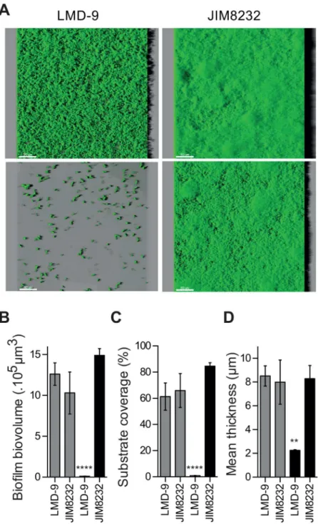
For validation of the CV assay and assessment of the spatial organization of the biofilms, we analyzed the biofilms formed by JIM8232 and LMD-9, used as a negative control, by CLSM. Rep-resentative 3D projections of the biofilm formed were obtained, before and after the washing steps used to remove loosely attached and sedimented bacteria (Fig 2Aupper and lower panels, respectively). These images were used for quantitative measurements of biofilm structure, includ-ing biovolume, substrate coverage and mean thickness (Fig2B–2D). No significant differences were found between the biofilms formed by strains LMD-9 and JIM8232 before washing. How-ever, all structural measurements were significantly lower after washing for strain LMD-9 (P<0.0001), whereas washing had no effect on the structural measurements for JIM8232
(P>0.05). CLSM images revealed that the cells in the washed LMD-9 biofilm formed very small
microcolonies on the polystyrene surface, corresponding mostly to surface-associated isolated cell chains (Fig 2A, lower panel). By contrast, the biofilm-producing strain JIM8232 formed a co-hesive, washing-resistant film of cells on the surface of the substrate. The JIM8232 biofilm had a relatively homogeneous structure and it covered the entire available surface. These observations confirm that strains JIM8232 and LMD-9 may be considered to be strong and poor biofilm pro-ducers, respectively. As mostS.thermophilusstrains behave like strain LMD-9, at least under our experimental conditions, this species can be classified as a poor biofilm producer.
The poor biofilm-formation ability of
Streptococcus thermophilus
is
correlated with a low capacity for adhesion
Biofilm development is a complex process with multiple steps following the attachment of the microbe to a surface [52]. We investigated whether the failure of most of theS.thermophilus
strains tested to form biofilms was due to an inability to adhere to the substrate. We counted the number of bacterial cells adhering to polystyrene after one hour of incubation in the wells of a microtiter plate (Fig 3). The ability of strains to form biofilms was correlated with the percentage of cells attaching to polystyrene (r2= 0.9769,P<0.0001, Pearson’s correlation). The strains
0.63%) adhesion, respectively. Our findings thus suggest that the poor ability of mostS. thermo-philusstrains to form biofilms is a consequence of their poor initial adhesion to surfaces.
Identification of genes involved in the formation of biofilms by strain
JIM8232
We used the pGhost9::ISS1vector [39] to generate a transposon mutant library for identifica-tion of the genetic factors involved in biofilm formaidentifica-tion. We screened over 4,000 inseridentifica-tion mu-tants for their ability to form biofilms in 96-well plate CV assays. Fifty mumu-tants reproducibly failed to form biofilms. The chromosomal DNA fragments flanking the pGhost9::ISS1insertion site of 23 of these mutants were rescued inE.coli, sequenced and compared with the full ge-nome sequence of JIM8232 [41]. The functions of the genes identified by this procedure were analyzed and we searched for homologs in the publicly availableS.thermophilusgenome se-quences. The transposons in these mutants had affected eight different genes (S2 Table).
Two of them, STH8232_1361 and STH8232_0714, were each inactivated by several inde-pendent transposition events, i.e. the transposon was found at different positions within these genes in different insertion mutants (14 mutants for STH8232_1361, 3 mutants for
STH8232_0714), which had lost their biofilm formation capability. This observation consti-tutes a strong indication for their potentially critical role in biofilm formation and these genes were therefore selected for further study. STH8232_1361 and STH8232_0714 encode a trans-membrane protein from the polysaccharide transporter (PST) family (TCDB database) and a cell surface-exposed protein with a MucBP domain (MUCin-Binding-Protein, Entry
PF06458), respectively. STH8232_0171 was also selected for further study because this gene is
Fig 1. Differences in biofilm formation between naturalS.thermophilusisolates.Biofilm formation was evaluated by quantitative microtiter plate assays based on the staining of 18-hour-old biofilms with crystal violet (CV). The columns show the mean values for CV absorbance (at 590 nm) of at least four independent experiments performed in duplicate, and the error bars indicate standard deviations. Statistical analyses included one-way ANOVA and the Bonferroni post hoc test, comparing the mean value for each strain with that of the strain with the lowest mean value (CNRZ1595). Asterisks denote statistically significant differences (P<0.0001).
Fig 2. Spatial organization ofS.thermophilusLMD-9 and JIM8232 biofilms, from series of confocal images.(A) Three-dimensional confocal projection of 18-hour-old biofilms produced by strains LMD-9 and JIM8232, before (upper panel) and after (lower panel) washing, obtained fromxyzconfocal image series with IMARIS software. The images show a representative and aerial view of biofilm structures, with the virtual shadow projection on the right. Scale bars correspond to 50 nm. (B) Mean biofilm biovolume, (C), substrate coverage, and (D) biofilm thickness of the 18-hour-old biofilms produced by strains LMD-9 and JIM8232 before (gray bars) and after (black bars) washing, extracted from confocal images with the PHLIP Matlab tool. The columns show the means of three independent experiments performed in triplicate, and the error bars indicate the standard deviation. Statistical analysis included Student’s pairedt-tests (****P<0.0001,**,
P<0.01 for comparisons with JIM8232 after washing).
present in only 3 of 18 currently sequencedS.thermophilusstrains and the one available for testing, JIM8232, formed biofilms. It encodes a predicted cytoplasmic protein (Psort predic-tion) with unknown function and containing a NACHT domain (nucleoside-triphosphatase domain, Entry PF05729). The remaining five genes were not studied further because they were targeted only once by the transposon and they are present in most sequencedS.thermophilus
strains, including those unable to form biofilms (S2 Table).
For independent confirmation of the involvement of STH8232_1361, STH8232_0714 and STH8232_0171 in JIM8232 biofilm formation, and to exclude the possibility of secondary muta-tions, we inactivated these genes in the wild-type strain and evaluated the biofilm-forming ability of the corresponding knockout mutants (Fig 4). The biofilm-forming capability of each of the three mutants was similar to that of the corresponding ISS1transposon mutants and was signifi-cantly lower (approximately 70%;P<0.0001) than that of the wild-type strain confirming that
these three genes directly contribute to biofilm formation. We further analyzed the correspond-ing genomic regions in sequencedS.thermophilusstrains and, using a PCR-based approach, in the 23 strains of our collection, to determine the functions and variability of these genes.
STH8232_1361 is responsible for a remnant function involved in surface
polysaccharide production
The STH8232_1361 gene is part of a large cluster of 16 open-reading frames (ORFs) flanked upstream bymetK(STH8232_1369) and downstream byendA(STH8232_1351) (Fig 5A). The STH8232_1361 locus appears to be variable inS.thermophilus. A complete or usable draft genome sequence is available for 12S.thermophilusstrains. The STH8232_1361 region
Fig 3. Initial adhesion of naturalS.thermophilusisolates to polystyrene microplates.The numbers of adherent bacteria were determined as colony-forming units on M17 solid agar plates and are expressed as percentages of the input inoculum. The columns show the mean percentages of adherent bacteria from at least three independent experiments performed in duplicate, and the error bars indicate standard deviations. Statistical analyses included one-way ANOVA and Bonferroni post hoc tests for comparisons of the mean value for each strain with that of the strain with the lowest mean value (CNRZ385). Asterisks denote statistically significant differences (P<0.001).
(from STH8232_1368 to STH8232_1356) is entirely absent from nine of these strains, includ-ing LMG18311 and CNRZ1066, and contains numerous pseudogenes and transposases in the others (Fig 5A). Conversely, a similar, intact region is found in the closely related species
S.salivariusJIM8777. Fifteen of our 23S.thermophilusstrains yielded a PCR product corre-sponding to themetK-murA1region and of similar size to that from CNRZ1066 and
LMG18311 (~3.5 kb). These strains thus contained a cluster with a large deletion encompass-ing STH8232_1361 (Fig 5AandS2 Fig). A PCR product with a size corresponding to that of STH8232_1361 was obtained only from LMD-9 (but as a pseudogene), JIM8232 and
JIM10010, confirming the absence of this gene from most of theS.thermophilusstrains stud-ied (Table 1). These findings indicate that, in mostS.thermophilusstrains, the
STH8232_1361-containing locus has undergone multiple inactivation events, some of which have resulted in gene loss, consistent with the regressive evolution occurring in this species [21–23].
This cluster contains genes encoding functions associated with extracellular activities, in-cluding polysaccharide synthesis in particular: STH8232_1361 (polysaccharide transporter), STH8232_1362 (glycosyltransferase I), STH8232_1355 (UDP-glucose 4-epimerase), and STH8232_1353 (UDP-N-acetylglucosamine 1-carboxyvinyltransferase). A search of complete genomes revealed the presence of homologs of STH8232_1361 in several phyla. In particular, homologs of STH8232_1361 and STH8232_1362, which are always organized in tandem in all STH8232_1361-containing bacteria, were found to be prevalent in host-associated bacterial families, including many Streptococcaceae, Ruminococcaceae, Bifidobacteriaceae, Clostridia-ceae, CoriobacteriaClostridia-ceae, Lachnospiraceae and Paenibacillaceae. All these families contain bac-teria from the intestinal flora. The homologs identified includePelGandPelFfrom
Pseudomonas aeruginosa; these genes encode proteins involved in the synthesis of the carbohy-drate-rich polymers playing a key role in initial interaction of the bacterium with solid surfaces and biofilm structure [53–55]. Thus, the STH8232_1361 locus in JIM8232 is involved in the production of surface polysaccharides likely to play a role in biofilm formation, and has been lost from otherS.thermophilusstrains.
Fig 4. Biofilm formation byS.thermophilusJIM8232 and its isogenic mutants.The biomass of 18-hour-old biofilms from the ISS1transposon mutants (STH8232_0171::ISS1, STH8232_0714::ISS1,
STH8232_1361::ISS1) and the corresponding reconstructed mutants (STH8232_0171 [JIM9171], STH8232_0714 [JIM9169], STH8232_1361 [JIM9170]) was measured in a crystal violet (CV) assay. The columns show mean CV absorbance values for at least four independent experiments performed in triplicate, and the error bars indicate standard deviations. The significance of the differences between JIM8232 and its isogenic mutants was determined in Student’s pairedt-tests (****,P<0.0001).
STH8232_0714 encodes a surface-exposed protein and is part of a
locus that has been subject to gene loss
Half the sequencedS.thermophilusgenomes contain an ortholog of STH8232_0714 (S2 Table), and this gene was detected by PCR amplification in seven of the 23 strains we tested for biofilm formation (Table 1). The STH8232_0714 genomic region was compared between eight
Fig 5. Schematic diagram of the STH8232_1361, STH8232_0714 and STH8232_0171 loci.(A) The STH8232_1361 regionin S.thermophilusLMD-9, JIM8232, ND03, CNRZ1066-like (CNRZ1066, TH985, TH982, TH1436, DGCC7710, 1F8CT), LMG18311, M17PTZA496-like (M17PTZA496, TH1435) and
S.salivariusJIM8777. (B) The STH8232_0714 region inS.thermophilusJIM8232, DGCC7710, LMD-9, ND03-like (ND03, MN_ZLW_002), CNRZ1066-like (CNRZ1066, LMG18311) and 1F8CT. (C) The STH8232_0171 region inS.thermophilusJIM8232, M17PTZA496 and TH1477. The orthologous genes in each panel are represented by pentagons of the same color, except for mobile elements, which are shown in white. The lengths of genes and intergenic regions are drawn to scale.
sequenced strains (Fig 5B): only two (LMD-9 and DGCC7710) contained a gene orthologous to STH8232_0714. In strain JIM8232, STH8232_0714 is followed by an ABC transporter and preceded by several genes and mobile elements. The transporter is conserved in the genomes of the other seven strains, but the upstream region is highly variable. The STH8232_0714 locus thus presents features of reductive evolution, including gene inactivation and almost total deletion.
A remarkable feature of STH8232_0714 and its ortholog in LMD-9 is their substantial di-vergence (91% identity), greater than the average for all the genomes considered (99.8% identi-ty, [21,22]). The divergent nucleotides are clustered in an ~1 kb inner region (positions 717 to 1856, displaying 15% nucleotide divergence, and 19% divergence at the amino-acid level), whereas the flanking regions contain only two mismatches. The sequence of this variable re-gion was determined in the other five strains and compared with the sequences of the LMD-9 and JIM8232 orthologs (S3 Fig). The sequences clustered into two well separated groups taining the JIM8232 and LMD-9 orthologs, respectively. However, these two groups each con-tained biofilm producers and non-producers. The variability of STH8232_0714 cannot therefore be considered to be directly related to biofilm formation.
STH8232_0714, which is flanked by a promoter and terminator sequence, is predicted to encode a cell surface protein containing a MucBP domain and belonging to a group of adhesins facilitating the attachment of bacteria to host cells. MucBP-containing proteins have been found in more than 315 bacterial species (Pfam analysis with PF06458 as the input,http:// pfam.xfam.org), mostly from the Streptococcaceae (211 species) and Lactobacillaceae (86 spe-cies), many found in association with animal mucosa. The putative protein encoded by STH8232_0714 thus has several features of proteins involved in adhesion to surfaces. This gene is highly polymorphic and is subject to gene loss inS.thermophilus.
STH8232_0171 was acquired by HGT
The STH8232_0171 gene, which is flanked by a promoter and terminator sequence, is sur-rounded by ORFs encoding transposases and maps to a 40-kb genomic island encoding pro-teins involved in the synthesis of a yellow pigment—an atypical trait of this species and of streptococci in general [41]. Moreover, its GC% (29.1%) differs significantly from the mean value for the JIM8232 chromosome (38.9%). This gene was also present in the genomes ofS.
thermophilusstrains M17PTZA496 and TH1477 (100% identity), which contain part of the JIM8232 40-kb genomic island. PCR-based tests showed this gene to be absent from the 23 strains studied (Table 1andS2 Table). BLAST searches of the genomes of other species for the STH8232_0171 gene product gave significant matches to putative proteins fromEnterococcus cecorumDSM 20682 (79%),S.oralisSK255 (78%), andS.mitisSK95 (78%). InE.cecorum, the corresponding gene is located in a genomic island flanked by an integrase and IS elements, whereas, inS.oralisandS.mitis, it is present in a variable region. These various findings indi-cate that the STH8232_0171 gene was acquired by HGT.
Role of STH8232_0171, STH8232_0714 and STH8232_1361 in biofilm
formation
cell-substratum interactions and, in particular, the initial adhesion occurring at the start of bio-film development. The STH8232_0171 mutant (JIM9171) presented a similar pattern, but with smaller clusters of cells. STH8232_0171 may therefore be involved in both primary adhesion and biofilm cohesion, through cell-cell and/or cell-matrix interactions. The STH8232_1361 mutant (JIM9170) cells displayed even higher levels of adhesion to the substratum than the other two mutants and it formed no clusters. This suggests that STH8232_1361 is principally involved in biofilm cohesion. These observations indicate that STH8232_0714, STH8232_1361 and STH8232_0171 play different roles (initial adhesion and biofilm cohesion) in the early stages of biofilm development.
We then studied the cell surface-related properties—such as adhesion to a polystyrene sur-face, hydrophobicity and cell morphology—of the STH8232_0171, STH8232_0714 and STH8232_1361 0171 mutants. The number of cells adhering to the polystyrene surface was much smaller for the STH8232_0714 and STH8232_0171 mutants (JIM9169 and JIM9171, re-spectively) than for the wild-type (approximately 1%,P<0.0001); the number of adherent
cells was intermediate for the STH8232_1361 mutant (JIM9170; 29% of the wild-type value,
P<0.0001;Fig 6C). These results were consistent with the CLSM analysis and confirmed the
requirement of STH8232_0171 and STH8232_0714 for surface attachment during the early stages of biofilm formation. They indicate that STH8282_1361 is involved in interactions of cells with the substrate, but to a lesser extent. Differences in hydrophobicity between the wild-type and mutant cells were estimated in MATH tests. JIM8232 displayed a high affinity for the solvent (>90%,Fig 6D), reflecting its hydrophobic nature. All the mutant strains had
signifi-cantly lower solvent affinities. However, the affinity for hexadecane of the STH8232_0171 and STH8232_1361 mutants was only moderately lower (61% to 68%), whereas that of the STH8232_0714 mutant was substantially lower and similar to that of hydrophilic strains. STH8232_0714 is therefore likely to be a cell surface protein determining the hydrophobicity of JIM8232 cells, possibly facilitating hydrophobic interactions with the substrate during adhe-sion. Transmission electron microscopy showed that strains JIM9169, JIM9171 and JIM8232 were indistinguishable in terms of their overall morphologies (data not shown). However, the external layer corresponding to cell wall polysaccharides was denser in the STH8232_1361 mu-tant than in the other strains (Fig 6E), consistent with a role for STH8232_1361 in polysaccha-ride synthesis.
The biofilm-associated genes of JIM8232 are involved in adhesion to
epithelial cells
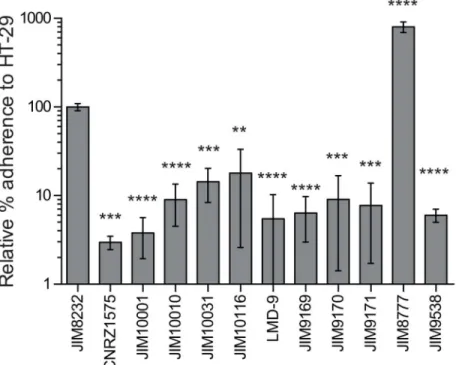
STH8232_0714 and STH8232_1361 display sequence similarity to genes present in many host-associated bacteria but absent from mostS.thermophilusstrains (see above). These genes may, therefore, be remnants from the ancestor ofS.thermophilus, believed to be closely related to theS.salivariusandS.vetibulariscommensals. This suggests a role for these genes in interac-tions between the bacterium and its host. We investigated this possibility, by evaluating the adhesion of wild-type and mutantS.thermophilusandS.salivariusstrains to HT-29 epithelial cells (Fig 7). JIM8232 was the natural isolate ofS.thermophilusdisplaying the strongest
columns show the mean values for five independent experiments, each performed in triplicate, and the error bars indicate the standard deviations. (D), Percentage of bacteria bound to hexadecane solvent. The columns show the mean values for four independent experiments performed in duplicate, and the error bars indicate standard deviations. (E), Scanning electron microscopy ofS.thermophilusJIM8232 and its isogenic mutants. Arrows indicate the cell wall polysaccharide layer. Scale bar, 0.2μm. The significance of the differences between JIM8232 and its isogenic mutants was determined in Student’s pairedt-tests (****,
P<0.0001;***,P<0.001;**,P<0.01).
adhesion to HT-29 cells (P<0.05), although its binding affinity was only one tenth that of the
S.salivariuscommensal strain JIM8777. The binding of STH8232_0171, STH8232_0714 and STH8232_1361 mutants was only 10% that of JIM8232, implicating these genes in adhesion to HT-29 cells. A mutant for theS.salivariusSTH8232_1361 ortholog (JIM9395) also bound sig-nificantly less strongly than the control to HT-29 cells, confirming the involvement of this gene in host cell interaction. Thus, unlike mostS.thermophilusstrains, JIM8232 can adhere to epi-thelial cells via a mechanism similar to that of one of its commensal relatives,S.salivarius.
Discussion
In this study, we explored the biofilm-forming ability of 23 representativeS.thermophilusisolates of diverse origins. We found that this species was a poor producer of biofilms, and that this prop-erty was associated with a lack of ability to adhere firmly to surfaces, the first step in biofilm de-velopment [52]. However, one strain, JIM8232, was a strong biofilm producer. Screening for JIM8232 mutants with impaired biofilm formation led to the identification of three genes—
STH8232_0714, STH8232_1361 and STH8232_0171—required for biofilm production byS.
thermophilus. Several observations provide compelling evidence that the biofilm negative pheno-type of the transposon insertion mutants is the result of the interruption of these genes rather than the result of secondary mutations or polar effects on downstream genes. Firstly, reconstruc-tion of mutareconstruc-tions for these genes in the wild-type strain rendered this strain biofilm negative. Secondly, both STH8232_0171 and STH8232_0714 are followed by putative rho-independent terminators and promoter regions indicating that transposon insertion should not significantly affect downstream gene expression. Lastly, the fact that 14 independent transposon insertions targeted STH8232_1361 and 3 STH8232_0714, but none the downstream genes indicates that
Fig 7. Adhesion to HT-29 cells of severalS.thermophilusandS.salivariusstrains.Adherent bacteria were counted as colony-forming units on M17 solid agar plates and the results are expressed as percentages of the input inoculum. The columns show the mean values for three independent experiments, each performed in triplicate, and the error bars indicate the standard deviations. Student’s pairedt-test was used for statistical analysis (****P<0.0001 for comparisons with JIM8232 or JIM8777).
their inactivation is specifically required to produce a biofilm negative phenotype. Biofilm devel-opment is a dynamic, complex multistep process involving the initial attachment of planktonic bacterial cells to a surface, production of the extracellular matrix, cluster formation and develop-ment and maturation of the biofilm architecture [52]. We show that the functions encoded by STH8232_0714, STH8232_1361 and STH8232_0171, which are absent from mostS. thermophi-lusstrains (Table 1), are related to early stages of biofilm development and are therefore crucial for biofilm formation. The surface-exposed STH8232_0714 protein may facilitate hydrophobic interactions with the substrate during primary adhesion, and the STH8232_1361-dependent polysaccharide matrix may trigger adhesion and biofilm cohesion [56]. Phenotypic analysis of the STH8232_0171 mutant indicated that it was also involved in these two steps of biofilm for-mation. However, the product of this gene is predicted to be cytoplasmic, and its precise role in adhesion and biofilm cohesion therefore remains unclear. The deduced amino-acid sequence of the STH8232_0171 gene product contains a domain predicted to be involved in signal transduc-tion mechanisms, suggesting a possible role in the regulatransduc-tion of biofilm formatransduc-tion.
The study of these three loci provides information about their evolutionary history. STH8232_0714 and STH8232_1361 are located within gene clusters presenting features of re-ductive evolution: in mostS.thermophilusstrains, there is evidence to suggest that major rear-rangements to these clusters have occurred, including their almost total deletion. Similar clusters are present in the closely related commensal streptococci, from whichS.thermophilus
recently emerged as a food bacterium. In commensal bacteria, the functions associated with the homologs of STH8232_0714 and STH8232_1361 relate to adhesion and interaction with the host mucosa (this work) [57–60], which may lead to biofilm formation, an important feature frequently associated with colonization capability [61–63]. Such functions are probably dis-pensable for growth in milk and loss of the functions of the STH8232_0714 homologs has been reported in other domesticated bacteria [23,57,64]. The STH8232_0714- and
STH8232_1361-dependent biofilm phenotype thus appears to be an ancestral trait that, like other features, has been lost during the genetic domestication ofS.thermophilus[21–23]. An analysis of STH8232_0171, the other biofilm-associated gene identified in this study, revealed a different story. STH8232_0171 was found in only three strains, all isolated from raw milk. In JIM8232, it maps to a 40-kb island also involved in the synthesis of a yellow pigment not typi-cally present in this species or other streptococci [41]. Its close homologs in commensal bacte-ria are also carried by genomic islands, suggesting that this gene may be exchanged through horizontal gene transfer. The function of this gene in these bacteria is unknown, but our find-ings indicate that it is associated with biofilm formation inS.thermophilusJIM8232. The bio-film formation phenotype displayed by JIM8232 therefore seems to be dependent on both the inheritance of ancestral functions and the acquisition of another gene, all related to the com-mensal lifestyle.
The poor biofilm production capacity ofS.thermophilusis intriguing. Association with a biofilm is the predominant lifestyle in bacteria and a key strategy for survival in harsh environ-ments. ManyS.thermophilusstrains have been isolated from equipment bearing multispecies biofilms, such as wood vats, used for the fermentation of traditional products [16,32,33]. Such biofilms may act as a reservoir for this species. Strain JIM10116 was isolated from traditional Brinza cheese. It has a moderate ability to form biofilms and may be representative of such strains. The development of multispecies biofilms, which probably predominate in natural con-ditions, results from cooperation and interactions between different microbial species [27,28, 65,66]. Our biofilm model, involving the attachment of a single strain to an abiotic surface, may therefore not be representative of the natural environment ofS.thermophilus.
whereas, in several traditional thermophilic dairy production processes, the acidification step is started by back-slopping. Moreover, current fermentation processes are based on the inocula-tion of milk with commercial starters selected on the basis of criteria such as rapid growth in fermenters. Biofilm producers may not be the most appropriate strains for these conditions. The propagation of commercial strains as planktonic cultures adapted to industrial processes may have led to the loss of genetic material, as demonstrated for natural isolates cultured under laboratory conditions [67–69]. Consequently, genetic determinants, such as those involved in bacterium-host interactions identified in this study, may have been lost spontaneously or even eliminated by counter-selection in most food-associatedS.thermophiluslineages. The 23 strains included in this study are representative of such strains because most are domesticated strains with a long history of use in dairy fermentations. JIM8232, a pigment producer isolated from a milk tank truck, may not therefore be representative of strains selected for
dairy fermentation.
Our study raises questions about the origins of such strains. The JIM8232 genome contains functional clusters that are probably vestiges of the commensal origin of theS.thermophilus
ancestor. It also contains islands, such as that carrying STH8232_0171, another feature consis-tent with a commensal origin. This strain has the PrtS island allowing casein assimilation, which was probably acquired by transfer from an animal-associated streptococcal strain [19]. Such repeated exchanges of genetic material betweenS.thermophilusand bacteria not thought to occupy the same ecological niche raise questions about how such transfers occur. One possi-bility is thatS.thermophilusoccupies an as yet undiscovered animal-associated niche on farms. The ability of JIM8232 to adhere to epithelial cells, and the isolation ofS.thermophilusfrom raw milk and cow udders are consistent with this possibility. However, the presence of such strains in animals has yet to be confirmed. Alternatively, such transfers may be facilitated by the frequent addition ofS.thermophilusto bacterial mixtures used as probiotics for animals, in-cluding pigs and chickens, or as starters for silage. This extensive release ofS.thermophilusin the farm environment would probably facilitated horizontal gene transfer events between this species and the animal gut microbiota, as proposed for antibiotic resistance genes [70,71]. The resulting strains could then contaminate milk, leading to their dissemination throughout the food chain. The selection ofS.thermophilusstrains with functional attributes other than those required for milk fermentation, and their use for new applications, such as animal probiotics, could therefore contribute to the emergence of new strains and the evolution of theS. thermo-philusgenome.S.thermophilusis currently considered to be innocuous and is consumed daily in massive amounts around the world with no known harmful effects on human health. How-ever, given the ability of this species to acquire new genes readily and its extended use at sites other than dairies, particular attention should be paid to the possibility of undesirable traits dis-seminating amongS.thermophilusstarter strains.
Supporting Information
S1 Fig. Final cell density ofS.thermophilusnatural isolates grown in microtiter plates. Col-umns represent means absorbance at 600 nm of at least four independent experiments per-formed in duplicate, and bar errors indicate standard deviations.
(TIFF)
7, JIM10001; 8, JIM10020; 9, JIM10031; 10, JIM10037; 11, JIM10050; 12, JIM10055; 13, JIM10 087; 14, JIM10100; 15, JIM10114; 16, JIM10117; 17, LMG18311; 18, CNRZ1066; 19, CNRZ759; 20, CNRZ1575; 21, CNRZ1595.
(TIFF)
S3 Fig. Phylogenic tree of STH8232_0714 orthologous genes inS.thermophilus.Blue, mod-erate and strong biofilm producing strains; Red, no or poor biofilm producing strains; Black, strains for which biofilm formation ability was not determined.
(TIFF)
S1 Table. Bacterial strains, plasmids and primers used in this study.
(TIFF)
S2 Table. Features of genes identified as involved in biofilm formation of strain JIM8232 by a genome-wide mutagenesis approach and their occurrence with percent identity among sequencedStreptococcus thermophilusstrains.
(TIFF)
Acknowledgments
We thank Sophie Chat for TEM observations and Julien Deschamps for MATH experiments at the MIMA2 microscopy platform (INRA) (http://www6.jouy.inra.fr/mima2). We thank Nico-las Lapaque and Alexandra Gruss for critical reading of the manuscript, and Bob Petersen and Alex Edelman and Associates (http://www.alexedelman.com) for improving English usage in this paper.
Author Contributions
Conceived and designed the experiments: PR RB EG. Performed the experiments: BC CG CT RB EG. Analyzed the data: BC CG CT RB EG. Contributed reagents/materials/analysis tools: BC CT CG RB EG. Wrote the paper: RB PR EG.
References
1. Wiedenbeck J, Cohan FM. Origins of bacterial diversity through horizontal genetic transfer and adapta-tion to new ecological niches. FEMS microbiology reviews. 2011; 35(5):957–76. doi: 10.1111/j.1574-6976.2011.00292.xPMID:21711367.
2. Francino MP. The ecology of bacterial genes and the survival of the new. International journal of evolu-tionary biology. 2012; 2012:394026. doi:10.1155/2012/394026PMID:22900231; PubMed Central PMCID: PMC3415099.
3. Makarova K, Slesarev A, Wolf Y, Sorokin A, Mirkin B, Koonin E, et al. Comparative genomics of the lac-tic acid bacteria. Proceedings of the National Academy of Sciences of the United States of America. 2006; 103(42):15611–6. doi:10.1073/pnas.0607117103PMID:17030793; PubMed Central PMCID: PMC1622870.
4. Lebreton F, van Schaik W, McGuire AM, Godfrey P, Griggs A, Mazumdar V, et al. Emergence of epi-demic multidrug-resistant Enterococcus faecium from animal and commensal strains. mBio. 2013; 4 (4). doi:10.1128/mBio.00534-13PMID:23963180; PubMed Central PMCID: PMC3747589.
5. Richards VP, Palmer SR, Pavinski Bitar PD, Qin X, Weinstock GM, Highlander SK, et al. Phyloge-nomics and the dynamic genome evolution of the genus streptococcus. Genome biology and evolution. 2014; 6(4):741–53. doi:10.1093/gbe/evu048PMID:24625962.
6. Marri PR, Hao W, Golding GB. Gene gain and gene loss in streptococcus: is it driven by habitat? Molec-ular biology and evolution. 2006; 23(12):2379–91. doi:10.1093/molbev/msl115PMID:16966682.
8. Lefebure T, Stanhope MJ. Evolution of the core and pan-genome of Streptococcus: positive selection, recombination, and genome composition. Genome biology. 2007; 8(5):R71. doi: 10.1186/gb-2007-8-5-r71PMID:17475002; PubMed Central PMCID: PMC1929146.
9. Ding F, Tang P, Hsu MH, Cui P, Hu S, Yu J, et al. Genome evolution driven by host adaptations results in a more virulent and antimicrobial-resistant Streptococcus pneumoniae serotype 14. BMC genomics. 2009; 10:158. doi:10.1186/1471-2164-10-158PMID:19361343; PubMed Central PMCID:
PMC2678160.
10. Ward PN, Holden MT, Leigh JA, Lennard N, Bignell A, Barron A, et al. Evidence for niche adaptation in the genome of the bovine pathogen Streptococcus uberis. BMC genomics. 2009; 10:54. doi:10.1186/ 1471-2164-10-54PMID:19175920; PubMed Central PMCID: PMC2657157.
11. Jans C, Follador R, Hochstrasser M, Lacroix C, Meile L, Stevens MJ. Comparative genome analysis of Streptococcus infantarius subsp. infantarius CJ18, an African fermented camel milk isolate with adapta-tions to dairy environment. BMC genomics. 2013; 14:200. doi:10.1186/1471-2164-14-200PMID: 23521820; PubMed Central PMCID: PMC3640971.
12. Papadimitriou K, Anastasiou R, Mavrogonatou E, Blom J, Papandreou NC, Hamodrakas SJ, et al. Comparative genomics of the dairy isolate Streptococcus macedonicus ACA-DC 198 against related members of the Streptococcus bovis/Streptococcus equinus complex. BMC genomics. 2014; 15 (1):272. doi:10.1186/1471-2164-15-272PMID:24713045.
13. Facklam R. What happened to the streptococci: overview of taxonomic and nomenclature changes. Clinical microbiology reviews. 2002; 15(4):613–30. PMID:12364372; PubMed Central PMCID: PMC126867.
14. Delorme C, Abraham AL, Renault P, Guedon E. Genomics of Streptococcus salivarius, a major human commensal. Infection, genetics and evolution: journal of molecular epidemiology and evolutionary ge-netics in infectious diseases. 2014. doi:10.1016/j.meegid.2014.10.001PMID:25311532.
15. Aponte M, Fusco V, Andolfi R, Coppola S. Lactic acid bacteria occurring during manufacture and ripen-ing of Provolone del Monaco cheese: Detection by different analytical approaches. Int Dairy J. 2008; 18 (4):403–13. doi:10.1016/J.Idairyj.2007.10.011PMID:WOS:000254686000009.
16. Settanni L, Di Grigoli A, Tornambe G, Bellina V, Francesca N, Moschetti G, et al. Persistence of wild Streptococcus thermophilus strains on wooden vat and during the manufacture of a traditional Cacioca-vallo type cheese. International journal of food microbiology. 2012; 155(1–2):73–81. doi:10.1016/j. ijfoodmicro.2012.01.022PMID:22336514.
17. Cruciata M, Sannino C, Ercolini D, Scatassa ML, De Filippis F, Mancuso I, et al. Animal rennets as sources of dairy lactic Acid bacteria. Applied and environmental microbiology. 2014; 80(7):2050–61. doi:10.1128/AEM.03837-13PMID:24441167.
18. Delorme C, Poyart C, Ehrlich SD, Renault P. Extent of horizontal gene transfer in evolution of Strepto-cocci of the salivarius group. Journal of bacteriology. 2007; 189(4):1330–41. doi:10.1128/JB.01058-06 PMID:17085557; PubMed Central PMCID: PMC1797340.
19. Delorme C, Bartholini C, Bolotine A, Ehrlich SD, Renault P. Emergence of a cell wall protease in the Streptococcus thermophilus population. Applied and environmental microbiology. 2010; 76(2):451–60. doi:10.1128/AEM.01018-09PMID:19915034; PubMed Central PMCID: PMC2805209.
20. Delorme C. Safety assessment of dairy microorganisms: Streptococcus thermophilus. International journal of food microbiology. 2008; 126(3):274–7. doi:10.1016/j.ijfoodmicro.2007.08.014PMID: 17822794.
21. Bolotin A, Quinquis B, Renault P, Sorokin A, Ehrlich SD, Kulakauskas S, et al. Complete sequence and comparative genome analysis of the dairy bacterium Streptococcus thermophilus. Nature biotechnolo-gy. 2004; 22(12):1554–8. doi:10.1038/nbt1034PMID:15543133.
22. Hols P, Hancy F, Fontaine L, Grossiord B, Prozzi D, Leblond-Bourget N, et al. New insights in the mo-lecular biology and physiology of Streptococcus thermophilus revealed by comparative genomics. FEMS microbiology reviews. 2005; 29(3):435–63. doi:10.1016/j.femsre.2005.04.008PMID: 16125007.
23. Goh YJ, Goin C, O'Flaherty S, Altermann E, Hutkins R. Specialized adaptation of a lactic acid bacterium to the milk environment: the comparative genomics of Streptococcus thermophilus LMD-9. Microbial cell factories. 2011; 10 Suppl 1:S22. doi:10.1186/1475-2859-10-S1-S22PMID:21995282; PubMed Central PMCID: PMC3231929.
24. Rasmussen TB, Danielsen M, Valina O, Garrigues C, Johansen E, Pedersen MB. Streptococcus ther-mophilus core genome: comparative genome hybridization study of 47 strains. Applied and environ-mental microbiology. 2008; 74(15):4703–10. doi:10.1128/AEM.00132-08PMID:18539806; PubMed Central PMCID: PMC2519362.
environmental microbiology. 2009; 75(12):4120–9. doi:10.1128/AEM.02898-08PMID:19395564; PubMed Central PMCID: PMC2698337.
26. Cefalo AD, Broadbent JR, Welker DL. Intraspecific and interspecific interactions among proteins regu-lating exopolysaccharide synthesis in Streptococcus thermophilus, Streptococcus iniae, and Lactococ-cus lactis subsp. cremoris and the assessment of potential lateral gene transfer. Canadian journal of microbiology. 2011; 57(12):1002–15. doi:10.1139/w11-090PMID:22107596.
27. Jefferson KK. What drives bacteria to produce a biofilm? FEMS microbiology letters. 2004; 236(2):163– 73. doi:10.1016/j.femsle.2004.06.005PMID:15251193.
28. Hall-Stoodley L, Costerton JW, Stoodley P. Bacterial biofilms: from the natural environment to infectious diseases. Nature reviews Microbiology. 2004; 2(2):95–108. doi:10.1038/nrmicro821PMID:15040259.
29. Hall-Stoodley L, Stoodley P. Evolving concepts in biofilm infections. Cellular microbiology. 2009; 11 (7):1034–43. doi:10.1111/j.1462-5822.2009.01323.xPMID:19374653.
30. Davies D. Understanding biofilm resistance to antibacterial agents. Nature reviews Drug discovery. 2003; 2(2):114–22. doi:10.1038/nrd1008PMID:12563302.
31. Marks LR, Reddinger RM, Hakansson AP. Biofilm formation enhances fomite survival of Streptococcus pneumoniae and Streptococcus pyogenes. Infection and immunity. 2014; 82(3):1141–6. doi:10.1128/ IAI.01310-13PMID:24371220; PubMed Central PMCID: PMC3957990.
32. Licitra G, Ogier JC, Parayre S, Pediliggieri C, Carnemolla TM, Falentin H, et al. Variability of bacterial biofilms of the "tina" wood vats used in the ragusano cheese-making process. Applied and environmen-tal microbiology. 2007; 73(21):6980–7. doi:10.1128/AEM.00835-07PMID:17720831; PubMed Central PMCID: PMC2074957.
33. Lortal S, Di Blasi A, Madec MN, Pediliggieri C, Tuminello L, Tanguy G, et al. Tina wooden vat biofilm: a safe and highly efficient lactic acid bacteria delivering system in PDO Ragusano cheese making. Inter-national journal of food microbiology. 2009; 132(1):1–8. doi:10.1016/j.ijfoodmicro.2009.02.026PMID: 19361876.
34. Maniatis T, Fritsch EF, Sambrook J, editor. Molecular cloning: a laboratory manual. NY: Cold Spring Harbor Laboratory Press, Cold Spring Harbor; 1982.
35. Guedon E, Serror P, Ehrlich SD, Renault P, Delorme C. Pleiotropic transcriptional repressor CodY senses the intracellular pool of branched-chain amino acids in Lactococcus lactis. Molecular microbiol-ogy. 2001; 40(5):1227–39. PMID:11401725.
36. Minic Z, Marie C, Delorme C, Faurie JM, Mercier G, Ehrlich D, et al. Control of EpsE, the phosphoglyco-syltransferase initiating exopolysaccharide synthesis in Streptococcus thermophilus, by EpsD tyrosine kinase. Journal of bacteriology. 2007; 189(4):1351–7. doi:10.1128/JB.01122-06PMID:16980450; PubMed Central PMCID: PMC1797369.
37. O'Toole GA. Microtiter dish biofilm formation assay. Journal of visualized experiments: JoVE. 2011;(47: ). doi:10.3791/2437PMID:21307833; PubMed Central PMCID: PMC3182663.
38. Bridier A, Dubois-Brissonnet F, Boubetra A, Thomas V, Briandet R. The biofilm architecture of sixty op-portunistic pathogens deciphered using a high throughput CLSM method. Journal of microbiological methods. 2010; 82(1):64–70. doi:10.1016/j.mimet.2010.04.006PMID:20433880.
39. Maguin E, Prevost H, Ehrlich SD, Gruss A. Efficient insertional mutagenesis in lactococci and other gram-positive bacteria. Journal of bacteriology. 1996; 178(3):931–5. PMID:8550537; PubMed Central PMCID: PMC177749.
40. Law J, Buist G, Haandrikman A, Kok J, Venema G, Leenhouts K. A system to generate chromosomal mutations in Lactococcus lactis which allows fast analysis of targeted genes. Journal of bacteriology. 1995; 177(24):7011–8. PMID:8522504; PubMed Central PMCID: PMC177576.
41. Delorme C, Bartholini C, Luraschi M, Pons N, Loux V, Almeida M, et al. Complete genome sequence of the pigmented Streptococcus thermophilus strain JIM8232. Journal of bacteriology. 2011; 193 (19):5581–2. doi:10.1128/JB.05404-11PMID:21914889; PubMed Central PMCID: PMC3187418.
42. Sperandio B, Gautier C, Pons N, Ehrlich DS, Renault P, Guedon E. Three paralogous LysR-type tran-scriptional regulators control sulfur amino acid supply in Streptococcus mutans. Journal of bacteriology. 2010; 192(13):3464–73. doi:10.1128/JB.00119-10PMID:20418399; PubMed Central PMCID: PMC2897675.
43. Fontaine L, Boutry C, de Frahan MH, Delplace B, Fremaux C, Horvath P, et al. A novel pheromone quo-rum-sensing system controls the development of natural competence in Streptococcus thermophilus and Streptococcus salivarius. Journal of bacteriology. 2010; 192(5):1444–54. doi: 10.1128/JB.01251-09PMID:20023010; PubMed Central PMCID: PMC2820839.
45. Sun Z, Chen X, Wang J, Zhao W, Shao Y, Wu L, et al. Complete genome sequence of Streptococcus thermophilus strain ND03. Journal of bacteriology. 2011; 193(3):793–4. doi:10.1128/JB.01374-10 PMID:21131489; PubMed Central PMCID: PMC3021231.
46. Kang X, Ling N, Sun G, Zhou Q, Zhang L, Sheng Q. Complete genome sequence of Streptococcus thermophilus strain MN-ZLW-002. Journal of bacteriology. 2012; 194(16):4428–9. doi:10.1128/JB. 00740-12PMID:22843572; PubMed Central PMCID: PMC3416250.
47. Prajapati JB, Nathani NM, Patel AK, Senan S, Joshi CG. Genomic analysis of dairy starter culture Streptococcus thermophilus MTCC 5461. Journal of microbiology and biotechnology. 2013; 23(4):459– 66. PMID:23568199.
48. Treu L, Vendramin V, Bovo B, Campanaro S, Corich V, Giacomini A. Genome Sequences of Strepto-coccus thermophilus Strains MTH17CL396 and M17PTZA496 from Fontina, an Italian PDO Cheese. Genome announcements. 2014; 2(1). doi:10.1128/genomeA.00067-14PMID:24526643; PubMed Central PMCID: PMC3924375.
49. Treu L, Vendramin V, Bovo B, Campanaro S, Corich V, Giacomini A. Whole-Genome Sequences of Streptococcus thermophilus Strains TH1435 and TH1436, Isolated from Raw Goat Milk. Genome an-nouncements. 2014; 2(1). doi:10.1128/genomeA.01129-13PMID:24435859; PubMed Central PMCID: PMC3894273.
50. Treu L, Vendramin V, Bovo B, Campanaro S, Corich V, Giacomini A. Genome Sequences of Four Ital-ian Streptococcus thermophilus Strains of Dairy Origin. Genome announcements. 2014; 2(2). doi:10. 1128/genomeA.00126-14PMID:24625867; PubMed Central PMCID: PMC3953188.
51. Guedon E, Delorme C, Pons N, Cruaud C, Loux V, Couloux A, et al. Complete genome sequence of the commensal Streptococcus salivarius strain JIM8777. Journal of bacteriology. 2011; 193(18):5024– 5. doi:10.1128/JB.05390-11PMID:21742871; PubMed Central PMCID: PMC3165664.
52. Costerton JW, Cheng KJ, Geesey GG, Ladd TI, Nickel JC, Dasgupta M, et al. Bacterial biofilms in na-ture and disease. Annual review of microbiology. 1987; 41:435–64. doi:10.1146/annurev.mi.41. 100187.002251PMID:3318676.
53. Vasseur P, Vallet-Gely I, Soscia C, Genin S, Filloux A. The pel genes of the Pseudomonas aeruginosa PAK strain are involved at early and late stages of biofilm formation. Microbiology. 2005; 151(Pt 3):985–97. doi:10.1099/mic.0.27410-0PMID:15758243.
54. Vasseur P, Soscia C, Voulhoux R, Filloux A. PelC is a Pseudomonas aeruginosa outer membrane lipo-protein of the OMA family of lipo-proteins involved in exopolysaccharide transport. Biochimie. 2007; 89 (8):903–15. doi:10.1016/j.biochi.2007.04.002PMID:17524545.
55. Ghafoor A, Jordens Z, Rehm BH. Role of PelF in pel polysaccharide biosynthesis in Pseudomonas aer-uginosa. Applied and environmental microbiology. 2013; 79(9):2968–78. doi:10.1128/AEM.03666-12 PMID:23435893; PubMed Central PMCID: PMC3623137.
56. Flemming HC, Wingender J. The biofilm matrix. Nature reviews Microbiology. 2010; 8(9):623–33. doi: 10.1038/nrmicro2415PMID:20676145.
57. Boekhorst J, Helmer Q, Kleerebezem M, Siezen RJ. Comparative analysis of proteins with a mucus-binding domain found exclusively in lactic acid bacteria. Microbiology. 2006; 152(Pt 1):273–80. doi:10. 1099/mic.0.28415-0PMID:16385136.
58. Du Y, He YX, Zhang ZY, Yang YH, Shi WW, Frolet C, et al. Crystal structure of the mucin-binding do-main of Spr1345 from Streptococcus pneumoniae. Journal of structural biology. 2011; 174(1):252–7. doi:10.1016/j.jsb.2010.10.016PMID:21055474.
59. Etzold S, Kober OI, Mackenzie DA, Tailford LE, Gunning AP, Walshaw J, et al. Structural basis for ad-aptation of lactobacilli to gastrointestinal mucus. Environmental microbiology. 2014; 16(3):888–903. doi:10.1111/1462-2920.12377PMID:24373178.
60. Jensen H, Roos S, Jonsson H, Rud I, Grimmer S, van Pijkeren JP, et al. Role of Lactobacillus reuteri cell and mucus-binding protein A (CmbA) in adhesion to intestinal epithelial cells and mucus in vitro. Mi-crobiology. 2014; 160(Pt 4):671–81. doi:10.1099/mic.0.073551-0PMID:24473252.
61. Munoz-Elias EJ, Marcano J, Camilli A. Isolation of Streptococcus pneumoniae biofilm mutants and their characterization during nasopharyngeal colonization. Infection and immunity. 2008; 76(11):5049–61. doi:10.1128/IAI.00425-08PMID:18794289; PubMed Central PMCID: PMC2573321.
62. Marks LR, Parameswaran GI, Hakansson AP. Pneumococcal interactions with epithelial cells are cru-cial for optimal biofilm formation and colonization in vitro and in vivo. Infection and immunity. 2012; 80 (8):2744–60. doi:10.1128/IAI.00488-12PMID:22645283; PubMed Central PMCID: PMC3434590.
64. Ventura M, O'Flaherty S, Claesson MJ, Turroni F, Klaenhammer TR, van Sinderen D, et al. Genome-scale analyses of health-promoting bacteria: probiogenomics. Nature reviews Microbiology. 2009; 7 (1):61–71. doi:10.1038/nrmicro2047PMID:19029955.
65. Costerton JW, Stewart PS, Greenberg EP. Bacterial biofilms: a common cause of persistent infections. Science. 1999; 284(5418):1318–22. PMID:10334980.
66. Yang L, Liu Y, Wu H, Hoiby N, Molin S, Song ZJ. Current understanding of multi-species biofilms. Inter-national journal of oral science. 2011; 3(2):74–81. doi:10.4248/IJOS11027PMID:21485311; PubMed Central PMCID: PMC3469880.
67. McLoon AL, Guttenplan SB, Kearns DB, Kolter R, Losick R. Tracing the domestication of a biofilm-form-ing bacterium. Journal of bacteriology. 2011; 193(8):2027–34. doi:10.1128/JB.01542-10PMID: 21278284; PubMed Central PMCID: PMC3133032.
68. Lawrence D, Fiegna F, Behrends V, Bundy JG, Phillimore AB, Bell T, et al. Species interactions alter evolutionary responses to a novel environment. PLOS biology. 2012; 10(5):e1001330. doi:10.1371/ journal.pbio.1001330PMID:22615541; PubMed Central PMCID: PMC3352820.
69. Rao C, Benhabib H, Ensminger AW. Phylogenetic reconstruction of the Legionella pneumophila Phila-delphia-1 laboratory strains through comparative genomics. PLOS one. 2013; 8(5):e64129. doi:10. 1371/journal.pone.0064129PMID:23717549; PubMed Central PMCID: PMC3661481.
70. Kazimierczak KA, Flint HJ, Scott KP. Comparative analysis of sequences flanking tet(W) resistance genes in multiple species of gut bacteria. Antimicrobial agents and chemotherapy. 2006; 50(8):2632–9. doi:10.1128/AAC.01587-05PMID:16870752; PubMed Central PMCID: PMC1538676.