106
Srp Arh Celok Lek. 2014 Jan-Feb;142(1-2):106-112 DOI: 10.2298/SARH1402106J
ПРЕГЛЕД ЛИТЕРАТУРЕ / REVIEW ARTICLE UDC: 616.8-009.613.7:612.81
Correspondence to:
Marina JOVANOVIĆ
Clinic of Dermatovenereology Diseases
Clinical Center of Vojvodina Hajduk Veljkova 1-9 21000 Nоvi Sad Serbia
SUMMARY
Discovery of pruritus-specific mediators and receptors facilitated the neurobiological concept of pruri-tus: itch-specific (histamine-dependent and histamine-independent C-fibers); itch-specific receptors on cutaneous and spinal neurons; “dialogue“ between the pruritus-specific neurons and cells in the skin; peripheral and central mediation of pruritus; functional “pruritus-specific matrix” in the brain with a role of pruritus center. In 10%–50% of persons without skin diseases, pruritus is considered the manifesta-tion of a systemic disorder. Identificamanifesta-tion of pruritus within autoimmune and inflammatory diseases in dermatology is based on the clinical picture and nature of the underlying disease, implying the develop-ment of pruritus on primarily and/or secondarily inflamed skin. In the internal medicine, pruritus com-monly presents on primarily non-inflamed skin., involvement of the skin and gastrointestinal tract are two independent risk factors of pruritus in systemic sclerosis, and of anal/vulvar pruritus. Classification combines etiological and clinical criteria and should be considered the only segment of a comprehensive approach to pruritus of unknown origin.
Keywords: pruritus; skin diseases; prevalence; comorbidity
Current Concepts of Pathophysiology, Epidemiology
and Classification of Pruritus
Marina Jovanović1,2
1Faculty of Medicine, University of Novi Sad, Novi Sad, Serbia;
2Clinic of Dermatovenereology Diseases, Clinical Center of Vojvodina, Novi Sad, Serbia
INTRODUCTION
As Liddell [1] wrote in his paper, pruritus vul-vae, senile pruritus and prurigo were described by Hippocrates of Cos (460-377 BC). In 1660, a German physician Samuel Hafenreffer [2] was the first to define pruritus (s. itch; kinesis) as a sensation that may provoke scratching. Nowa-days, pruritus is considered a sensation that, if sufficiently strong, provokes reflex/conscious scratching or desire to scratch [3]. Previously, pruritus was considered a mild pain. Both sen-sations share common features: receptors in the form of unmyelinated sensory nerve endings with axons entering the dorsal horn of the spi-nal cord; secondary transmission neurons that ascend within the contralateral spinothalamic tract. However, pruritus cannot be transformed to pain and stimulation of fascicles that results in pain cannot evoke pruritus, thus two distinct sensations probably interact [4].
If peripheral, itch and pain are induced by activation of receptors, namely, free unmyeli-nated sensory nerve endings (C-fibers) in the epidermis and epithelia. These fibers are ana-tomically identical, but functionally distinct. It was believed that itch occurred after stimu-lation of non-specialized free unmyelinated nerve endings [5]. However, Schmelz et al. [6] demonstrated a small subset (5%) of itch-specific histamine-sensitive slow conducting unmyelinated C-fibers, which only transmit itch (and temperature) but not pain. These are: mechano-insensitive and termed CMi-fibers; selectively activated demonstrating spontane-ous activity in chronic pruritic dermatoses. However, mechano- and heat-sensitive
C-fibers (CMH), called polymodal nociceptors, are histamine-insensitive and transmit pain. Identification of the second-order neurons, an itch-specific subclass of lamina I spinothalamic tract neurons, has demonstrated that specific neurons also transmit itch centrally [5, 6, 7].
PATHOPHYSIOLOGY
Different C-fibers are involved in pruritus. There are sensory nerve fibers, present both in peripheral and ascending sensory neurons in the spinal cord and thalamus that are hista-mine-insensitive but transmit itch, mechani-cal stimuli and pain. Moreover, the presence of the heat-sensitive receptor TPRV1 (transient receptor potential vanilloid ion channel-1) on the CMi-fibers is needed in histamine-mediat-ed pruritus. These receptors are also expresshistamine-mediat-ed on keratinocytes, dendritic and mast cells, and are important for pruritus in the inflammatory skin conditions [8-11].
depletes substance P (SP) from the C-fibers which, with destruction of epidermal sensory fibers, reduces pain and pruritus. Neuropeptides (SP, calcitonin gene-related pep-tide – CGRP, vasoactive intestinal peppep-tide – VIP, endothe-lin-1 – ET-1, bradykinin), that are potential chemical me-diators of pruritus, are released after thermal or chemical activation of TRPV1, producing pruritus e.g., in atopic der-matitis, tissue acidosis activates TRPV1 on its own [8, 11]. Histamine plays primary pruritic role in urticaria and mastocytosis, via histamine receptors 1 and 4 (H1, H4). In the skin, H1 is expressed on sensory nerves and endothe-lia, H4 on Th2 cells, mast cells, fibroblasts, keratinocytes and hematopoietic cells. When H4 is activated on Th2 cells, these cells produce pruritic IL-31. This production is enhanced by the Staphylococcus aureus superantigens. In the skin, receptor for IL-31 is expressed on sensory C-fibers, keratinocytes, and dorsal spinal ganglia. In atopic dermatitis, IL-31 in the peripheral blood and skin is el-evated and correlates with the severity of disease. Elel-evated IL-31 in the peripheral blood was demonstrated in the chronic spontaneous urticaria [14, 15].
Among the external/environmental pruritogens, only chloroquine, opioids and plant cysteine proteases have their own receptors on sensory neurons. The protease-activated receptor (PAR) family comprises G protein-coupled receptors (GPCRs), found on primary sensory neurons and dorsal root ganglia (DRG) neurons. Activated PARs can sensitize TRPV1. Initially, PARs were linked to inflammation: the elevated expression was detected in the lesional skin in atopic dermatitis. During inflamma-tion, there is a cross-talk between dermal mast cells and C-fibers: mast cell-derived tryptase activates PAR2 on neuron terminals triggering secretion/release of CGRP and SP, which bind to the CGRP-receptor (CGRPr) and neurokinin-1 receptor (NKR1), respectively. Neurokinin receptors 1 to 3 are expressed on keratinocytes, endothe-lium, mast cells and spinal dorsal horn neurons. If it binds to NKR1 on mast cells, SP activates (degranulates) them. In the therapy of individual patients with the renal failure, prurigo nodularis, Sézary syndrome, paraneoplastic, drug-induced pruritus, an antipruritic effect was obtained with aprepitant, a NKR1 antagonist [16, 17].
Activation of specific opioid receptors is responsible for opioid-induced pruritus [5], which includes mast-cell independent mechanisms: when injected intradermally, morphine and some opiates induce local pruritus which is not opioid receptor-mediated, it can be relieved by H1-antihistamines but not with opioid receptor antagonists; pruritus induced by opioids administered systemically or spinally cannot be relieved by antihistamines, but opioid receptor antagonists. Peripheral- and central-specific µ-opioid and κ-opioid receptors are identified in the skin (µ- on sensory nerve fibers, κ- on keratinocytes, mast cells fibroblasts) and CNS. In the skin and sera of patients with cholestatic pruritus, an increased level of opioid peptides has been detected due to an increased intrahepatic syn-thesis. An opioid agonist-inducing pruritus acts on central rather than peripheral opioid µ-receptors, e.g., in order to produce analgesia (labor pain or peri-operatively),
in-traspinal administration of opioids produces pruritus (in more than 10% of patients) via central µ-opioid receptors. Opioid receptors exhibit different effect in the skin and the CNS: activation of central µ-receptors induces pruri-tus; stimulation of the central κ-opioid receptors leads to suppression of pruritus. Thus, antagonists of the µ-opioid receptors (naloxone, naltrexone, nalmefene), are effective in cholestasis, chronic renal failure, prurigo nodularis and opioid-induced pruritus. The κ-opioid receptor agonist nalfurafine exhibited antipruritic activity in hemodialysis-related pruritus [18-21]. Nalbuphine, a mixed µ-receptor antagonist and κ- receptor agonist, appears more effective than naltrexone, since generalized pruritus reflects an im-balance between κ- and µ-opioid receptors [5].
Cannabinoid receptors are present in the CNS, periph-eral nervous system (PNS), skin and on immune cells. Nerve cells and keratinocytes release endogenous can-nabinoids. Cannabinoids suppress pruritus when bind to specific receptors [22]. Topical application of cannabinoids (e.g., N-palmitoylethanolamine) can reduce pruritus.
Thus, different receptors coexist on a single fiber, con-cerning the polymodal nature of many primary neurons e.g., mechano-insensitive C-fibers express H1 and TRPV1 receptors and transmit itch; mechano-sensitive C-fibers ex-press TRPV1 receptors (mediating pain and itch) and PAR2 receptors (mediate itch) and transmit pain and itch [5].
Pruritus can originate in the PNS, CNS or spinal cord. A specific GPCR, called mass-related G-protein coupled re-ceptor X1 (mrgpr X1) that mediates chloroquine-induced pruritus and responds to PAR-2 agonists, was detected in a subset of human neurons in the DRGs. These neurons express gastrin-releasing peptide which is important in the transmission of itch [23].
Itch-specific pathways exist in the spinal cord [7]. Like C-fibers, the primary and secondary sensory neurons in the spinal ganglia are sensitive and histamine-insensitive [8]. From lamina 1 of the dorsal horn to the thalamic cortex, pruritus-specific sensory spinal neurons form the specific pathway. No pruritus-specific brain cent-er/area has been detected. There are multiple sites in the brain with overlapping of pain and itch: anterior cingulate cortex, supplementary motor area, inferior parietal lobe. Functional differences were detected in pruritus: left hemi-spheric dominance; no activation of thalamic and somato-sensory cortex on parietal brain; predominant activation of ipsilateral motor areas; no subcortical activation [5].
EPIDEMIOLOGY
108
Jovanović M. Current Concepts of Pathophysiology, Epidemiology and Classiication of Pruritus
pruritus during 24 months prior to the survey was 12.4%, and the estimated current prevalence was 5.4% [25]. In a German pilot study which objective was to develop and validate an instrument that will measure prevalence and characteristics of chronic pruritus in general popula-tion, the point prevalence was 13.9%, annual 16.5%, and lifetime 22.6% [26]. In an African cross-sectional multi-country study made in order to assess the public-health importance of the onchocercal skin disease, 42% of popu-lation older than 20 years in highly endemic communities suffered from pruritus. The prevalence strongly correlated with the endemicity of cutaneous onchocerciasis which was measured by the prevalence of nodules in the studied population. Pruritus was more prevalent among those with lower household income and socio-economic status [27]. Patients with skin diseases exhibit a significant negative correlation between the level of psychosocial well-being and the prevalence of itch. Among depressed people the prevalence is significantly higher than in non-depressed, 18% versus 9%, respectively. Depressed patients have an el-evated corticotrophin releasing factor in the cerebrospinal fluid, a major physiologic regulator of pro-opiomelanocor-tin-derived peptides, e.g. beta-endorphin [24, 28].
In age-specific populations, the following prevalences of chronic itch were estimated: 8.8% among adolescents [29]; 9% in pediatric dialysis patients; 22.2% in children on peritoneal dialysis [30]; 19.5% in patients older than 85 years. Chronic pruritus in elderly arises from the fol-lowing: xerosis; increased mast cell degranulation; skin sensitivity to histamine, higher rate of comorbidity and drug-induced adverse effects that make them more sus-ceptible [31]. We should substitute the term “senile “, with an “inappropriately diagnosed” chronic pruritus.
Pruritus occurs in 18% of all pregnancies. Pruritus leads in intrahepatic cholestasis of pregnancy (ICP), being more prevalent in women on oral contraceptives, with personal history of cholestasis, multiple gestations, advanced ma-ternal age. After viral hepatitis, ICP represents the second most common cause of gestational jaundice: the preva-lence ranges from 1% (Europe) to 27.6% (Chile) [32].
PRURITUS IN DERMATOLOGIC DISEASES
In general practice, 50% of dermatological patients report pruritus as their main problem. Skin diseases are present in 57% of patients consulting dermatologists for chronic pruritus [33]. Pruritus represents a characteristic symptom not only in parasitic skin infestations, contact reactions, e.g., dermatitis [34], erythema multiforme [35] urticaria [36], atopic dermatitis [37], but also in psoriasis [38], ve-nous insufficiency, bacterial, fungal, viral infections [39]; bullous diseases, neoplastic, e.g., cutaneous T-cell lympho-ma. Drug-induced pruritus may start during the treatment of pruritus [40].
In one report, 87% of patients with the atopic dermatitis reported daily itching; in 65% of them pruritus was most frequent at night [41]. In atopic dermatitis, pathophysiol-ogy includes: free nerve endings; keratinocytes expressing
neuropeptides; Th2-lymphocytes; mast cells; keratinoc-ytes and eosinophils expressing neurotrophin NGF (nerve growth factor); ECP (eosinophil cationic protein); IL-31, and receptors H4, IL-31 and PAR2 [8].
Generalized pruritus was reported in 84% of patients with widespread psoriasis; daily occurrence was reported in 77% [42]. Szepietowski et al. [43] reported pruritus in 80% of patients: Psoriasis Severity Index (PASI) was sig-nificantly higher among patients with pruritus; in 81%, pruritus was confined to psoriatic lesions; it also involved non-lesional skin in 19%. Local lesion- limited inflamma-tion implies peripheral origin of pruritus, whereas changes in neuropeptide distribution and in epidermal nerve fiber density suggest neuropathic origin! Vulvar psoriasis is significantly more common among women with vulvar pruritus [44]. In a questionnaire-based study in patients with the chronic plaque psoriasis, drying of the skin (80%) and stress (66%) were major pruritus-aggravating factors, while sleep, sun and holidays alleviated chronic pruritus [45]. Depression was the only significant predictor of pruritus in psoriasis and anger in chronic spontaneous urticaria [46]. Apart from histamine acting via H1 recep-tor, IL-31 and H4 receptors are important mediators in chronic urticaria [8].
Pruritus may follow several months post-burn in 87% of patients. Post-burn represents a scar and a consequence of nerve regeneration (NGF). In a recent investigation, the most significant predictors of pruritus were deep dermal injury and early post-injury stress-symptoms [47].
Pruritus may arise during bacterial, viral, and fungal infections and is usually acute. One third of patients with secondary syphilis have pruritus [4].
PRURITUS IN SYSTEMIC DISEASES
In dermatology departments, 10-50% of patients with pruritus, without obvious dermatologic cause, have an underlying systemic disease. However, if true relation-ship between the onset/course of both the pruritus and the disease was previously elucidated, such an association would vary between 4.3% and 22%: the most severe pru-ritus was in patients who had multiple systemic diseases; assessment of patients with chronic pruritus seldom re-veals a preexisting unknown systemic disease responsible for pruritus [48, 49].
Investigations of patients with generalized chronic pru-ritus in France, Germany and Uganda revealed the preva-lence of underlying systemic diseases of 40%, 36% and 0%, respectively. In France, the most common systemic disease was toxocariasis (8.4%) [33, 50].
PRURITUS IN INFECTIOUS DISEASES
Severe pruritus occurs in advanced HIV disease, and correlates with faster decline of CD4+ T-lymphocyte counts, thus it can be regarded as a cutaneous marker of disease progression [39]. In HIV-related diseases, pruritus may occur without skin disease, but it can frequently be associated with: scabies, seborrheic dermatitis, eczema; renal or hepatic disease, lymphoma; eosinophilic follicu-litis, pruritic papular eruption, and prurigo nodularis. In Uganda 28% of dermatological patients have chronic pru-ritus, 70% are HIV-positive; among all HIV-tested patients with prurigo, 88% were positive [52]. Pruritus is mediated with cytokine-induced prostaglandin E2, and the effect of
the HIV-1 coat protein gp 120 on C-fibers in the skin, and on inflammatory axonal damage [53].
PRURITUS IN CERTAIN INTERNAL DISEASES
Due to improved technology (polysulphone membrane), pruritus affects not 85%, as a decade ago, but 38-55% of patients receiving dialysis. Pruritus is unrelated to sex, age, duration, cause of dialysis, a list of unproven suggestions: dry skin; mast cell proliferation; Th1 cytokines; secondary hyperparathyroidism; disturbed balance between µ-opioid and κ-opioid receptors; substance P; increased skin ions Ca++, Mg++, PO
4
--; abnormal transmission via the spine
(gabapentin represents the first-line systemic therapy); se-rotonin, (mirtazapine represents the second-line therapy) [54, 55].
In cholestasis pruritus is generalized, migratory, typi-cally triggered by tight clothing, not relieved with scratch-ing: affecting 70% of patients with primary biliary cirrho-sis, and 15% of hepatitis-C virus-positive patients. Since the prevalence of HCV-positivity in patients with chronic pruritus did not significantly differ from that in general population, it was proposed not to perform routine testing in the absence of risk factors [56].
In diabetes mellitus, generalized pruritus occurs in less than 10% of patients, which is not significantly higher than in non-diabetic patients. Localized pruritus in the peri-anal/genital region affects diabetic women more frequent-ly and is significantfrequent-ly associated with poor diabetes con-trol: in some, it may be the consequence of an increased predisposition to fungal infections [48, 57].
The relation between iron deficiency/replacement and chronic pruritus has to be questioned: no control trials have been conducted. There is no evidence supporting the routine determination of iron level in patients with vulvar/perianal pruritus. In polycythemia rubra vera, the history of chronic pruritus has been documented in 48% of patients: water-produced pruritus may precede devel-opment of disease by years; pruritus was significantly as-sociated with high leukocyte count and lower blood cell mean corpuscular volume [57]. The exact pathomecha-nism includes: increased basophils; JAK2 617V>F-mutat-ed cutaneous mast cells [8]. Platelet aggregation has been proposed as a possible mechanism, leading to release of serotonin, and histamine. The treatment includes aspirin, paroxetine, H1-, H2-antihistamines.
Generalized pruritus affects 30% of patients with Hodg-kin’s lymphoma, 10% with non-HodgHodg-kin’s lymphoma (usually with cutaneous lesions). Localized pruritus af-fects 5% of patients with leukemia. Chronic pruritus pre-cedes Hodgkin’s lymphoma by years, if severe/persistent, predicts a poor prognosis, if recurs, it announces tumor recurrence. Pruritus in Hodgkin’s disease is mediated by histamine, since it responses to cimetidine, but may follow cholestasis and disturbed central neurotransmission, since its beneficial response to mirtazapine [57, 58].
Chronic pruritus as a paraneoplastic sign, is present in 10% of patients exhibiting pruritus of “undetermined ori-gin” (previously “idiopathic”), with lymphoma and leuke-mia being the most common malignancies. It is defined as pruritus that: precedes clinical appearance or occurs early during the natural course of the malignancy; is not provoked by mass invasion/compression; subsides after re-moval of the tumor. Pruritus is common in skin paraneo-plastic syndromes: erythroderma (97%), dermatomyositis (18-32%), malignant acanthosis nigricans (41%); aquagenic pruritus (can precede development of T cell lymphoma or myelodysplasia by years). Some known/suggested factors include: histamine release from basophils; eosinophilia. As-sessing the roles of IL-6, -8, -31 is a “timely topic” [49, 58].
DRUG-INDUCED PRURITUS
Drug-induced pruritus without skin lesions accounts for 5% of drug-induced cutaneous side-effects among hospi-talized patients: it can start with the first dose or be de-layed for several weeks/months: it resolves shortly after the drug has been discontinued or lasts for months/years after drug withdrawal. A clear time-relation has been esti-mated for some drugs: pruritus usually lasts in this group less than 6 weeks. Proposed pathomechanisms include: cholestasis, hepatotoxicity, sebostasis/xerosis, phototox-icity, neurologic mechanisms, deposition (pruritus after hydroxyethyl-starch deposition in the skin lasts 15 months without skin lesions) [4, 31, 58].
There are no epidemiological studies in long-term treat-ment with morphine and anti-malarials, including chronic drug-induced pruritus in pregnancy. The list of drugs in-ducing chronic pruritus includes: antihypertensives, an-tiarrhythmics, anticoagulants, antidiabetics, hypolipemics, antibiotics, chemotherapeutics, psychotropic drugs, antie-pileptics, cytostatics, cytokines, growth factors, monoclonal antibodies, plasma volume expanders and others [31, 57].
PRURITUS IN NEUROLOGICAL DISEASES
110
neurogenic pruritus represents a consequence of circulating metabolic or chemical (neurochemical pruritus) pathogens, such as pruritus in cholestasis or in response to intraspinal morphine injection [3, 5, 31]. It has been postulated that any damage along the afferent pathway of the nervous system can elicit chronic pruritus. Neuropathic pruritus is related to primary lesion/dysfunction located at any level along the afferent pathway of the nervous system. It is characterized by the association with other sensory symptoms-signs (pain, paresthesia/hyperesthesia) in a dermatome, or motor or au-tonomic damage. Pathomechanisms of neuropathic itch are still incompletely elucidated, but several are proposed: lo-cal nerve damage; central neuronal deprivation of afferent input, when central itch neurons fire due to increased tone in descending pathways that leads to diminishing of itch traffic; central sensitization; long-term changes in cortical somatosensory pathways. Sensitization of nerve fibers may be peripheral and central. Central sensitization (alloknesis) of nerve fibers is implicated in the majority of cases of neu-ropathic itch. The phenomenon of central sensitization is ex-plained by the excitation of secondary transmission neurons in the dorsal root spinal ganglia due to continuing activation of C-fibers. Since abnormal transmission via the spine has been proposed, antiepileptic drug – gabapentin, a structural analogue of the inhibitory transmitter γ-aminobutyric acid, represents the first-line systemic therapy in almost all cases of neuropathic itch [5, 13].
Neuropathic pruritus may arise due to peripheral nerve lesions and the central nervous system lesions [5, 13]. More than a half of all patients with postherpetic neuralgia, which is a prototype of peripheral neuropathy, have pru-ritus. Apart from postherpetic, other types of peripheral sensitization may provoke pruritus such as brachioradial pruritus (compression of the cervical C2-C8 nerve root); notalgia paresthetica (trauma/entrapment of the thoracal T2-T6 nerve root); cheiralgia paresthetica (entrapment of the radial nerve); trigeminal trophic syndrome (ablation of the Gasserian ganglion); pruritus in keloids, burn and postmastectomy scars; anogenital pruritus in lumbosacral radiculopathy. Central nervous system lesions include spinal cord and brain pathologies (tumors, strokes abscesses, multi-ple sclerosis). The paroxysmal spontaneous pruritus or that triggered by movement, which often awakes patient, occurs in 5% of cases with multiple sclerosis: mediated by impaired synaptic conductivity, rather than by demyelination [13].
According to Jeffrey D. Bernhard [3], terms neurogenic and neuropathic should be considered synonymous.
PRURITUS IN PSYCHIATRIC/PSYCHOSOMATIC DISEASES – SOMATOFORM PRURITUS
If onset of pruritus is temporally connected with significant psychopathology (anxiety, depression, schizophrenia), it is a psychogenic or somatoform pruritus. It represents a psy-chosomatic definition of “pruritus sine materia”, a diagnosis of exclusion. It can occur with another type of pruritus or/ and along with a preexisting psychopathology. It is present in 2-12% of patients visiting dermatologists without
pri-mary skin disease [48, 59]. Depression, psychogenic para-sitosis, obsessive-compulsive disorder, anxiety, somatoform disorders, psychosis, substance abuse were reported with pruritus: somatoform pruritus may accompany comorbidi-ties of psychiatric and psychosomatic diseases. Diagnostic criteria that do not include obligatory psychiatric disease have been proposed: 3 compulsory (all must be met) and 7 optional (at least 3 must be present). Compulsory crite-ria include localized/generalized pruritus: without primary skin lesions; lasting for more than 6 weeks; without somatic cause. Optional criteria are: chronologic relationship with one/several life events that could have psychologic repercus-sions; variations in intensity associated with stress; nocturnal variations; predominance during rest/inaction; association with psychologic disorders; improvement by psychotropic drugs; improvement by psychotherapy [60-63].
Depression may be the primary condition in somato-form pruritus. Depressed patients have elevated levels of corticotrophin-releasing factor in cerebrospinal fluid that may decrease the threshold for pruritus by increasing the central nervous opiate levels [28].
The secondary skin lesions are found on areas accessible to the hands. There are three types of psychogenic excoria-tions: obsessive-compulsive (neurotic and those in delusions of parasitosis, affecting middle-aged women, fully aware of what they are doing; impulsive, in anxious and depressive disorders; and mixed. According to Arnold, psychogenic pruritus is a prototype of impulsive excoriations, however, could also develop without any skin signs [13, 59].
Beside many advances in our comprehension of patho-physiology and treatment of pruritus, inconsistencies are still present “when thinking about the most prominent and Zen-like of dermatological symptoms – itch, an invisible sensation in the most visual of medical specialties“. [3].
CLASSIFICATION
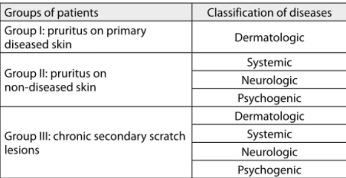
Clinically-based classification of pruritus is proposed by the International Forum for the Study of itch [60]. The first part classifies the underlying conditions according to groups I-III (Table 1). Primary skin lesions may be con-founded by secondary [60, 64]. Primary skin lesions and secondary (reactive to scratching/rubbing) must be dif-ferentiated in, for example, atopic dermatitis. The second part classifies the underlying conditions according to etiol-ogy in different categories I-IV (Table 1). Category V or
Table 1. Classification of chronic pruritus
Groups of patients Classification of diseases
Group I: pruritus on primary
diseased skin Dermatologic
Group II: pruritus on non-diseased skin
Systemic Neurologic Psychogenic
Group III: chronic secondary scratch lesions
Dermatologic Systemic Neurologic Psychogenic
“mixed” includes patients with more than one underlying disease that may be responsible. Category VI or “pruritus of undetermined origin” includes chronic pruritus without finding of underlying origin.
In management of patients with chronic pruritus, first they should be grouped according to clinical picture/his-tory. Second, they should be classified on the basis of labo-ratory/histological/radiological findings (Table 1) [60].
CONCLUSION
Pruritus is prevalent in the general population. Pruritus and pain have their own neuronal pathways with wide
interactions. The proposed classification distinguishes between pruritus with and without primary or second-ary skin lesions and should be considered as the only seg-ment of a comprehensive approach to pruritus of unknown origin. Thus, persistent pruritus without obvious derma-tologic pathology should encourage investigation for the underlying systemic cause.
ACKNOWLEDGMENT
The author acknowledges the Ministry of Education, Sci-ence and Technological Development of the Republic of Serbia, Project: 172058.
1. Liddell K. Hippocrates of Cos (460-377 BC). Clin Exp Dermatol. 2000; 25:86-8.
2. Hafenreffer S. De pruriti, in Nosodochium, in quo cutis, eiqe adhaerentium partium, affectus omnes singulari methodo et cognoscendi et curandi fidelissime traduntur. Ulm: B Kuehn; 1660. p.98-102.
3. Bernhard JD. Itch and pruritus: what are they and how should itches be classified? Dermatol Ther. 2005; 18:288-91.
4. Weisshaar E, Kucenic MJ, Fleischer JR. Pruritus a review. Acta Derm Venereol Suppl. 2003; (213):5-32.
5. Greaves MW. Pathogenesis and treatment of pruritus. Curr Allergy Asthma Rep. 2010; 10:236-42.
6. Schmelz M, Schmidt R, Bickel A, Handwerker HO, Torebjork HE. Specific C-receptors for itch in human skin. J Neurosci. 1997; 17:8003-8.
7. Andrew D, Craig AD. Spinothalamic lamina I neurons selectively sensitive to histamine: a central neural pathway for itch. Nat Neurosci. 2001; 4:72-7.
8. Ständer S, Raap U, Weisshaar E, Schmelz M, Mettang T, Handwerker H, et al. Pathogenesis of pruritus. J Dtsch Dermatol Ges. 2011; 9:456-63.
9. Davidson S, Zhang X, Yoon CH, Khasabov SG, Simone DA, Giesler GJ Jr. The itch-producing agents histamine and cowhage activate separate populations of primate spinothalamic tract neurons. J Neurosci. 2007; 27:10007-14.
10. Imamachi N, Park GH, Lee H, Anderson DJ, Simon MI, Basbaum AI, et al. TRPV1-expressing primary afferents generate behavioral responses to pruritogens via multiple mechanisms. Proc Natl Acad Sci U S A. 2009; 106:11330-5.
11. Steinhoff M, Biro T. A TR(I)P to pruritus research: role of TRPV3 in inflammation and itch. J Invest Dermatol. 2009; 129:531-5. 12. McNeil B, Dong X. Peripheral mechanisms of itch. Neurosci Bull.
2012; 28(2):100-10.
13. Yosipovitch G, Samuel L. Neuropathic and psychogenic itch. Dermatol Ther. 2008; 21:32-41.
14. Cowden JM, Zhang M, Dunford PJ, Thurmond RL. The histamine H4 receptor mediates inflammation and pruritus in Th2-dependent dermal inflammation. J Invest Dermatol. 2010;
130:1023-33.
15. Gutzmer R, Mommert S, Gschwandtner M, Zwingmann K, Stark H, Werfel T. The histamine H4 receptor is functionally expressed on T(H)2 cells. J Allergy Clin Immunol. 2009; 123:619-25.
16. Soh UJ, Dores MR, Chen B, Trejo J. Signal transduction by protease-activated receptors. Br J Pharmacol. 2010; 160(2):191-203.
17. Ständer S, Siepmann D, Herrgott I, Sunderkötter C, Luger TA. Targeting the neurokinin receptor 1 with aprepitant: a novel antipruritic strategy. PLOS One. 2010; 5:e10968.
18. Metz M, Gundmann S, Stander S. Pruritus: an overview of current concepts. Vet Dermatol. 2011; 22:121-31.
19. Phan NQ, Bernhard JD, Luger TA, Ständer S. Antipruritic treatment with systemic μ-opioid receptor antagonists: a review. J Am Acad Dermatol. 2010; 63:680-8.
20. Reich A, Szepietowski JC. Opioid-induced pruritus: an update. Clin Exp Dermatol. 2010; 35:2-6.
21. Kumagai H, Ebata T, Takamori K, Muramatsu T, Nakamoto H, Suzuki H. Effect of a novel kappa-receptor agonist, nalfurafine hydrochloride, on severe itch in 337 haemodialysis patients: a phase III, randomized, double-blind, placebo- controlled study. Nephrol Dial Transplant. 2010; 25:1251-7.
22. Kupczyk P, Reich A, Szepietowski JC. Cannabinoid system in the skin – a possible target for future therapies in dermatology. Exp Dermatol. 2009; 18:669-79.
23. Sun YG, Chen ZF. A gastrin-releasing peptide receptor mediates the itch sensation in the spinal cord. Nature. 2007;
448(7154):700-3.
24. Dalgard F, Svensson A, Holm JO, Sundby J. Self-reported skin morbidity in Oslo: Associations with sociodemographic factors among adults in a cross-sectional study. Br J Dermatol. 2004; 151:452-7.
25. Wolkenstein P, Grob JJ, Bastuji-Garin S, Ruszczynski S, Roujeau JC, Revuz J. French people and skin diseases: results of a survey using a representative sample. Arch Dermatol. 2003; 139:1614-9.
26. Matterne U, Strassner T, Apfelbacher CJ, Diepgen TL, Weisshaar E. Measuring the prevalence of chronic itch in the general population: a pilot study. Acta Derm Venereol. 2009; 89:250-6.
27. Murdoch ME, Asuzu MC, Hagan M, Makunde WH, Ngoumou P, Ogbuagu KF, et al. Onchocerciasis: the clinical and epidemiological burden of skin disease in Africa. Ann Trop Med Parasitol. 2002; 96:283-96.
28. Roy A, Pickar D, Paul S. Doran A, Chrousos GP, Gold PW. CSF corticotropin-releasing hormone in depressed patients and normal control subjects. Am J Psychiatry. 1987; 144:641-5.
29. Halvorsen JA, Dalgard F, Thoresen M, Bjertness E, Lien L. Itch and mental distress: a cross-sectional study among late adolescents. Acta Derm Venereol. 2009; 89:39-44.
30. Senturk N, Ozkaya O, Aytekin S, Bek K, Acikgoz Y, Aydin F, et al. Characteristics of pruritus in children on peritoneal dialysis. Nephron Clin Pract. 2008; 109:168-72.
31. Reich A, Stander S, Szepietowski JC. Pruritus in elderly. Clin Dermatol. 2011; 29:15-23.
32. Black MM, Ambros-Rudolph C, Edwards L, Lynch P, editors. Obstetric and Gynaecologic Dermatology. 3rd ed. London: Mosby Elsevier; 2008.
33. Weisshaar E, Apfelbacher C, Jager G, Zimmermann E, Bruckner T, Diepgen TL, et al. Pruritus as a leading symptom: clinical characteristics and quality of life in German and Ugandan patients. Br J Dermatol. 2006; 155:957-64.
34. Jovanović M, Poljački M, Mimica-Dukić N, Boža P, Vujanović Lj, Djuran V, et al. Sesquiterpene lactone mix patch testing supplemented with dandelion extract in patients with allergic contact dermatitis, atopic dermatitis and non-allergic chronic inflammatory skin diseases. Contact Dermatitis. 2004; 51:101-10.
35. Jovanović M, Mimica-Dukić N, Poljački M, Boža P. Erythema multiforme due to contact with weeds: a recurrence after patch testing. Contact Dermatitis. 2003; 48:17-25.
36. Jovanović M, Oliwiecki S, Beck MH. Occupational contact urticaria from beef associated with hand eczema. Contact Dermatitis. 1992; 27:188-9.
112
37. Jovanović M , Poljački M, Djuran V, Vujanović Lj, Sente R, Stojanović S. Kontaktna senzibilizacija izazvana Compositae biljkama kod obolelih od atopijskog dermatitisa. Med Pregl. 2004; 57:209-18.
38. Jovanović M, Boža P, Karadaglić Dj, Brkić S, Petrović A, Mimica-Dukić N, et al. Contact sensitivity in patients with psoriasis in Vojvodina. Int Arch Allergy Immunol. 2009; 148:311-20.
39. Dlova NC, Mosam A. Inflammatory noninfectious dermatoses of HIV. Dermatol Clin. 2006; 24:439-48.
40. Jovanović M, Karadaglić Dj, Brkić S. Contact urticaria and allergic contact dermatitis to lidocaine in a patient sensitive to benzocaine and propolis. Contact Dermatitis. 2006; 54:124-6.
41. Yosipovitch G, Goon AT, Wee J, Chan YH, Zucker I, Goh CL. Itch characteristics in Chinese patients with atopic dermatitis using a new questionnaire for the assessment of pruritus. Int J Dermatol. 2002; 41:212-6.
42. Yosipovitch G, Goon A, Wee J, Chan YH, Goh CL. The prevalence and clinical characteristics of pruritus among patients with extensive psoriasis. Br J Dermatol. 2000; 143:969-73.
43. Szepietowski JC, Reich A, Wisnicka B. Pruritus and psoriasis. Br J Dermatol. 2004; 151:1284.
44. Zamirska A, Reich A, Berny-Moreno J, Salomon J, Szepietowski JC. Vulvar pruritus and burning sensation in women with psoriasis. Acta Derm Venereol. 2008; 88:132-5.
45. Amatya B, Wennersten G, Nordlind K. Patients’ perspective in chronic plaque psoriasis: a questionnaire-based study. J Eur Acad Dermatol Venereol. 2008; 22:822-6.
46. Conrad R, Geiser F, Haidl G, Hutmacher M, Liedtke R, Wermter F. Relationship between anger and pruritus perception in patients with chronic idiopathic urticaria and psoriasis. J Eur Acad Dermatol Venereol. 2008; 22:1062-9.
47. Van Loey NE, Bremer M, Faber AW, Middelkoop E, Nieuwenhuis MK. Itching following burns: epidemiology and predictors. Br J Dermatol. 2008; 158:95-100.
48. Ferm I, Magnus S, Wallengren J. Somatic and psychiatric comorbidity in patients with chonic pruritus. Acta Derm Venereol. 2010; 90:395-400.
49. Sommer F, Hensen P, Bockenholt B, Metze D, Luger TA, Stander S. Underlying diseases and co-factors in patients with severe chronic pruritus: a 3-year retrospective study. Acta Derm Venereol. 2007; 87:510-6.
50. Afifi Y, Aubin F, Puzenat E, Degouy A, Aubrion D, Hassam B, et al. Enquête étiologique d’un prurit sine materia: étude prospective d’une série de 95 patients. Rev Med Interne. 2004; 25:490-3. 51. Özdemir M, Tuzun Y. Herpes zoster and pruritus. Int J Dermatol.
2004; 43:779-80.
52. Schmidt E, Rose C, Mulyowa GK, Jager G. Dermatologie an der Universitätsklinik Mbarara, Uganda. J Dtsch Dermatol Ges. 2004; 2:920-7.
53. Herzberg U, Sagen J. Peripheral nerve exposure to HIV viral envelope protein gp120 induces neuropathic pain and spinal gliosis. J Neuroimmunol. 2001; 116:29-39.
54. Razeghi E, Eskandari D, Ganji MR, Meysamie AP, Togha M, Khashayar P. Gabapentin and uremic pruritus in haemodyalisis patients. Ren Fail. 2009; 31:85-90.
55. Davis MP, Frandsen JL, Walsh D, Andersen S, Taylor S. Mirtazapine for pruritus. J Pain Symptom Manage. 2003; 25:288-91.
56. Cribier B, Santinelli F, Schmitt C, Stoll-Keller F, Grosshans E. Should patients with pruritus be tested for hepatitis C virus infection? A case controlled study. Br J Dermatol. 2000; 142:1249-50.
57. Weisshaar E, Dalgard Florence. Epidemiology of itch: adding to the burden of skin morbidity. Acta Derm Venereol. 2009; 89:339-50. 58. Yosipovitch G. Chronic pruritus: a paraneoplastic sign. Dermatol
Ther. 2010; 23:590-6.
59. Arnold LM, Auchenbach MB, McElroy SL. Psychogenic excoriation. Clinical features, proposed diagnostic criteria, epidemiology and approaches to treatment. CNS Drugs. 2001; 15:351-9.
60. Stander S, Weisshaar E, Mattang T, Szepietowski JC, Carstens E, Ikoma A, et al. Clinical classification of itch: a position paper of the international forum for the study of itch. Acta Derm Venereol. 2007;87:291-4.
61. Tey HL, Wallengren J, Yosipovitch G. Psychosomatic factors in pruritus. Clin Dermatol. 2013;31:31-40.
62. Misery L, Wallengren DJ, Weisshaar E, Zalewska A. Validation of diagnostic criteria of functional itch disorder or psychogenic pruritus. Arch Derm Venereol. 2008; 88:503-4.
63. Misery L, Alexandre S, Dutray S, Chastaing M, Consoli SG, Audra H, et al. Functional itch disorder or psychogenic pruritus: suggested diagnosis criteria from the French psychodermatology group. Acta Derm Venereol. 2007; 87:341-4.
64. Jovanović M. Manifestations of contact allergy. Srp Arh Cel Lek. 2012; 140(11-12):786-91.
КРАТАК САДРЖАЈ
От кри ва њем ме ди ја то ра и ре цеп то ра спе ци фич них за пру-ри тус по ста вљен је не у ро би о ло шки кон цепт пру пру-ри ту са ко-јег од ли ку ју: Ц не у ро ни спе ци фич ни за свраб (хи ста мин и хи ста мин-не за ви сни); ре цеп то ри спе ци фич ни за свраб на ку та ним и спи нал ним не у ро ни ма; функ ци о нал ни „ди ја лог“ из ме ђу не у ро на спе ци фич них за пру ри тус и ће ли ја стал но или при вре ме но (ин фла ма ци ја) за сту пље ни у ко жи; пе ри-фер но и цен трал но пре но ше ње пру ри ту са; функ ци о нал но ак тив ни „пру ри тус-спе ци фич ни ма трикс“ у мо згу у уло зи цен тра за пру ри тус. Пру ри тус је нај че шће симп том дер ма-то за, али код 10–50% осо ба ко је не ма ју обо ље ње ко же он је ма ни фе ста ци ја си стем ског по ре ме ћа ја. Из два ја ње пру ри-ту са у окви ру ауто и мун ских и упал них бо ле сти у дер ма
то-ло ги ји за сни ва се на кли нич кој сли ци и при ро ди основ ног обо ље ња и под ра зу ме ва по ја ву пру ри ту са на при мар но и/ или се кун дар но ин фла ми ра ној ко жи. Пру ри тус у ин тер ној ме ди ци ни пр вен стве но под ра зу ме ва пру ри тус у при мар но не ин фла ми ра ној ко жи: за хва та ње ко же и га стро ин те сти нал-ног трак та су не за ви сни фак то ри ри зи ка за по ја ву пру ри ту-са у си стем ској скле ро зи, анал ном и вул вар ном пру ри ту су. Кла си фи ка ци ја пру ри ту са об је ди њу је ети о ло шке кри те ри-ју ме, на ко ји ма је за сно ва на не у ро а на том ска по де ла, и кли-нич ке кри те ри ју ме, на ко ји ма се за сни ва кли кли-нич ка по де ла. Кла си фи ка ци ју пру ри ту са тре ба схва ти ти као део све о бу-хват ног при сту па пру ри ту су нео д ре ђе ног узро ка.
Кључ не ре чи: пру ри тус; ко жне бо ле сти; пре ва лен ци ја; ко-мор би ди тет
Актуелни концепти патофизиологије, епидемиологије и класификације
пруритуса
Марина Јовановић1,2
1Медицински факултет, Универзитет у Новом Саду, Нови Сад, Србија;
2Клиника за дерматовенеролошке болести, Клинички центар Војводине, Нови Сад, Србија
Примљен • Received: 18/07/2013 Прихваћен • Accepted: 07/10/2013