Efect of pH on Optic and Structural Characterization of Chemical Deposited AgI Thin Films
Fatma Meydaneri Tezela*, İshak Afşin Kariperb
Received: March 26, 2017; Revised: May 29, 2017; Accepted: June 20, 2017
AgI thin ilms were grown on amorphous commercial glass substrates with chemical bath deposition (CBD) at diferent pH values (2, 3, 4, 5, 6), 6 hours deposition time and 60 °C. The structure of the nanocrystals was characterized by X-ray difraction (XRD). The ratio of Ag+ and I- ions changed the crystalline structures. The presence of the Ag+ ions produces the γ-phase of AgI and excess of iodine concentration produces β-phase of AgI. The pH: 4 was like a transition pH for these phases. The number of crystallites per unit area has maximum value at pH: 5, as the structure is re-crystallization to hexagonal phase. Also, the thicknesses of produced thin ilms were decreased with increased pH values. Therefore, transmission, relection, extinction coeicients and refractive index of the materials were afected by thicknesses, and calculated to be 32, 35, 3, 11, 9 (%) - 27, 25, 61, 45, 49 (%) - 0.036, 0.032, 0.067, 0.107, 0.075 and 3.21, 3.02, 5.16, 8.35, 5.70 in 550 nm at pH: 2-3-4-5-6 values, respectively. The exciton peaks of AgI were observed at between 320 and 420 nm. Surface properties were investigated by using scanning electron microscopy (SEM).
Keywords: AgI, crystal growth, pH efect, chemical bath deposition, optical and surface properties.
*e-mail: [email protected]
1. Introduction
Semiconductor industry has signiicantly developed and will develop next years. The chalcogenides are very useful in the semiconductor industry, for instance, they are used in computers. These materials are used in two terminal devices which have two diferent resistance states1,2. This situation was investigated widely in the optoelectronic world. AgI synthetic particles were also known as well semiconductors due to their photosensitivity and superionic conductivity3-7.
The crystallite size and crystal structure of AgI have been afected by growth mechanism which is important in terms of photosensitivity. Therefore, we have to give much attention to characterization and preparation of AgI8-16.
According to the literature, it has been observed several polymorphic forms depending on the experimental conditions and the temperature of formation17. AgI occurred in two polymorphic forms; a hexagonal wurtzite type β-AgI structure and cubic zinc blend type γ-AgI structure. Moreover, AgI changed several phases from cubic γ phase to the rhombohedral, tetragonal and rock salt modiications with magnitude of pressure, also ZnS-type structural system is observed18,19.
Many researchers attempted to produce AgI ilms according to these diferent properties of AgI. Someone investigated γ-AgI formation, its' memory efect and someone searched its' structural and morphological properties. The
resistance switching of AgI was obtained to control the current or the voltage of the devices. This property and the other properties of AgI depend on its structure and electrical properties. For instance, the resistivity of AgI was 1 Ωcm at 200 °C for α-AgI structure, but AgI had β and α phase at 147 °C. So, the structure of AgI crystal changes depending on preparation conditions.
The research related to the efects of pH with chemical bath on AgI structure and optical properties for the solar cell substrates can be rarely found in literature. Therefore, in this study, our purpose is to investigate the efects of pH and doping parameters on AgI thin ilms growth with CBD. The characterizations of the surface, structural, optical properties depending on crystal growth were examined.
2. Experimental Procedure
2.1 Crystal growth and characterizations
All chemicals were purchased from Merck and Aldrich and they were used without further puriication. High purity reagents were used in preparing all of the solutions. The stock solutions with several concentrations were diluted daily in order to be used as solutions. The other stock solutions were prepared from highly pure compounds (99.9%, E. Merck, Darmstadt, Sigma Aldrich). All plastic and glassware
aDepartment of Metallurgy and Materials Engineering, Faculty of Engineering, Karabük
University,78050, Karabük, Turkey
equipment were cleaned by soaking them in dilute HNO3 and were rinsed with deionizer water before they were used. Substrates were cleaned and dipped into alcohol and HNO3. Baths with concentrations AgCl, 1×10⁻³ M; KI 1×10⁻³ M;
HNO3 0.1 M, were used. In order to adjust the pH value of the solution as 2, 3, 4; 1100 µl, 80 µl, 8 µl of nitric acid (HNO3), respectively, and14µL 0.1 M KOH solution for pH: 6. But, nothing was added to the solution of pH: 5. The pH meter was used to measure the pH values of the chemical baths (Lenko mark 6230N). The samples were left in the oven at 60 ºC for 6 hours.
The chemical reactions for the deposition of the silver iodide crystals taking place on the glass substrate in the bath are as below. Silver ions (Ag+) don't dissolve with iodine
(I-) in the bath and form an insoluble AgI.
(1)
(2)
Silver nitrate and potassium iodide were used for formation of AgI (2), but the chemical bath was in acidic area at pH: 2-6 (1). Hydrogen iodide occurred in reaction. When the pH of bath is decreased by adding nitric acid, iodine concentration decreases. So, the formation AgI depended on HI (1) reaction and pH of the bath. The pH of the bath afected the ilm thickness grown on glass substrate and structure of AgI crystals.
The crystalline structure of AgI was determined by X-ray difraction (XRD) with a CuKα1 radiation source (Rikagu RadB model, λ=1.5406 Å) over the range 10° < 2θ < 90° at a speed of 3° min-1 with a step size of 0.02°. We investigated
the surface properties of all of formed crystals by using an EVO40-LEO computer controlled digital scanning electron microscope (SEM). Ratio of chemical compositions was measured by an EDX spectrometer attached to the SEM. We used Lange 500 Spectrophotometer at room temperature to make optical measurements by placing an uncoated identical glass substrate in the reference beam. The optical spectrum of the thin ilms grown on glass substrate was recorded in the wavelength at the range of 200-1100 nm. The thicknesses of the thin ilms were measured with a Veeco Multi Mode AFM (Controller=NanoScope 3D).
3. Results and Discussion
3.1 Structural properties
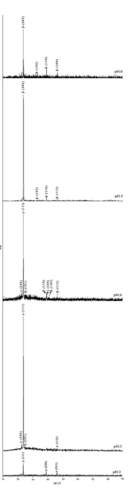
It was seen that XRD patterns changed with pH of the chemical bath. The peaks of γ-AgI and β-AgI can be observed at the difraction angle of 10°<2θ<90° on the XRD spectrum. These difraction patterns have cubic (γ-AgI) and hexagonal (β-AgI) structures and preferential orientation
in the (111), (220) and (311) plane at pH: 2, (220), (111), (101) and (112) at pH: 3, (220), (111), (101), (110), (220), (103) and (112) at pH: 4, (002), (102), (110) and (112) at pH: 5, (002), (102), (110) and (112) at pH: 6. The intensity of XRD peaks, preferential orientation of atoms and crystal structures changed with deposition pH, were shown in Fig. 1 and Table 1. According to Kennedy17, Ves and et. al18 and Goswami19, γ- and β-AgI phase depends on the high temperature, not only the temperature but also pH changed the phase in this study. The pH of bath directly afected the iodine concentration, and the iodine concentration decreased below pH:5-6. While γ-AgI phase was observed at the lower pH (pH=2-4), iodine concentration increased above pH: 5-6 and γ-AgI phase is transformed to β-AgI phase depend on XRD spectrum. The XRD patterns showed that iodized Ag ilm contained β-phase of AgI which was conirmed by the (102) and (103) peaks when XRD data of γ-AgI determined superposition with the (002) and (112) peaks by Liang20, Kumar21 and Validźić22.
Crystal lattice parameters along the (111) at pH: 2, (300) at pH: 3, (300) at pH: 4, (001) at pH: 5 and (001) at pH: 6 plane were calculated by using the formulas given below,
(3)
(4) crystallite size (D), dislocation density (δ) and the number of crystallization per unit area (N) were obtained by using Scherrer's formula23-25:
(5)
(6)
(7)
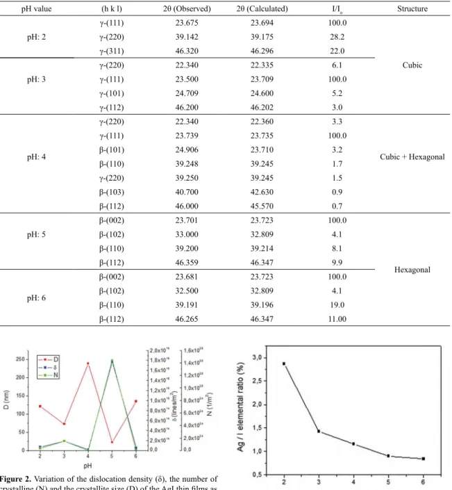
where t is the formed crystal layer thickness, λ is wavelength of X-ray (1.5406 Å), β is FWHM of the peak, θ is Bragg's angle, δ is dislocation density which is deined as the length of dislocation lines per unit volume of the crystal. Higher δ values indicate lower crystalline levels and the amount defects in the structure. In this study, a higher δ value was calculated at pH: 4, since the cubic structure of AgI converted to hexagonal phase. So, the pH: 4 was like a transition pH for these phases. The number of crystallites per unit area has maximum value at pH: 5, as the structure is re-crystallization to hexagonal phase. The crystallites obtained at pH: 2-4-6 had the best crystallinity. The changes in dislocation density, the number of crystallites per unit area and the average crystallite sizes are presented in Fig. 2.
H O
3 aq+
I
( )aq*
H O
2+
HI
aq+
-Q V Q V
Ag
+( )aq+
I
-( )aq$
AgI
( )kCubic Structure d
h k l
a 2
2 2 2
2
= + + Q V
Hexagonal Structure
d a
h hk k
c l 1 34 2 2 2 2 2 2
= T + + Y+ R W
.
cos
D
0 9
Figure 1. XRD spectrum of AgI thin ilms (a) pH: 2, (b) pH: 3, (c) pH: 4, (d) pH: 5, (e) pH: 6.
3.2 Concentration distribution and surface
morphology
Energy-dispersive X-ray analysis (EDX) was used in order to understand the efect of the ion concentrations on the structure. Theoretically, the Ag+ and I- ions are 45.94%, 54.06% and atomic ratio is (Ag/I) 0.84979 respectively. As seen in Fig. 3, the ratio of Ag+ and I- ions decrease with increasing pH values. The presence of Ag+ ions gets the γ-phase of AgI and excess of iodine concentration produces β-phase of AgI. The iodine concentration increases at pH:5 and 6, and the structure transforms from γ-phase to β-phase, and γ-phase observes at pH: 2-4. The atomic ratio is about 0.83 at pH: 6. The hexagonal wurtzite type (β-AgI) is observed while the silver and iodine atomic ratio is below 0.90. The cubic zinc blend type (γ-AgI) forms while the atomic ratio of silver and iodine is over 0.90.
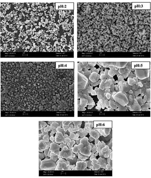
The SEM images of the thin ilms with same magniication factor for pH:2-6 are depicted in Fig. 4. As seen from SEM images, the thin ilms are very dense except for pH:2. It is seen that crystallization is rare for pH:2, but any broke are not observed on the surface. As seen from igures, crystals bonded substrate. Small particles are obtained for pH:2-4 and both big and small particles are obtained for pH:5 and 6. For each pH value, speciic accumulate type of the crystals are observed for some regions. This is a result of preferred growth of atoms. This situation can be explained by speed of reaction in solution, amorphous structure of substrate, temperature diference of particles inside solution, pH value and composition of solution. Crystallization number for unit surface area and dislocation density are also luctuating with the efect of the phase transitions.
3.3 Optical properties
The transmittance (T) for AgI thin ilms formed on glass substrates can be obtained by using absorbance (A) and relectivity (R) spectra from the expression26:
(8) Transmission and the relectivity measurements are performed at room temperature at the range of 200-1100 nm, and absorption and transmission-relectivity curves versus wavelengths are shown in Figs. 5-6, respectively. The absorption coeicient,α determines efective penetration distance of light with a particular wavelength inside the material. As seen from Fig. 5, the absorption coeicients (α) are changed to be 0.497, 0.447, 1.478, 0.935 and 1.041 at pH: 2, 3, 4, 5, 6 in the visible region (550 nm), respectively. Optical transmission was the largest at pH: 4; then the transmission values decreased at pH 5-6. The transmission and relection values of AgI thin crystalline layers occurred on glass substrates are obtained to be 32, 35, 3, 11, 9% and 27, 25, 61, 45, 49% in 550 nm at pH: 2, 3, 4, 5, 6 values, respectively. The exciton peaks
T
1
R e
2 A-Figure 2. Variation of the dislocation density (δ), the number of crystalline (N) and the crystallite size (D) of the AgI thin ilms as a function of the pH of the chemical bath.
Figure 3. The ratio of Ag+ and I- ions.
Table 1. The observed and literature peaks (ASTM Data) and crystal orientations at the difraction angle for the crystals grown at diferent pH values.
pH value (h k l) 2θ (Observed) 2θ (Calculated) I/Io Structure
pH: 2
γ-(111) 23.675 23.694 100.0
Cubic
γ-(220) 39.142 39.175 28.2
γ-(311) 46.320 46.296 22.0
pH: 3
γ-(220) 22.340 22.335 6.1
γ-(111) 23.500 23.709 100.0
γ-(101) 24.709 24.600 5.2
γ-(112) 46.200 46.202 3.0
pH: 4
γ-(220) 22.340 22.360 3.3
Cubic + Hexagonal
γ-(111) 23.739 23.735 100.0
β-(101) 24.906 23.710 3.2
β-(110) 39.248 39.245 1.7
γ-(220) 39.250 39.245 1.5
β-(103) 40.700 42.630 0.9
β-(112) 46.000 45.570 0.7
pH: 5
β-(002) 23.701 23.723 100.0
Hexagonal
β-(102) 33.000 32.809 4.1
β-(110) 39.200 39.214 8.1
β-(112) 46.359 46.347 9.9
pH: 6
β-(002) 23.681 23.723 100.0
β-(102) 32.500 32.809 4.1
β-(110) 39.191 39.196 19.0
β-(112) 46.265 46.347 11.00
of AgI were observed at between 320 and 420 nm with pH: 2, 3, 4, 5 to be similar with the indings in literature, so the transmission and relection curves are touching each other in these wavelengths4,27-29. In terms of optical properties;
if you produced AgI thin crystalline layers with diferent solvents with spray pyrolysis technique, exciton peak of AgI was observed around 420 nm dispersion in water when 360-495 nm aged in 2-propanol. The preparation of AgI with diferent solvents afected absorption wavelength with spray pyrolysis technique20-22.
The refractive index and extinction coeicient for the materials are given as26:
(9)
(10)
n
R
R
R
R
k
1
1
1
4
2 2=
-
+
+
Q
-
V
-k
4
r
am
Figure 4. SEM imagines of AgI thin ilms.
The changes of the refractive indexes and extinction coeicients versus wavelength are shown in Fig. 7. The refractive index formed a shape similar to the relection. The refractive indexes are changed to be 3.21, 3.02, 5.16, 8.35 and 5.70 at pH: 2-6, respectively. The refractive index has the highest at pH: 5. The ilm thickness afected the refractive index and extinction coeicient. The thicknesses of the thin ilms are decreased with increased pH values as shown in Fig. 8.
The optical constants are parameters which deine the reaction of a matter excited by an electromagnetic radiation at a given wavelength. These optical constants can be expressed as a complex dielectric function by the following formula30,31:
(11)
or as a complex refractive index by the following formula:
i
1 2
Figure 5. The absorbance change versus wavelength of AgI thin ilms grown at diferent pH values.
Figure 6. Optical transmission and relectance spectra of AgI thin ilms grown at diferent pH values of the chemical bath.
Figure 7. Refraction index (n) and extinction coeicient (k) of AgI thin ilms grown at diferent pH values.
Figure 8. The ilm thickness changed with pH of the AgI thin ilms.
Figure 9. Dielectric coeicients (ε) of AgI thin ilms grown at diferent pH values.
(12)
(13)
(14)
The real part or refraction index (nr) deines the phase velocity of light in material. The imaginary part or extinction coeicient (k) determines attenuation into material. Thus, the optical constants represent the optical properties of a material in terms of electromagnetic propagation in the material. The values of dielectric constants versus the wavelength for the thin ilms are shown in Fig. 9. The dielectric coeicients of the crystals are changed to be 2.11, 1.80, 16.92, 6.15 and 7.63 at pH: 2-6, respectively.
Light extinction and scattering by gold and silver nanoparticles by using Mie calculations and size-dependence of the extinction coeicient for silver and gold nanoparticles were given by Julien and Martinus32. They compared available
experimental data concerning the molar extinction coeicients for gold and silver nanoparticles and the results obtained by Mie calculations and developed simple power-law deinition for the dependence of the maximum extinction coeicient on particle diameter. Mie calculations have already been applied to obtain the extinction spectrum of gold nanoparticles in solution33,34, but silver has received less attention.
n
=
n
r+
ik
n
rk
1 2 2
f
=
-nk
2
2The experimental measurements of the extinction coeicients of silver nanoparticles for a range of sizes are obtained by Evanof and Chumanov35. The agreements
between the experimental data and the results obtained by Mie calculations are not very good. A quadrupole resonance was observed at short wavelengths for larger particle sizes (The spectral region of interest chosen was 320-420 nm), whose peak extinction coeicient is larger, but this band is neglected.
The optical response of silver is signiicantly afected by surface efects. It can be concluded that Mie calculations are actually most reliable method for silver nanoparticles, if these nanoparticles are mono-disperse and perfectly spherical. Also, the angular distribution of scattered light is of interest for the right-angle. Asymmetry parameter, which is the average value of the cosine of the scattering angle, can be estimated by the angular distribution of scattered light. The asymmetry parameter is zero for very small particles. Deviations from zero may be explained as changes in the angular distribution32. Also, high-index materials are used
to make the ilters and mirrors used in lasers with special relecting by optimal designs, and these kind of structures based on heavy metal oxide have practical applications in the ield of relecting windows, glass ceramics, thermal and mechanical sensors, layers for optical and electronic devices, etc36.
4. Conclusıons
AgI thin ilms have been grown on glass substrates by the CBD method at diferent pH values. XRD studies depict the phase transformation of AgI crystals. This efect can be explained by increasing of crystallite sizes and pH efect. SEM images show that increasing crystallite size and the grain structures turn into hexagonal from cubic when the pH values are increased. The results of EDX analysis depict the presence of Ag and I in the crystals. UV-VIS spectrophotometric analysis showed that the optical response of silver is much more sensitive to surface efects. According to optical properties, AgI thin ilms with high-index are appropriate to make the ilters and mirrors used in lasers with special relecting by optimal designs, layers for optical and electronic devices, relecting windows and mechanical and thermal sensors.
5. Acknowledgement
This work was supported by the Karabük University Scientific Research Project Unit under Contract No: KBÜ-BAP-13/1-YL-033, KBÜ-BAP-14/2-DS-042 and KBÜ-BAP-16/1-YD-016. The authors would like to thank to the Karabük University Scientiic Research Project Unit for their inancial support.
6. References
1. Ovshinsky SR. Reversible Electrical Switching Phenomena in Disordered Structures. Physical Review Letters. 1968;21(20):1450-1452.
2. Suh DS, Lee E, Kim KHP, Noh JS, Shin WC, Kang YS, et al. Nonvolatile switching characteristics of laser-ablated Ge2Sb2Te5
nanoparticles for phase-change memory applications. Applied Physics Letters. 2007;90(2):023101.
3. Otterwill RH, Woodbridge RF. The preparation of monodisperse silver bromide and silver iodide sols. Journal of Colloid Science. 1961;16(6):581-594.
4. Berry CR. Structure and Optical Absorption of AgI Microcrystals.
Physical Review. 1967;161(3):848.
5. Tanaka T, Iwasaki M. The multistage process of formation of ultrafarine silver bromide particles as revealed by multichannel spectrophotometry. J. Imaging Sci. 1985;29(3):86-92. 6. Hayes D, Schmidt KH, Miesel D. Growth mechanisms of silver
halide clusters from the molecule to the colloidal particle. The Journal of Physical Chemistry. 1989;93(16):6100-6109. 7. Mulvaney P. Nucleation and stabilization of quantized AgI clusters
in aqueous solution. Colloids and Surfaces A: Physicochemical and Engineering Aspects. 1993;81:231-238.
8. Vucemilovic MI, Micic OI. Electron-transfer reactions of extremely small AgI colloids. International Journal of Radiation Applications and Instrumentation. Part C. Radiation Physics and Chemistry. 1988;32(1):79-83.
9. Micic OI, Nenadovic MT, Rajh T, Nedeljkovic JM, Vucemilovic MI. Kinetic and spectroscopic properties of small colloids of metal iodide semiconductors. In: Degiorgio V, ed. Progress in Colloid and Polymer Science, Volume 76 - Trends in Colloid and Interface Science II. New York: Springer; 1988. p. 20-23.
10. Schmidt KH, Patel R, Meisel D. Growth of silver halides from the molecule to the crystal. A pulse radiolysis study. Journal of the American Chemical Society. 1988;110(15):4882-4884. 11. Henglein A, Gutiérrez M, Weller H, Fojtik A, Jirkovský J.
Photochemistry of colloidal semiconductors 30. Reactions and luorescence of AgI and AgI - Ag2S colloids. Berichte der
Bunsengesellschaft für physikalische Chemie. 1989;93(5):593-600.
12. Mićić OI, Meglić M, Lawless D, Sharma DK, Serpone N. Semiconductor photophysics. 5. Charge carrier trapping in ultrasmall silver iodide particles and kinetics of formation of silver atom clusters. Langmuir. 1990;6(2):487-492.
13. Chen SH, Ida T, Klimura K. Thiol-Derivatized AgI Nanoparticles:? Synthesis, Characterization, and Optical Properties. The Journal of Physical Chemistry B. 1998;102(32):6169-6176.
14. Brelle MC, Zhang JZ. Femtosecond study of photo-induced electron dynamics in AgI and core/shell structured AgI/Ag2S and AgBr/Ag2S colloidal nanoparticles. The Journal of Chemical Physics. 1998;108(8):3119.
16. Wang Y, Mo J, Cai W, Yao L, Zhang L. Synthesis of nano-AgI arrays and their optical properties. Journal of Materials Research. 2001;16(4):990-992.
17. Kennedy JH. Thin ilm solid electrolyte systems. Thin Solid Films. 1977;43(1-2):41-92.
18. Ves S, Glötzel D, Cardona M, Overhof H. Pressure dependence of the optical properties and the band structure of the copper and silver halides. Physical Review B. 1981;24(6):3073-3085. 19. Goswami A. Thin Film Fundamentals. 1st ed. New Delhi: New
Age International; 1996. p. 51-71.
20. Liang XF, Chen Y, Shi L, Lin J, Yin J, Liu ZG. Resistive switching and memory efects of AgI thin ilm. Journal of Physics D: Applied Physics. 2007;40(16):4767-4770. 21. Kumar PS, Dayal PB, Sunandana CS. On the formation mechanism
of γ-AgI thin ilms. Thin Solid Films. 1999;357(2):111-118. 22. Validzic IL, Jokanpvic V, Uskokovic DP, Nedeljkovic JM.
Inluence of solvent on the structural and morphological properties of AgI particles prepared using ultrasonic spray pyrolysis. Materials Chemistry and Physics. 2008;107(1):28-32.
23. Mamazza R Jr., Morel DL, Ferekides CS. Transparent conducting oxide thin ilms of Cd2SnO4 prepared by RF magnetron
co-sputtering of the constituent binary oxides. Thin Solid Films. 2005;484(1-2):26-33.
24. Ferro R, Rodríguez JA, Vigil O, Morales-Acevedo A. Chemical composition and electrical conduction mechanism for CdO:F thin ilms deposited by spray pyrolysis. Materials Science and Engineering: B. 2001;87(1):83-86.
25. Callister WD. Materials Science and Engineering: An Introduction. Hoboken: John Wiley and Sons; 1997. p. 102.
26. Pejova B, Grozdanov I, Tanuševski A. Optical and thermal band gap energy of chemically deposited bismuth(III) selenide thin ilms. Materials Chemistry and Physics. 2004;83(2-3):245-249.
27. Benramdane N, Murad WA, Misho RH, Ziane M, Kebbab Z. A chemical method for the preparation of thin ilms of CdO and ZnO. Materials Chemistry and Physics. 1997;48(2):119-123. 28. Kondo S, Itoh T, Saito T. Strongly enhanced optical absorption
in quench-deposited amorphous AgI ilms. Physical Review B. 1998;57(20):13235-13240.
29. Cardona M. Optical Properties of the Silver and Cuprous Halides. Physical Review. 1963;129(1):69-78.
30. Meydaneri Tezel F, Kariper IA. Synthesis, surface tension, optical and dielectric properties of bismuth oxide thin ilm.
Materials Science-Poland. 2017;35(1):87-93.
31. Meydaneri Tezel F, Özdemir O, Kariper IA. The efects of pH on structural and optical characterization of iron oxide thin ilms. Surface Review and Letters. 2017;24(4):1750051. DOI: 10.1142/S0218625X17500512
32. Navarro JRG, Werts MHV. Resonant light scattering spectroscopy of gold, silver and gold-silver alloy nanoparticles and optical detection in microluidic channels. Analist. 2013;138(2):583-592
33. Haiss W, Thanh NTK, Aveyard J, Fernig DG. Determination of Size and Concentration of Gold Nanoparticles from UV-Vis Spectra. Analytical Chemistry. 2007;79(11):4215-4221. 34. Khlebtsov NG. Determination of Size and Concentration of Gold
Nanoparticles from Extinction Spectra. Analytical Chemistry. 2008;80(17):6620-6625.
35. Evanof DD, Chumanov G. Size-Controlled Synthesis of Nanoparticles. 2. Measurement of Extinction, Scattering, and Absorption Cross Sections. The Journal of Physical Chemistry
B. 2004;108(37):13957-13962.