SYMMETRY OF TYMPANOMETRIC MEASUREMENTS AND
TRANSIENT EVOKED OTOACOUSTIC EMISSIONS
IN NEONATES
Simetria das medidas timpanométricas e as respostas
das emissões otoacústicas transientes em neonatos
Liliane Aparecida Fagundes Silva(1), Kilza de Arruda Lyra e Silva(1), Seisse Gabriela Gandoli Sanches(1), Renata Mota Mamede de Carvallo(1)
(1) Faculdade de Medicina da Universidade de São Paulo,
FMUSP, São Paulo, SP, Brasil.
Study conducted at the Department of Physical Therapy, Spe-ech Therapy and Occupational Therapy, FMUSP - São Paulo (SP), Brazil.
Conlict of interest: non-existent
For TEOAE recording to be effective, it is important that the middle ear is in a suitable
condition and that the external auditory canal does not contain residual amniotic luid. The size of the external auditory canal is also an aspect to
be considered, given that responses of greater amplitude are observed in neonates because the volume of the canal is small7,9-13.
Acoustic immittance measurements are used to evaluate the general condition of the middle ear. They can be used to obtain a tympanogram, which is a record of the mobility of the tympanic-ossicular system in response to pressure variation, which allows abnormalities in the middle ear to be ruled
out or conirmed14.
As such abnormalities may hinder the recording of TEOAEs, tympanogram analysis can be used as a complementary method for hearing evaluation15
because it enables the identiication of middle ear
impairment that can affect TEOAE results. For this INTRODUCTION
Hearing impairment early in life can affect various aspects of child development, including linguistic, cognitive, psychosocial and academic aspects. For this reason, methods for the early evaluation of auditory system integrity are needed1-4.
The recording of transient-evoked otoacoustic emissions (TEOAEs) is a method for evaluating the pre-neural function of the cochlea, especially the
outer hair cells. Thus, this method is an excellent
tool for detecting moderate to severe cochlear hearing loss5-8.
ABSTRACT
Purpose: to determine the occurrence of symmetry equal or greater than to 70% between ears comparing the results of 226 and 1000 Hz tympanograms to the otoacoustic emissions in neonates.
Methods: participants were 39 neonates (20 female and 19 male) with 60 hours post birth on average. Each newborn underwent 226 and 1000 Hz probe tone timpanometry and transient evoked otoacoustic emissions in both ears. Data were submitted to statistical analysis. Results: an occurrence of symmetry <70% was observed for the otoacoustic emissions test in 74.4% of the neonates. The incidence of
symmetry ≥70% for the timpanometry for both probe tone of 226 and 1000Hz was 76.9% and 84.6 %, respectively. The comparison between gender and ear did not indicate a signiicant difference,
although larger otoacoustic emissions amplitude was observed in the right ear. Conclusion: the results suggest a symmetry in the peripheral portion of auditory system, and the onset of asymmetries arising from the cochlea.
males, and the infants exhibited no risk factors for
hearing loss according to the Joint Committee on Infant Hearing criteria19.
Equipment
The following equipment was used:
• Transient-evoked otoacoustic emissions module, included in the MEPA3 middle ear power analyzer (Mimosa Acoustics Inc., Champaign, Illinois, USA) with an ER-10C acoustic probe (Etymotic Research, Elk Grove Village, Illinois, USA) having two output and one input (micro-phone) transducers;
• MADSEN OTOFLEX 100 middle ear analyzer
(GN Otometrics, Taastrup, Denmark) with output
tympanometry for 226- and 1000-Hz probe tones for tympanometry research.
Procedures
Each infant was assessed for both ears. For tympanometry, a neonatal-sized rubber tip was
introduced into the external auditory canal. After
sealing the canal, the 226- and 1000-Hz tympa-nograms were obtained, with two measurements being taken in each ear for each probe tone to
conirm the results. The 226-Hz tympanogram was obtained from the established baseline, excluding the equivalent volume of the external auditory canal.
This measurement was obtained in milliliters (ml) in the tympanometric mode of compensated admit-tance at the height of the tympanic membrane (YTM). For the 1000-Hz probe tone, the tympanogram was
obtained without external auditory canal volume
compensation in the Acoustic Admittance mode (Ya), and the unit of measurement was the millimho (mmho).
For the evaluation of TEOAEs, an ER-10C probe with a suitably sized rubber tip was inserted into
the external auditory canal of the newborn, with a
microphone and a miniature stimulus generator. Using this probe, an acoustic stimulus was emitted by the generator to stimulate the cochlear hair cells, which in turn emitted a response that was recorded by the microphone.
TEOAEs were considered present when the
following results were obtained: reproducibility ≥ 50%, probe stability ≥ 70% and signal to noise ratio responses greater than 3 dBNPS in the irst two
bands and 6 dBSPL in the last three bands.
Analysis of Results
The results were analyzed by descriptive statis-tical tests to characterize the study population. Inferential analyses were performed using paired Student t-tests, ANOVA and tests for the Equality of Two Proportions. The agreement between the
tests with respect to the symmetry of responses ≥
reason, acoustic immittance assessment should always precede TEOAEs8.
The literature suggests that 226-Hz acoustic immittance measurements in neonates can provide normal tympanogram data even in infants with abnormalities in the outer and middle ear. It is therefore necessary to also apply a 1000-Hz probe tone to acquire more reliable results16,17. Acoustic
immittance using a 1000-Hz probe tone produces better results than using 226- and 678-Hz probe tones 1.
The relationship between the two tests described above notwithstanding, there is evidence in the literature that the auditory system works asymmetri-cally. In the central auditory nervous system, the left hemisphere is dominant for language skills. Regarding the peripheral auditory system, there is also evidence that TEOAE responses are stronger in the right ear7,18. Given that middle ear
condi-tions interfere with TEOAE responses and that an asymmetry in tympanometry could indicate a difference in this structure, the study of symmetry in tympanometric measurements, even within normal limits, will assist in understanding whether this fact is related to failures or differences in the otoacoustic emission records between the ears. This infor-mation will make it possible to understand whether
differences between tympanograms can explain
the differences observed in otoacoustic emission records.
This study aimed to analyze tympanometry measurements in newborns with 226- and 1000-Hz probe tones using the variables Tympanometric Peak Pressure (TPP), peak compensated admit-tance (Ytm) and ear canal volume (ECV) along with
TEOAEs to ascertain whether there is symmetry ≥
70% between the right and left ears for the TEOAE and tympanometry results.
METHODS
This work was a cross-sectional study conducted in the Human Hearing Investigation Laboratory, Faculty of Medicine, University of São Paulo (Faculdade de Medicina da Universidade de São Paulo – FMUSP). The research project was approved by the institution’s Ethics Committee (CEP HU/USP 917/08).
The parents of the study’s participants were informed of the research objectives and signed informed consent forms.
Case sample
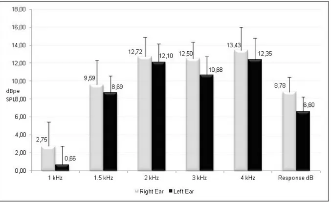
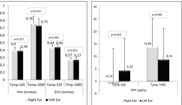
peak compensated admittance (Ytm) and ear canal volume (ECV) (Figure 2). The paired Student’s t-test was used to analyze these measurements. Although the TEOAE response amplitude was higher in the right ears than in the left ears, the differences were
not signiicant (Figure 1). There was a general trend
for the right ear response to be greater than the left ear response. The differences observed for the
tympanometry parameters were also not signiicant
(Figure 2)
70% was also analyzed using the Kappa index. The signiicance level adopted was 0.05.
RESULTS
The TEOAE responses of the right and left ears were compared (Figure 1), as were the results of 226- and 1000-Hz tympanometry regarding the variables Tympanometric Peak Pressure (TPP),
Legend: CI = conidence interval
was a difference between the types of probe used. The 226-Hz probe produced a Double Peak (DP) curve in 93.6% of cases, whereas the 1000-Hz probe produced a type A curve in 94.9%of cases (Figure 3).
The test of Equality of Two Proportions was used to statistically compare the tympanogram curve type in the two studied frequencies (226 and 1000 Hz) and in the left and right ears. The results showed no difference regarding the ear, but there
-0,74
13,95
4,33
8,74
-5 0 5 10 15 20 25 30
Tymp 226 Tymp 1000
TPP (daPa)
Right Ear Left Ear
0,4
0,76
0,44
0,27 0,39
0,73
0,44
0,27
0 0,1 0,2 0,3 0,4 0,5 0,6 0,7 0,8 0,9 1
Tymp 226 Tymp 1000 Tymp 226 Timp 1000 Ytm (mmho) ECV (mmho)
Right Ear Feft Ear
p=0.757
p=0.510
p=0.560
p=0.811
p=0.563
p=0.466
The left panel shows comparison for the variables Ytm and ECV, and the right panel shows the results for TPP.
Legend: CI = Conidence interval; TPP = tympanometric peak pressure; Ytm = admittance peak; ECV = ear canal volume
Figure 2- Comparison of tympanometry responses between right and left ears (mean and CI).
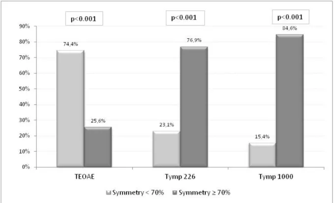
The test for the Equality of Two Proportions was
used to compare the percentages of symmetry ≥
70% with the values below 70%. The three
statis-tical tests with p-value < 0.001 showed signiicant
differences, with tympanometry always showing a
higher percentage of symmetry ≥ 70%, whereas
TEOAE showed a higher occurrence of symmetry < 70%. The p-value of the comparison among the three tests was < 0.001.
Tympanometry showed a high occurrence of
symmetry (≥ 70%) between the ears at both 226 Hz
and 1000 Hz. The TEOAE results, however, showed a higher occurrence of < 70% symmetry (Figure 5). The degree of agreement regarding the occurrence of symmetry between the tests was obtained using
the Kappa index. The statistical analysis showed
no correlation between these procedures, with the
index for the 226 Hz probe being 2.5% (p-value 0.789) and the index for the 1000 Hz probe being
-11.2% (p-value 0.137).
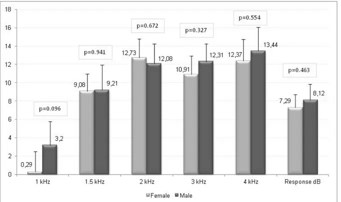
The same measurements discussed above (TEOAE as well as TPP, Ytm and ECV for 226- and 1000-Hz tympanometry) were also compared with respect to gender using the ANOVA test.
The differences found in the comparison between genders for both the TEOAEs (Figure 4) and the
tympanometry parameters were not signiicant
(Table 1).
Figure 4 - Comparison of transient-evoked otoacoustic emissions according to gender
Table 1 – Difference between genders in the evaluation of transient-evoked otoacoustic emissions and tympanometry according to ANOVA statistical test
Test p-value
Tympanometry
226 Hz
TPP 0.841
Ytm 0.962
ECV 0.458
1000 Hz
TPP 0.920
Ytm 0.603
Regarding the tympanometric indings in this
study, namely, the TPP, Ytm and ECV measure-ments for 226- and 1000-Hz tympanometry, no
statistically signiicant difference was observed
between ears or genders. Tympanometry revealed
a pattern of symmetry (≥ 70%) between the ears, which agrees with the indings described in the
literature21. Regarding the type of tympanogram,
we found a high incidence of DP curve at 226 Hz, representing 93.6% of responses, while at 1000 Hz, this type of curve was observed in only 5.1% of ears tested. This high percentage of DP curves at 226 Hz and of type A curves at 1000 Hz is in agreement with
the indings in the literature1. In a previous study
that analyzed tympanometric measurements using 226-Hz probes, the authors found an equivalent number of single-peak curves - which are similar to the type A curve described in this study - and DP curves at 226 Hz, while at 1000 Hz, they found a higher incidence of single-peak curves. Regarding
TPP, Ytm and ECV, a signiicant effect was observed
on the ECV measurement for the 226-Hz probe tone and on the Ytm measurements at 1000 Hz, both of which were higher for males. In another study analyzing data from 1000-Hz tympanometry in neonates, the authors found type A curves in 92.2% of cases and DP curves in 1.2% of cases as well as statistically greater Ytm measurements for the right ear22. The data observed in these two studies
DISCUSSION
This study aimed to compare data obtained from the analysis of tympanometry and TEOAE responses for the variables gender and ear.
Initially, the TEOAE response levels from each ear were compared. Analysis of the symmetry pattern revealed a predominance of symmetry < 70% between the ears. There was a general trend for the right ear response to be greater. This
“advantage” relative to the left ear has also been
observed in other studies7,20. It has been argued
that the difference in TEOAE records between the ears is associated with the right ear having a slight advantage in aural sensitivity. This advantage could be a factor favoring left hemispheric dominance for language7, suggesting that the cochlear portion of
the peripheral auditory system may also play an important role in the right ear advantage that is observed when evaluating the auditory processing of speech sounds.
Regarding the comparison of TEOAE responses between genders, the difference was also not
signiicant. One previous studyfound higher levels of responses in females5. The present study had
a small number of participants, which may have
inluenced the results. Studies with small numbers of participants usually do not show a signiicant
difference between genders.
normal middle ear conditions, there is no inluence
of possible differences in tympanometry that could
explain the differences in TEOAE records. The
difference in TEOAE responses between the ears does not occur due to anatomical and physiological differences between the right and left middle ears but due to differences between the right and left cochlea.
CONCLUSION
Signiicant symmetry was observed only in
tympanometry regardless of the probe used, suggesting that there is symmetry in the more peripheral portion of the auditory system and that asymmetries begin to occur from the cochlea. only agree with the present research in regard to the
higher incidence of type A curves found at 1000 Hz. DP curves usually occur at the resonance frequency of the middle ear9. It is known that the resonance
frequency in newborns is often lower than that in adults. Previous studies described resonance frequencies of 200 Hz in neonates, 380 Hz at three months and 1000 Hz in adults. The occurrence of DPs may be related to the resonance frequency of
approximately 200 Hz in neonates23.
Symmetry in tympanometry was observed
based on the agreement between symmetry ≥ 70%
in the 226- and 1000-Hz tympanograms and the TEOAE responses in neonates. However, there was a higher occurrence of symmetry < 70% in
the TEOAEs. These indings demonstrate that in
REFERENCES
1. Silva KAL, Novaes BACC, Lewis DR, Carvallo RMM. Achados timpanométricos em neonatos com emissões otoacústicas presentes: medidas e interpretações. Rev. Bras. Otorrinolaringol. 2007;73(5):633-9.
2. Russo ICP & Santos TMM. – Audiologia infantil. SP, Ed. Cortez,1994. 231p.
3. Volkweis MD. Avaliação auditiva no primeiro ano
de vida. [Monograia]. Porto Alegre (RS): Centro de
Especialização em Fonoaudiologia Clínica; 1999.
4. Pereira PKS, Martins AS, Vieira MR, Azevedo MF. Programa de triagem auditiva neonatal: associação entre perda auditiva e fatores de risco. Pró-Fono R Atual. Cient. 2007;19(3):267-78.
5. Prieve BA, Calandruccio L, Fitzgerald T, Mazevski A, Georgantas LM. Changes in transient-evoked otoacoustic emission levels with negative tympanometric peak pressure in infants and toddlers. Ear Hear. 2008;29(4):533-42.
6. Silveira JR, Durante AS, Almeida K, Taguchi CK, Greco MC. Emissões otoacústicas em lactentes
expostos a infecção intra-útero. Rev. Soc. imp.
Fonoaudiol. 2010;15( 2 ):184-90.
RESUMO
Objetivo: veriicar a ocorrência de simetria maior ou igual à 70%, entre as orelhas, comparando os resultados da timpanometria nas frequências de 226 e 1000Hz com as respostas das emissões
otoacústicas em neonatos. Métodos: foram avaliados 39 neonatos, em média com 60 horas de vida,
sendo 20 do sexo feminino e 19 do sexo masculino. Cada recém-nascido foi submetido à avaliação
timpanométrica com as sondas de 226 e 1000 Hz, e avaliação das emissões otoacústicas transientes em ambas as orelhas. Os resultados foram submetidos a testes estatísticos. Resultados: na análise
da amostra pode-se observar ocorrência de simetria <70% nas respostas das emissões otoacústicas em 74,4% do total de neonatos. Por outro lado, na timpanometria, houve uma maior ocorrência de simetria ≥ 70%, tanto para sonda de 226 quanto para 1000Hz (76,9% e 84,6%, respectivamente). No que diz respeito ao gênero e orelha, as diferenças encontradas em cada teste não foram signiican -tes, embora tenha sido observada maior amplitude de respostas de emissões otoacústicas na orelha direita. Conclusão: os resultados sugerem haver simetria no sistema auditivo em sua porção mais periférica, e início de assimetrias a partir da cóclea.
audiometry on infant hearing screening test with otoacoustic emissions. Acta Otorrinolaringol Esp. 2001;52(2):96-100.
16. Carvallo RMM. Imitância Acústica. In: Renata Mota Mamede Carvallo. (Org.). Fonoaudiologia Informação para a Formação. Rio de Janeiro: Guanabara Koogan, 2003, p. 1-22.
17. Couto CM, Carvallo RMM. O efeito das orelhas
externas e médias nas emissões otoacústicas. Rev.
Bras. Otorrinolaringol. 2009;75(1):15-23.
18. Khalfa S, Collet L. Functional asymmetry of medial olivocochlear system in humans. Towards a peripheral auditory lateralization. NeuroReport. 1996;7:993-6.
19. Joint Committee on Infant Hearing. Year 2007 Position Statement: Principles and Guidelines for Early Hearing Detection and Intervention Programs. Pediatrics. 2007;106(4):898-921.
20. Bassetto MCA, Chiari BM, Azevedo MF. Emissões otoacústicas evocadas transientes (EOAET): amplitude da resposta em recém-nascidos a termo e pré-termo. Rev Bras Otorrinolaringol. 2003;69(1):84-92.
21. Tazinazzio TG, Diniz TA, Marba STM, Colella-Santos MF. Emissões otoacústicas e medidas de imitância acústica com tons de sonda de 226 e 1000 hz em lactentes. Rev CEFAC. 2011;13(3):479-88. 22. Kei J, Allison-Levick J, Dockray J, Harrys R, Kirkegard C, Wong J et al. High-frequency (1000Hz) timpanometry in normal neonates. J Am Acad Audiol 2003;14:20-8.
23. André KD, Sanches SGG, Carvallo RMM. Ressonância da orelha média em lactentes: efeito da idade. Int. Arch. Otorhinolaryngol. 2012;16(3):353-7. 7. Durante AS, Carvallo RMM, Costa FS, Soares
JC. Características das emissões otoacústicas por transientes em programa de triagem auditiva neonatal. Pró-Fono R Atual. Cient. 2005;17(2):133-40.
8. Castro RM. Avaliação audiológica infantil 0 a 1
ano de idade. [Monograia]. Goiânia (GO): Centro
de Especialização em Fonoaudiologia Clínica; 1999.
9. Carvallo RMM. Emissões Otoacústicas: Conceitos Básicos e Aplicações. In: Renata Mota Mamede Carvallo. (Org.). Fonoaudiologia Informação para a Formação: Procedimentos em Audiologia. 1ª i. Rio de Janeiro: Guanabara Koogan, 2003, p. 22-41. 10. Fuzetti CB. Emissões otoacústicas espontâneas e evocadas por estímulo transiente em
recén-nascidos. [Dissertação]. São Paulo (SP): Pontifícia
Universidade Católica; 2002.
11. Pinto VS, Lewis DR. Emissões otoacústicas: produto de distorção em lactentes até dois meses de idade. Pró-Fono R Atual. Cient. 2007;19(2):195-204. 12. Linares AE, Carvallo RMM. Medidas
imitanciométricas em crianças com ausência de
emissões otoacústicas. Rev. Bras. Otorrinolaringol 2008;74(3):410-6.
13. Durante AS, Carvallo RMM. Mudanças das emissões otoacústicas por transientes na supressão contralateral em lactentes. Pró-Fono R Atual. Cient. 2006;18(1):49-56.
14. Carvallo RMM, Ravagnani MP, Sanches
SGG. Inluência dos padrões timpanométricos na
captação de emissões otoacústicas. Acta AWHO. 2000;19(1):18-25.
15. Mata J, Rando I, Shepherd M, Miguélez J, Jiménez F, Delgado F. Importance of impedance
Received on: November 07, 2013 Accepted on: February 21, 2014
Mailing address:
Renata Mota Mamede de Carvallo. R. Cipotânea, 51, Cidade Universitária São Paulo – SP – Brasil
CEP: 05360-160