ARTIGO ORIGINAL
125
SUMMARY
Objective: Methylprednisolone pulses are used in a variety of disease conditions, both for acute and chronic therapy. Although well tolerated, they increase glucose levels in both non-diabetic and diabetic patients. hey may also be considered a signiicant risk for acute metabolic alterations. he purpose of this report is to determine the metabolic changes in blood glucose levels in non-diabetic patients receiving methylprednisolone pulses and identify the presence of predictive factors for its development. Methods:
Observational, prospective study in 50 non-diabetic patients receiving 1 g intravenous methylpredniso-lone pulses for three consecutive days as an indication for diverse autoimmune disorders. Demographic, anthropometric, and metabolic variables were analyzed, and glucose, insulin and C-peptide levels ater each steroid pulse were identiied. Diferent variables and the magnitude of hyperglycemia were analyzed using Pearson’s correlation. Results: 50 patients were included, predominantly women (66%, n = 33). he average age was 41 ± 14 years with a BMI of 26 ± 3 kg/m2. Baseline glucose was 83 ± 10 mg/dL. Ater each steroid pulse, glucose increased to 140 ± 28, 160 ± 38 and 183 ± 44, respectively (p < 0.001). C-peptide and insulin concentrations increased signiicantly (p < 0.001). he prevalence of fasting hyper-glycemia ater each pulse was 68%, 94% and 98%, respectively. We found no correlation between the mag-nitude of hyperglycemia and the studied variables. Conclusion: Methylprednisolone pulses produced signiicant increases in fasting glucose in most patients without diabetes. Further studies are needed to deine its role in long-term consequences.
Keywords: Methylprednisolone; diabetesmellitus; hyperglycemia.
©2012 Elsevier Editora Ltda. All rights reserved.
RESUMO
Distúrbios de glicose em pacientes não diabéticos que recebem tratamento agudo com pulsos de metilprednisolona
Objetivo: Pulsos de metilprednisolona são usados em diversas doenças, tanto para tratamento agudo quanto crônico. Embora bem tolerados, eles aumentam os níveis de glicose em ambos os pacientes, não diabéticos e diabéticos. Eles também podem ser considerados um risco signiicativo para alterações me-tabólicas agudas. O propósito deste estudo é determinar as alterações meme-tabólicas nos níveis de glicose no sangue de pacientes não diabéticos que recebem pulsos de metilprednisolona e identiicar a presença de fatores preditivos para seu desenvolvimento. Métodos: Estudo observacional prospectivo em 50 pacien-tes não diabéticos que recebem pulsoterapia com 1 g de metilprednisolona intravenosa por três dias con-secutivos como tratamento para diversas doenças auto imunes. Variáveis demográicas, antropométricas e metabólicas foram analisadas, e glicose, insulina e níveis de peptídeo C foram identiicados após cada pul-so de esteroide. Diferentes variáveis e a magnitude da hiperglicemia foram analisadas utilizando a corre-lação de Pearson. Resultados: 50 pacientes foram incluídos, predominantemente mulheres (66%, n = 33). A idade média foi de 41 ± 14 anos com um IMC de 26 ± 3 kg/m2. A glicose de base foi de 83 ± 10 mg/dL. Após cada pulso de esteroide, a glicose aumentou para 140 ± 28, 160 ± 38 e 183 ± 44, respectivamente (p < 0,001). Peptídeo C e concentrações de insulina aumentaram signiicativamente (p < 0,001). A pre-valência de hiperglicemia em jejum após cada pulso foi de 68%, 94% e 98%, respectivamente. Não en-contramos nenhuma correlação entre a magnitude da hiperglicemia e as variáveis estudadas. Conclusão:
Os pulsos de metilprednisolona produziram aumentos signiicativos na glicemia de jejum na maioria dos pacientes sem diabetes. Mais estudos são necessários para deinir o seu papel nas consequências em longo prazo.
Unitermos: Metilprednisolona; diabetes mellitus; hiperglicemia.
©2012 Elsevier Editora Ltda. Todos os direitos reservados.
Study conducted at Subdivision of Investigation, College of Medicine and Hospital Universitario “Dr. José Eleuterio González”, UANL, Unidad Médica de Altas Especialidades (UMAE) No. 25, Instituto Mexicano Del Seguro Social, Universidad Juárez del Estado de Durango, Mexico
Submitted on: 06/23/2011
Approved on: 11/01/2011
Correspondence to: Hector Eloy Tamez Perez Dr. Aguirre Pequeño - s/n 64460 Monterrey, Mexico [email protected]
Conlict of interest:None.
Glucose disturbances in non-diabetic patients receiving acute
treatment with methylprednisolone pulses
HECTOR ELOY TAMEZ PEREZ1, MARÍA DOLORES GÓMEZDE OSSIO2, DANIA LIZET QUINTANILLA FLORES3, MAYRA IVONNE HERNÁNDEZ CORIA4, ALEJANDRA LORENA TAMEZ PEÑA5, GISSÉN JAZMÍN CUZ PÉREZ2, STEPHANIE LISSETTE PROSKAUER PEÑA3
1 MD, Endocrinology Service, Subdivision of Investigation, College of Medicine, Universidad Autónoma de Nuevo León (UANL), Monterrey, NL, Mexico 2 MD, Division of Internal Medicine, Hospital de Especialidades n° 25, Instituto Mexicano Del Seguro Social (IMSS), Monterrey, Mexico
HECTOR ELOY TAMEZ PEREZETAL.
126 Rev Assoc Med Bras 2012; 58(1):125-128
INTRODUCTION
Glucocorticoids are compounds that have long been used for a variety of diseases, both as chronic therapy and in acute cases. Methylprednisolone pulses are recommend-ed for critical events that require urgent treatment for an exacerbation of a known disease or when vital organs are compromised1.
Although generally well tolerated, they are not free of complications such as glucose intolerance, urinary tract infections, gastritis, luid retention, nausea, vomiting, in-somnia, altered consciousness, joint efusion, abnormal taste, hypertension and electrolyte disturbances such as hypokalemia2.
Glucocorticoids can worsen known diabetes and pre-cipitate previously unidentiied diabetes3. In all cases, it
transiently increases up to 50% baseline glucose levels4. An
OR of 1.36 to 2.31 of de novo diabetes has been reported in patients treated with steroids with an incidence of 12%5.
Steroids can also trigger a severe hyperosmolar hypergly-cemic decompensation and, in rare cases, death, especially in patients with preexisting diabetes6.
he aim of this paper is to present the changes in blood glucose levels in non-diabetic Mexican patients undergo-ing methylprednisolone pulses, and identify factors that correlate with the development post-bolus hyperglycemia.
METHODS
We performed an observational, longitudinal, prospective study in the Medical Specialties “Hospital No. 25 IMSS” in Monterrey, Nuevo Leon, from September to November 2010. We included 50 patients referred by specialists from the neurology, rheumatology and hematology services and hospitalized in the internal medicine department with the therapeutic indication of 1 g pulses of methylprednisolone for 3 consecutive days. Inclusion criteria were: individuals of both sexes, Mexican, aged 18 years or more and with no previous history of DM. he study was approved by the institutional ethics committee.
Demographic, anthropometric and metabolic variables were analyzed. Glucose measurements were performed at baseline and ater each 1 g pulse of intravenous methyl-prednisolone. We also determined insulin and C-peptide levels ater each pulse. All examinations were performed ater 8 hours of fasting. Glucose levels were determined by the glucose oxidase method, insulin by electrochemilu-minescence, and C-peptide levels by chemiluminescence.
he statistical analysis of all data was performed using SPSS version 19. he prevalence of post-bolus hypergly-cemia was calculated. Continuous variables are expressed as measures of central tendency and dispersion, and com-parisons were made using Student’s t-test. Correlations
be-tween quantitative variables (age, BMI, fasting glucose and post-bolus glucose levels) were made with Pearson’s cor-relation coeicient. A p ≤ 0.05 was considered signiicant.
RESULTS
We included 50 patients with no previous diagnosis of dia-betes mellitus (DM)5. Gender distribution was 33 women
(66%) and 17 men (34%). Average age was 41 ± 14 years with a BMI of 26 ± 3 kg/m2. Overweight was found in 22
patients (44%) and obesity in 11 (22%). A family history of DM was present in 42%.
he main diagnoses for patients referred by the attend-ing physician (blinded to the objectives of the study), were: systemic lupus erythematosus (22%), idiopathic thrombo-cytopenic purpura (24%), multiple sclerosis (22%), auto-immune hemolytic anemia (8%) and others (18%).
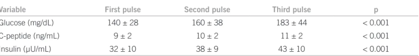
he baseline glucose level was 83 ± 10 mg/dL. Ater each pulse of methylprednisolone, glucose levels increased to 140 ± 28 mg/dL, 160 ± 38 mg/dL and 183 ± 44 mg/dL, respectively (p < 0.001). C-peptide and insulin concen-trations also showed statistically signiicant increases (p < 0.001) (Table 1).
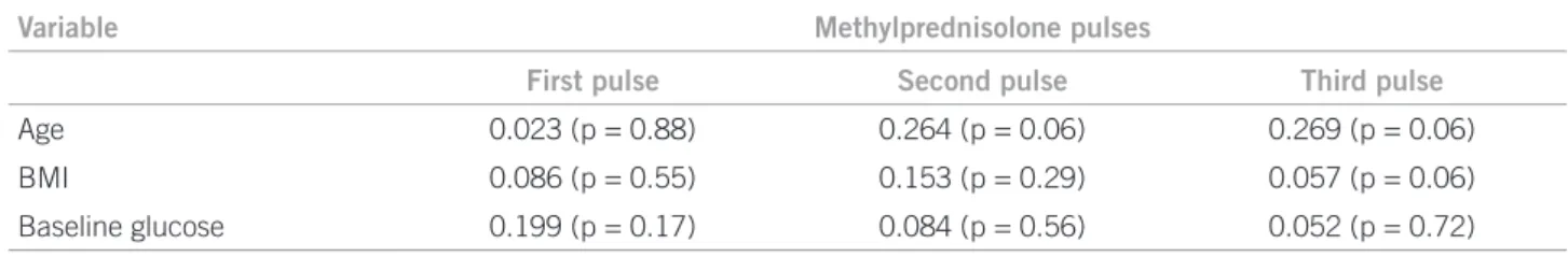
he prevalence of post-bolus hyperglycemia (glucose ≥ 126 mg/dL) was: 68% ater the irst pulse, 94% ater the second, and 98% ater the third pulse. When performing Pearson’s correlation we found no predictive factor with statistical signiicance for the development of post-bolus hyperglycemia (Table 2).
DISCUSSION
Pulsed methylprednisolone is highly efective in autoim-mune diseases due to its anti-inlammatory and immuno-suppressive efect. It reduces pain and active disease and provides acute symptomatic relief, while the beneicial efect of other drugs occurs1.he symptomatic beneit
of-fered by steroid pulses causes them to be frequently used despite their adverse efects. Our group recently published the changes in serum electrolytes in a cohort of patients. Without being the objective of that study, we identi-ied a change in glucose levels7, which had already been
observed in speciic groups of patients in the absence of
Table 1 – Metabolic changes after methylprednisolone pulses
Variable First pulse Second pulse Third pulse p
Glucose (mg/dL) 140 ± 28 160 ± 38 183 ± 44 < 0.001
C-peptide (ng/mL) 9 ± 2 10 ± 2 11 ± 2 < 0.001
Insulin (µU/mL) 32 ± 10 38 ± 9 43 ± 10 < 0.001
GLUCOSEDISTURBANCESINNON-DIABETICPATIENTSRECEIVINGACUTETREATMENTWITHMETHYLPREDNISOLONEPULSES
127
Rev Assoc Med Bras 2012; 58(1):125-128
diabetes8,9. Mignogna et al. reported that this represents
the most common complication10, and Feldman-Billard et
al. identiied increases of up to 50% compared to baseline levels prior to treatment4.
In our study, we found a signiicant increase in post-bolus glucose levels, similar to what has previously been reported for non-diabetic patients. his increase was more evident ater the irst pulse with an elevation of about 40 mg/dL, an increment up to 68% with respect to the bas-al level. At the end of the third pulse, 98% of the patients developed diagnostic criteria for diabetes mellitus, which could be explained by a loss of pancreatic islet adaptive phenomenon due to an acute and supra-physiological ste-roid load. In this phenomenon secretion disorders, insulin resistance and counterregulatory hormones are involved, together with alterations in the secretion and action of incretins11. he only patient that did not develop
diabe-tesmellitus criteria presented baseline glucose level below
54 mg/dL, however the metabolic changes were similar in proportion to the total group.
Although the increase is transitory and some authors feel that it does not have clinical relevance, there is evi-dence that identiies acute hyperglycemia as a cardiovas-cular risk factor, independently of the presence of previous diabetes. It has been associated with an increase in LDL cholesterol oxidation, impaired endothelial function, ac-tivation of the coagulation cascade, increased production of pro-inlammatory cytokines and oxidative stress3,5,12-14.
he magnitude of the hyperglycemic response has been previously associated with age, time and steroid dose, obe-sity, and in patients with type 2 diabetes mellitus, to poor
glycemic control5,15. In contrast with previous reports, our
work shows no link between the diferent variables stud-ied. his diference could be explained by genetic and en-vironmental factors related to the high prevalence of dia-betes and insulin resistance in our country, conirmed in our work by the signiicant increases in insulin levels.
As an observational non randomized study, we can-not exclude factors that could inluence our results such as stress hyperglycemia as well as the inpatient condition. However, considering the fact that glycemic levels in-creased progressively ater each pulse, we highly suggest that these modiications were due to a cause-efect phe-nomenon.
Some authors consider it unnecessary to monitor glu-cose levels in non-diabetic patients because these chang-es are transient and well tolerated4. We do not know the
long-term evolution of this expression in our population. We suggest monitoring blood glucose levels in all non-diabetic patients scheduled for methylprednisolone pulse therapy, because it is a simple, safe, and inexpensive pro-cedure. Additionally, we do not know whether these incre-ments in glucose levels, although transient, could predict future diabetes as well as cardiovascular co-morbidities. It is important to recall that our results need to be conirmed in further observational and randomized studies.
CONCLUSION
In conclusion, we found that the glucose proile changes signiicantly ater the administration of high doses of methylprednisolone. BMI, age and baseline glucose levels in non-diabetic patients do not correlate with the magni-tude of hyperglycemia. his alteration requires long-term studies to identify the clinical signiicance of these indings in our population.
ACKNOWLEDGEMENTS
We thank Sergio Lozano-Rodriguez, MD, for his help in translating the manuscript.
REFERENCES
1. Roubenof R, Roubenof RA, Ward LM, Stevens MB. Catabolic efects of high-dose corticosteroids persist despite therapeutic beneit in rheumatoid arthritis. Am J Clin Nutr. 1990;52:1113-7.
2. McDonough AK, Curtis JR, Saag KG. he epidemiology of glucocorticoid-associated adverse events. Curr Opinion Rheumatol. 2008;20:131-7. 3. Saigi I, Pérez A. Manejo de la hiperglucemia inducida por esteroides. Rev
Clin Esp. 2010;210:397-403.
4. Feldman-Billard S, Kassaei R, Benrabah R, Lissak B, Heron E. Glucose toler-ance of high-dose intravenous methylprednisolone therapy in ophthalmol-ogy. J Fr Ophtalmol. 2004;27:160-1.
5. Clore JN, hurby-Hay L. Glucocorticoid-induced hyperglycemia. Endocr Pract. 2009;15:469-74.
6. F-Vázquez SM. Manejo de la hiperglucemia secundaria al tratamiento con corticoides. Av Diabetol. 2006;22:194-9.
7. Tamez-Perez HE, Cisneros-Perez V, Cedillo-Rodriguez JA, Diaz-De-Leon-Gonzalez E, Torres-Valenzuela M, Tamez-Pena AL, et al. Prevalence of hy-pokalemia in patients with methylprednisolone pulse therapy. Rev Invest Clin. 2009;61:194-7.
8. Pandit MK, Burke J, Gustafson AB, Minocha A, Peiris AN. Drug-induced disorders of glucose tolerance. Ann Intern Med. 1993;7:529-39.
9. Abdelmannan DA, Tahboub R, Genuth S, Ismail-Beigi F. Efect of dexa-methasone on oral glucose tolerance in healthy adults. Endocr Pract. 2010;15:770-7.
10. Mignogna MD, Muzio LO, Ruoppo E, Fedele S, Russo LO, Bussi E. High-dose intravenous “pulse” methylprednisolone in the treatment of severe oropharyngeal pemphigus: a pilot study. J Oral Pathol Med. 2002;31:339-44.
Variable Methylprednisolone pulses
First pulse Second pulse Third pulse
Age 0.023 (p = 0.88) 0.264 (p = 0.06) 0.269 (p = 0.06)
BMI 0.086 (p = 0.55) 0.153 (p = 0.29) 0.057 (p = 0.06)
Baseline glucose 0.199 (p = 0.17) 0.084 (p = 0.56) 0.052 (p = 0.72)
BMI, body mass index.
HECTOR ELOY TAMEZ PEREZETAL.
128 Rev Assoc Med Bras 2012; 58(1):125-128
11. Hansen KB, Vilsboll T, Bagger JI, Holst JJ, Knop FK. Reduced glucose toler-ance and insulin resisttoler-ance induced by steroid treatment, relative physical in-activity, and high-calorie diet impairs the incretin efect in healthy subjects. J Clin Endocrinol Metab. 2010;3309-17.
12. Peralta FG, Padin CA. Glucemia postprandial y variabilidad glucémica: nuevos objetivos para conseguir el control glucémico óptimo en los pacien-tes con diabepacien-tes tipo 2. Av Diabetol. 2009;25:419-21.
13. Yang Z, Laubach VE, French BA, Kron IL. Acute hyperglycemia enhances oxidative stress and exacerbates myocardial infarction by activating nicotin-amide adenine dinucleotide phosphate oxidase during reperfusion. J horac Cardiovasc Surg. 2009;137:723-9.
14. Lemkes BA, Hermanides J, Devries JH, Holleman F, Meijers JC, Hoekstra JB. Hyperglycemia: a prothrombotic factor? J hromb Haemost. 2010;8:1663-9. 15. Hans P, Vanthuyne A, Dewandre PY, Brichant JF, Bonhomme V. Blood