BONE-CONDUCTED BRAINSTEM AUDITORY EVOKED
RESPONSE: AN INTEGRATIVE REVIEW
Potencial evocado auditivo de tronco encefálico por condução óssea:
uma revisão integrativa
Nathália Raphaela Pessôa Vaz Curado(1), Lilian Ferreira Muniz(1), Ana Karollina da Silveira(1),
Adriana Ribeiro de Almeida e Silva(1), Silvana Maria Sobral Griz(1)
(1) Universidade Federal de Pernambuco, UFPE, Recife,
Brasil.
Funding source: Propesq
Conlict of interest: non-existent
between the stimulus and response and positioning of the electrodes. However, the most commonly used classiication is the relation to latency, in which these potentials are known as short, medium or long latency 3.
The Brainstem Auditory Evoked Potential (BAEP) is an objective, non-invasive, short latency exam that assesses the electrophysiological activity of an auditory system as far as the brainstem, in response to an acoustic stimulus characterized by a rapid, short-duration burst, eliciting bioelectric responses that result from the successive activation of the cochlea and nerve ibers of this pathway2,4.
The responses generated by these potentials are a series of seven waves generated at a number of anatomic sites by an external auditory stimulus: wave I, the portion distal to the brainstem of the auditory nerve; wave II, the portion proximal to the
INTRODUCTION
The most widely used and clinically recognized short latency potential is the Brainstem Auditory Evoked Potential (BAEP), owing to its reproduc
-ibility and well-deined generators. This potential is obtained between 0 and 10 milliseconds (ms) after presentation of the acoustic stimulus, and its presence or not enables assessment of the integrity of the auditory pathway to the brainstem 1,2. Auditory Evoked Potential (AEP) exams are classiied according to latency, anatomic origin, relationship
ABSTRACT
The aim of this study was to conduct a integrative review about the procedures used in the acquisition criteria of the exam Auditory Brainstem bone conduction purposes to aid in the diagnosis of hearing problems. Latin American and Caribbean Literature on Health Sciences (LILACS), Medical Literature Analysis and Retrieval System Online (MEDLINE) and Scientiic Electronic Library Online (SciELO): a search of the following databases was performed. We used the following keywords: AEPs, Electrophysiology and Bone Conduction, found via Descriptors in Health Sciences Headings (MeSH) . The results shown are for the 35 selected studies. Most studies have opted for the use of click stimuli, with air conduction transducers supra-aural headphones , as the TDH – 39 for stimulation by bone conduction vibrator Radioear B-71, with a pressure of 425+/- 25g . It was observed that the mastoid was positioned over the region where more bone vibrator. Most studies report using alternating polarity, with diverse presentation rate 57.7/s most used and 30-3000 Hz ilter with a window of 15 ms duration. To rate the stimulus most studies used 2048, and a total of 2 records stimuli. The Evoked Auditory Brainstem Response is an exam that has been researched for many years and much has been described in the literature on aspects of acquisition and analysis, and highlights the importance of their use in the neonatal population.
of malformation of the auricular pavilion and/or the
middle ear8. Even though bone-conducted BAEP
has been routinely used in clinical practice for years, there are few studies regarding standardization and the procedures used to obtain responses with tone-burst stimuli in newborns, especially when this stimulus is presented by bone conduction at frequencies of 1000 and 4000 Hz, making it dificult to classify the alteration 9-11.
Thus, the knowledge obtained in this review article is important due to the fact that it collected information on the procedures used in bone-conducted BAEP, showing the patterns observed in a newborn population.
METHODS
An integrative review of the literature was conducted in 6 phases: 1) creating the guiding question, 2) literature search, 3) collection of article data, 4) critical analysis of study variables, 5) discussion of results, and 6) presentation of the integrative review.
In order to achieve the objective of this review (conduct an integrative review of procedures used for the acquisition criteria of the bone-conducted Brainstem Auditory Evoked Potentials test, whose purpose is to help diagnose auditory problems), the following guiding question was created: how have the procedures used in bone-conducted BAEP been described in the literature?
Literature articles were surveyed between April and August 2013, using the following methodology: conduct a search in BIREME and PUBMED as well as in the databases of the Latin American and Caribbean Literature in Health Sciences (Lilacs), Medical Literature Analysis and Retrieval System Online (Medline) and Scientiic Electronic Library Online (SciELO).
To search for the articles, all possible combi
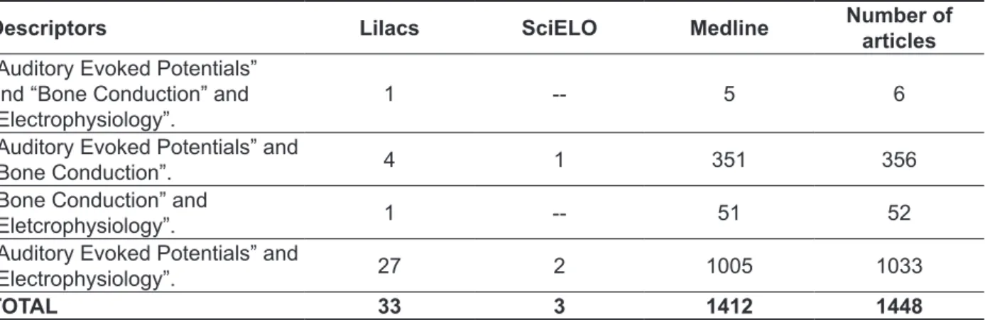
-nations between the controlled descriptors were used as follows: “Auditory Evoked Potential”, “Electrophysiology” and “Bone Conduction”, found by means of Health Sciences Descriptors (DeCS). The search results from combining descriptors (“Auditory Evoked Potential” and “Electrophysiology” and “Bone Conduction”, “Auditory Evoked Potential” and “Bone Conduction”, “Auditory Evoked Potential” and “Electrophysiology”, “Electrophysiology” and “Bone Conduction”), according to the data base, are shown in Table 1.
brainstem of the auditory nerve; wave III, cochlear nucleus; wave IV, superior olivary complex; wave V, lateral lemniscus; wave VI, inferior colliculus; and wave VII, medial geniculate body 1,4,5.
With respect to the manner of presenting the stimulus, this examination can be performed by air or bone conduction. However, although air conducted BAEP is the most widely used in clinical practice, when bone conduction is employed, there is an additional resource to help in audiologic diagnosis, characterizing hearing loss 4,6,7. In order to assess an individual that exhibits inconsistent or unreliable responses on pure-tone behavioral audiometry, it is recommended that BAEP be used by both air and bone conduction to obtain accurate electrophysi
-ological thresholds 4,6.
In relation to the type of stimulus used, the acoustic stimulus should activate numerous nerve ibers at the same time (synchronically) to capture electrical activity 3. Thus, brainstem electrical responses (BAEP) can be triggered by acoustic stimuli, such as clicks, tone pips, tone bursts or even speech, as long as presentation is transient. Clicks are the most commonly used since they are fast and exhibit a wide frequency range, allowing 5 stimulation of a larger amount of ibers. One of their disadvantages is that a wide frequency range precludes frequency selectivity, and the electric responses captured represent the region between 1000 and 4000 Hz. Responses with greater frequency selectivity are obtained on the BAEP test when acoustic stimuli such as tone bursts and tone
pips are used 1,3.
Tone burst acoustic stimulation allows obtaining relatively narrow frequency range responses, mainly at low frequencies. The use of tone-burst stimuli in BAEP is a reliable technique that is useful in clinical practice for estimating auditory sensi
-tivity at frequencies between 500 and 4000 Hz in children and adults. This is because electrophysi
-ological thresholds obtained with this stimulus are compatible with auditory thresholds for pure tones obtained in audiometry, despite being higher for a frequency of 500 Hz than for 4000 Hz1,3. Given that it assesses hearing threshold at lower frequencies, this type of stimulus, in conjunction with clicks, facilitates diagnosis of hearing loss with a ski-slope coniguration, thereby helping in the adaptation of individual sound application devices, especially in small children.
Table 1 – Articles found using the combination of descriptors, according to the database. Recife, 2014
Descriptors Lilacs SciELO Medline Number of
articles “Auditory Evoked Potentials”
and “Bone Conduction” and
“Electrophysiology”. 1 -- 5 6
“Auditory Evoked Potentials” and
“Bone Conduction”. 4 1 351 356
“Bone Conduction” and
“Eletcrophysiology”. 1 -- 51 52
“Auditory Evoked Potentials” and
“Electrophysiology”. 27 2 1005 1033
TOTAL 33 3 1412 1448
The search identiied 1448 publications, 33 in Lilacs, 3 in SciELO, and 1412 in Medline via BVS/
PubMed.
Articles that met the following criteria were included: those published in Portuguese, English and Spanish; those not within the required publication date range and articles on the bone-conducted BAEP procedure in newborns.
Literature articles were selected in three stages. The irst involved reading the titles of the articles. Those mentioning the bone-conducted BAEP test in the title were included. Articles that did not clearly comply with the inclusion criteria of this study proceeded to the second stage, which consisted of analyzing the abstracts. These were also included if the procedures described included bone-conducted
BAEP. Finally, the methodologies of articles that did
not mention bone-conducted BAEP in either their title or abstract were read to determine if they should
be included.
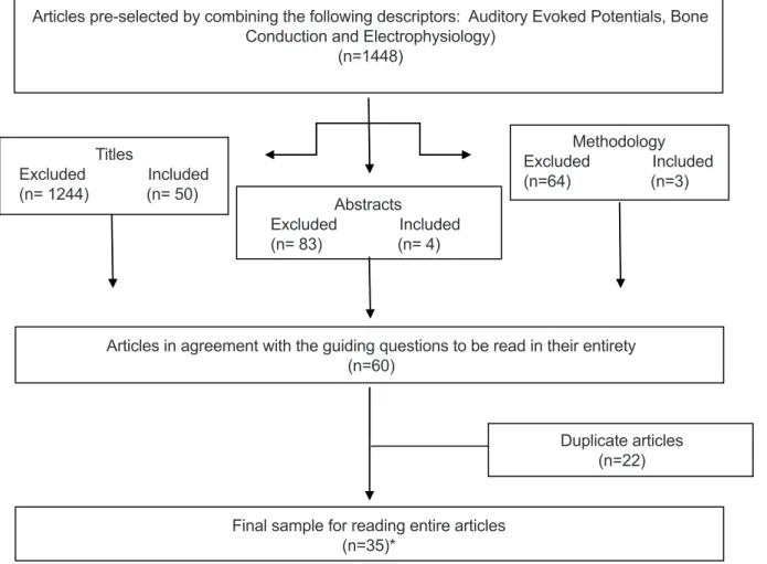
A total of 35 articles were selected (Figure 1). The reading of these articles focused on the following acquisition criteria: (a) transducer; (b) vibrator pressure; (c) vibrator position; (d) stimulus; (e) speed/frequency of the stimulus; (f) intensity of the stimulus; (g) polarity of the stimulus; (h) use of masking; (i) positioning of electrodes; (j) ilter; (k) window; (l) number of stimuli; (m) and number of reproductions.
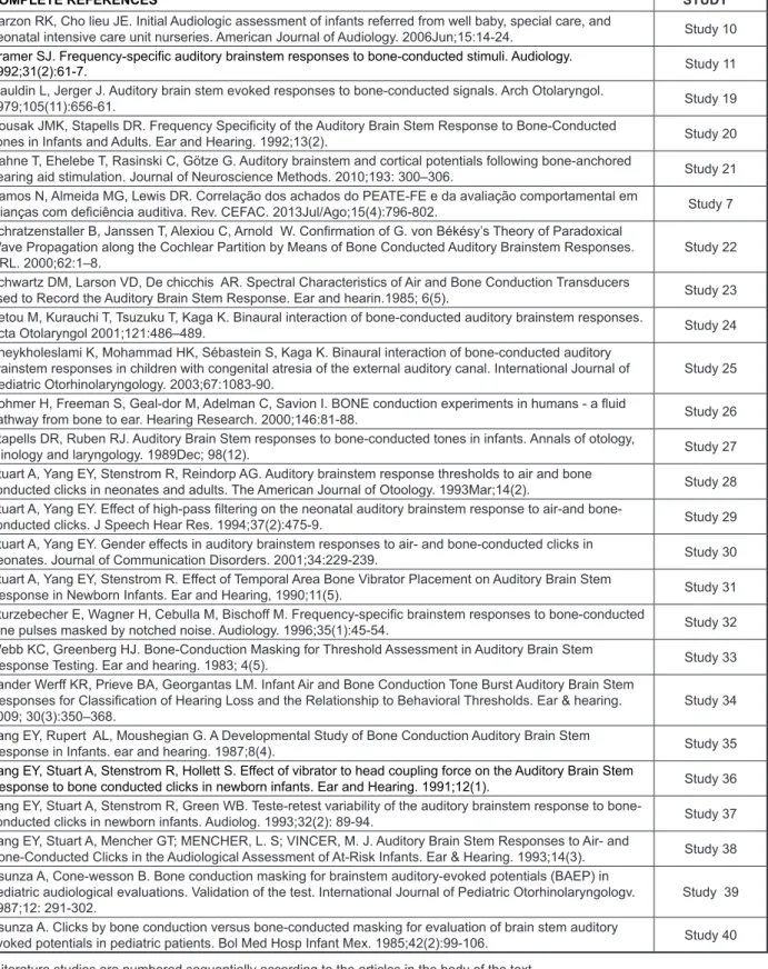
COMPLETE REFERENCES STUDY** Beattie RC. Normative Wave V Latency–Intensity Functions Using the EARTONE 3A Insert Earphone and the
Radioear B-71 Bone Vibrator. Scand Audiol1998;27:120–6 Study12
Boezeman EHJF, Kapteyn TS, Visser SL, Snel AM. Comparison Of The Latencies Between Bone And Air Conduction In The Auditory Brain Stem Evoked Potential. Electroencephalography and clinical Neurophysiologv,
Elsevier Scientiic Publishers Ireland, Ltd.1983;56:244-247. Study 13 Boezeman EHJF, Bronkhorstt AW, Kapteynb TS, Houffelaar A, Snel AM. Phase relationship between bone and
air conducted impulse signals in the human head. Acoustical Society of America. 1984July;76(1). Study 14 Cornacchia L, Martini A, Morra B. Air and bone conduction brain stem responses in adults and infants.
Audiology. 1983;22(5):430-7. Study 15
Fichino SN, Lewis DR, Fávero ML. Estudo dos limiares eletroisiológicos das vias aérea e óssea em crianças
com até 2 meses de idade. Rev. Bras. Otorrinolaringol. 2007Mar/Apr;73(2). Study 6 Fox JJ, Stapells DR. Normal infant and adult auditory brainstem responses to bone-conducted tones. Audiology.
1993;32:95-109. Study 16
Freitas VS, Alvarenga KF, Morettin M, Souza EF, Costa ilho OA. Bone conduction auditory brainstem responses in normal hearing individuals (original title: Potenciais evocados auditivos do tronco encefálico por condução
óssea em indivíduos normais). Pró-Fono Revista de Atualização Cientíica. 2006Set/Dez;18(3): 323-330. Study 17
Freitas VS, Alvarenga KF, Morettin M, Souza EF, Costa ilho OA. Potenciais Evocados Auditivos do Tronco Encefálico por condução óssea em crianças com malformação de orelha externa e/ou média. Distúrbios da
Comunicação. 2006Abr; 18(1):9-18. Study 8
Gorga MP, Kaminski JR, Beauchaine KL, Bergman BM. A Comparison of Auditory Brain Stem Response
Thresholds and latencies Elicited by Air- and Bone-Conducted Stimuli. Ear & Hearing. 1993;14(2). Estudo 9 Kaga K, Tanaka Y. Auditory air and bone conduction brainstem responses and damped rotation test for young
children with bilateral congenital atresia of the ears. International Journal of Pediatric Otorhinolaryngology.
1995;32:13-21. Study 18
Figure 1 – Sampling of the integrative review
Artigos pré-selecionados pelo cruzamento dos descritores: Potenciais Evocados Auditivos (Auditory Evoked Potentials), Condução Óssea (Bone Conduction)
e Eletrofisiologia (electrophysiology) (n=1448)
Artigos em consonância com a perguntadora condutora a serem lidos na íntegra (n=60)
Amostra final para leitura na íntegra dos artigos (n=35)*
Títulos
Excluídos Incluídos (n= 1244) (n= 50)
Metodologia Excluídos Incluídos (n=64) (n=3)
Artigos duplicados (n=22) Resumos
Excluídos Incluídos (n= 83) (n= 4)
Articles pre-selected by combining the following descriptors: Auditory Evoked Potentials, Bone Conduction and Electrophysiology)
(n=1448)
Titles
Excluded Included
(n= 1244) (n= 50)
Methodology Excluded Included
(n=64) (n=3)
Abstracts
Excluded Included (n= 83) (n= 4)
Duplicate articles
(n=22)
Final sample for reading entire articles
(n=35)*
Articles in agreement with the guiding questions to be read in their entirety
COMPLETE REFERENCES STUDY** Karzon RK, Cho lieu JE. Initial Audiologic assessment of infants referred from well baby, special care, and
neonatal intensive care unit nurseries. American Journal of Audiology. 2006Jun;15:14-24. Study 10 Kramer SJ. Frequency-speciic auditory brainstem responses to bone-conducted stimuli. Audiology.
1992;31(2):61-7. Study 11
Mauldin L, Jerger J. Auditory brain stem evoked responses to bone-conducted signals. Arch Otolaryngol.
1979;105(11):656-61. Study 19
Nousak JMK, Stapells DR. Frequency Speciicity of the Auditory Brain Stem Response to Bone-Conducted
Tones in Infants and Adults. Ear and Hearing. 1992;13(2). Study 20
Rahne T, Ehelebe T, Rasinski C, Götze G. Auditory brainstem and cortical potentials following bone-anchored
hearing aid stimulation. Journal of Neuroscience Methods. 2010;193: 300–306. Study 21 Ramos N, Almeida MG, Lewis DR. Correlação dos achados do PEATE-FE e da avaliação comportamental em
crianças com deiciência auditiva. Rev. CEFAC. 2013Jul/Ago;15(4):796-802. Study 7 Schratzenstaller B, Janssen T, Alexiou C, Arnold W. Conirmation of G. von Békésy’s Theory of Paradoxical
Wave Propagation along the Cochlear Partition by Means of Bone Conducted Auditory Brainstem Responses.
ORL. 2000;62:1–8. Study 22
Schwartz DM, Larson VD, De chicchis AR. Spectral Characteristics of Air and Bone Conduction Transducers
used to Record the Auditory Brain Stem Response. Ear and hearin.1985; 6(5). Study 23 Setou M, Kurauchi T, Tsuzuku T, Kaga K. Binaural interaction of bone-conducted auditory brainstem responses.
Acta Otolaryngol 2001;121:486–489. Study 24
Sheykholeslami K, Mohammad HK, Sébastein S, Kaga K. Binaural interaction of bone-conducted auditory brainstem responses in children with congenital atresia of the external auditory canal. International Journal of
Pediatric Otorhinolaryngology. 2003;67:1083-90. Study 25
Sohmer H, Freeman S, Geal-dor M, Adelman C, Savion I. BONE conduction experiments in humans - a luid
pathway from bone to ear. Hearing Research. 2000;146:81-88. Study 26 Stapells DR, Ruben RJ. Auditory Brain Stem responses to bone-conducted tones in infants. Annals of otology,
rhinology and laryngology. 1989Dec; 98(12). Study 27
Stuart A, Yang EY, Stenstrom R, Reindorp AG. Auditory brainstem response thresholds to air and bone
conducted clicks in neonates and adults. The American Journal of Otoology. 1993Mar;14(2). Study 28 Stuart A, Yang EY. Effect of high-pass iltering on the neonatal auditory brainstem response to air-and
bone-conducted clicks. J Speech Hear Res. 1994;37(2):475-9. Study 29
Stuart A, Yang EY. Gender effects in auditory brainstem responses to air- and bone-conducted clicks in
neonates. Journal of Communication Disorders. 2001;34:229-239. Study 30 Stuart A, Yang EY, Stenstrom R. Effect of Temporal Area Bone Vibrator Placement on Auditory Brain Stem
Response in Newborn Infants. Ear and Hearing, 1990;11(5). Study 31
Sturzebecher E, Wagner H, Cebulla M, Bischoff M. Frequency-speciic brainstem responses to bone-conducted
tone pulses masked by notched noise. Audiology. 1996;35(1):45-54. Study 32 Webb KC, Greenberg HJ. Bone-Conduction Masking for Threshold Assessment in Auditory Brain Stem
Response Testing. Ear and hearing. 1983; 4(5). Study 33
Vander Werff KR, Prieve BA, Georgantas LM. Infant Air and Bone Conduction Tone Burst Auditory Brain Stem Responses for Classiication of Hearing Loss and the Relationship to Behavioral Thresholds. Ear & hearing.
2009; 30(3):350–368. Study 34
Yang EY, Rupert AL, Moushegian G. A Developmental Study of Bone Conduction Auditory Brain Stem
Response in Infants. ear and hearing. 1987;8(4). Study 35
Yang EY, Stuart A, Stenstrom R, Hollett S. Effect of vibrator to head coupling force on the Auditory Brain Stem
Response to bone conducted clicks in newborn infants. Ear and Hearing. 1991;12(1). Study 36 Yang EY, Stuart A, Stenstrom R, Green WB. Teste-retest variability of the auditory brainstem response to
bone-conducted clicks in newborn infants. Audiolog. 1993;32(2): 89-94. Study 37 Yang EY, Stuart A, Mencher GT; MENCHER, L. S; VINCER, M. J. Auditory Brain Stem Responses to Air- and
Bone-Conducted Clicks in the Audiological Assessment of At-Risk Infants. Ear & Hearing. 1993;14(3). Study 38 Ysunza A, Cone-wesson B. Bone conduction masking for brainstem auditory-evoked potentials (BAEP) in
pediatric audiological evaluations. Validation of the test. International Journal of Pediatric Otorhinolaryngologv.
1987;12: 291-302. Study 39
Ysunza A. Clicks by bone conduction versus bone-conducted masking for evaluation of brain stem auditory
evoked potentials in pediatric patients. Bol Med Hosp Infant Mex. 1985;42(2):99-106. Study 40 **Literature studies are numbered sequentially according to the articles in the body of the text.
was diversiied, such as the Radioear B-70, B-71, B-70B, B-70A, B-72, among others. However, the most commonly used were B-716,7,9,12-14,23,26,33,34
B70-A 11,15,16,19,20,23,27,35,40. Diversiication was also
observed for the pressure used in the bone vibrators, such as: 225+/-25g36; 325+/-25g36; 350-450g16;
375-425g20; 400+/-25g6,7,34; 400 and 450g17; 408-612g14; 250 to 350g27; 612g12 and 525+/-25g36. However, the most frequently used bone pressure was 425+/-25g27-30,35-37.
The most widely used position for the bone vibrator was the temporal area, speciically on the mastoid 7, 11, 12, 18, 21, 23-25, 32,34,35.
LITERATURE REVIEW
The results presented refer to the 35 studies selected for this systematic review, which discuss aspects related to acquisition criteria of the BAEP test.
Table 2 shows the distribution of literary production according to the type of transducer used, the position of the bone vibrator and the pressure used. The most widely used transducers to capture the air-conducted stimulus were the supra-aural headphones, such as the TDH-39 8,13-15,18,19,33,35.
The transducer for the bone- conducted stimulus
Table 2 – Description of air-conducted and bone-conducted stimuli.
Air Conduction
Bone Conduction
Studies 6, 7, 8, 9, 10, 11, 12, 13, 14, 15, 16, 17, 18, 19, 20, 21, 22, 23, 24, 25, 26, 27, 28, 29, 30, 31, 33, 35, 36, 37 and 38, 39 and 40.
SUPRA INSERT PRESSURE POSITION
Studies 8, 13, 14, 15, 18, 19, 21, 22, 23, 33, 35, 39 and 40
Studies 6, 7, 9, 10, 12, 17, 28, 29, 30, 31, 34,
36 and 37.
FRONT TEMPORAL OCCIPITAL
Studies 6, 7, 12, 14, 16, 17, 20, 28, 29, 30, 31, 35, 36, 37 and
38.
Studies 14, 15,19, 22, 26,
33 and 39.
Studies 7, 11, 16, 18, 20, 21, 23, 24, 25, 26, 27, 28, 29, 30, 31, 32, 34, 35,
36 and 37.
Study 31
For the selected publications, Table 3 shows that the most frequently used electrode positions are Fz, Fpz, M1 and M2, followed by Cz, M1 and M2.
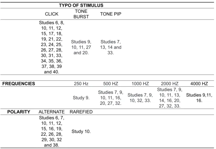
Analysis showed that most of the studies found used the click stimulus, followed by tone bursts at 500 and 2000 Hz or tone pips at frequencies of 1000, 2000 and 4000 Hz. Intensities ranged between 100 dB Nan and 10 dB Nan for the click stimulus, 70 dB Nan and dB Nan for the tone burst and 80 dB Nan and 10 dB Nan for the tone pip. Most studies used alternate polarity with different presentation rates, the most widely adopted being 57.7/s 26,29,30,31,36,38,
followed by 27.7/s 6,8,17 and 21.1/s 8,17,26.
Masking was used in a variety of ways. In studies using click stimuli it was 10 dB above the bone-conducted stimulus intensity12; -30 dB of test intensity 8; 10 dB above the bone-conducted click
intensity; and upper limit of 50 dB above normal hearing level22; applied only at intensity levels of
more than 35 dB Nan28; 40 dB Nan of noise between
20-20000 Hz35; ixed at 60 dB SPL39.
For studies that used the tone burst stimulus, masking of 80 dB of contralateral noise 9; 59 dB20; 60 dB SPL11 were employed, the last article also using the click stimulus.
Finally, for studies that employed the tone pip stimulus, the masking used was 70 dB SPL14 and 5 dB above the ABR value33.
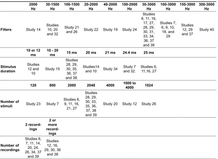
Table 4 shows the ilter, stimulus duration, number of stimuli and number of recordings. The literature review revealed that most studies used a 30-3000 Hz ilter, followed by a 100-3000 Hz ilter. The majority of studies used a 15ms window and a rate of 2048, followed by 2000.
diagnosis, primarily in characterizing hearing
loss 4,6,28,34.
Furthermore, performing the test only by bone conduction may result in a large number of false positives due to transitory pathologies of the middle
ear 33. A study conducted by Yang et al., in 1993 37 showed that bone-conducted BAEP tests exhibited high sensitivity and speciicity in detecting children with neurosensory hearing loss.
In relation to the transducer, 13 of the articles that reported using air and bone-conducted BAEP used The Brainstem Auditory Evoked Potential test
has been studied since the 1980s, and scientiic articles contain a number of aspects related to the procedures used, such as systems to record poten
-tials and the characteristics of the stimulus for these evoked potentials.
Analysis of the 35 articles included in the present study shows that 27 used air and bone conduction and seven employed bone conduction alone. Although the vast majority used air-conducted BAEP, bone conduction is a valuable ally in audiologic
Table 3 – Placement of surface electrodes used in the reviewed studies.
ELECTRODE PLACEMENT
Fz, Fpz, M1 and M2
Cz, Fpz, A1 and A2
Fz, A1 and A2
Cz, Fpz, M1 and M2
Cz, Fz, M1 and M2
Cz, A1 and A2
Cz, M1 and M2
Fz, M1 and M2 Studies 6,
7, 8, 14, 18, 21, 32 and
34
Study 15 Study 17 Studies 11, 19, 20 and 24
Study 25. Studies 26 and 40. Studies 26, 31, 33, 36, 39 and 40.
Studies 28, 29, 30, 36, 37 and 38.
A1 (left ear lobe); A2 (right ear lobe); Cz (vertex); Fpz (ground electrode); Fz (source); M2 (right mastoid); M1 (left mastoid).
Table 4 – Characteristics of stimuli to generate brainstem auditory evoked potentials used in the reviewed studies.
TYPO OF STIMULUS
CLICK BURSTTONE TONE PIP
Studies 6, 8, 10, 11, 12, 15, 17, 18, 19, 21, 22, 23, 24, 25, 26, 27, 28, 30, 31, 33, 34, 35, 36, 37, 38, 39
and 40.
Studies 9,
10, 11, 27 and 20.
Studies 7,
13, 14 and 33.
FREQUENCIES 250 Hz 500 HZ 1000 HZ 2000 HZ 4000 HZ
Study 9. Studies 7, 9, 10, 11, 16, 20, 27, 32.
Studies 7, 9,
10, 32, 33.
Studies 7, 9,
10, 11, 13, 14, 16, 20, 27, 32, 33.
Studies 9,11, 16.
POLARITY ALTERNATE RAREFIED
Studies 6, 7, 10, 11, 12, 15, 16, 19, 22, 26, 28, 29, 30, 32
and 38.
Table 5 – Brainstem auditory evoked potential recording system used in the reviewed studies. 2000 Hz 30-1500 Hz 100-1500 Hz 20-2000 Hz 40-2000 Hz 100-2000 Hz 30-3000 Hz 100-3000 Hz 150-3000 Hz 300-3000 Hz
Filters Study 14
Studies 10, 20 and 32
Study 21
and 26 Study 22 Study 19 Study 24
Studies 8, 11, 16,
17, 27, 28, 29, 30, 31, 33, 34, 36, 37 and 38 Studies 7, 6, 9, 10, 18, and 29 Studies 12, 29 and 37 Study 40
10 or 12 ms
10 - 20
ms 15 ms 20 ms 21 ms 24.4 ms 25 ms
Stimulus duration Studies 12 and 10 Study 15 Studies 28, 29, 30, 35, 36, 37 and 38. Studies14
and 10 Study 34
Study 7 and 32.
Studies 6, 11,16, 27
128 800 2000 2048 4000 1000 to
4000 1024
Number of
stimuli Study 23 Study 7
Studies 6, 9, 11, 16,
21, 27 Studies 28, 29, 30, 33, 35, 36, 37, 38 and 39
Study 20 Study 12 Study 26
2 record-ings 2 or more record-ings Number of recordings Studies 6, 7, 11, 14,
20, 24, 26, 34, 37
and 39
Studies 12, 16, 29, 30, 36
and 38
surface headphones (supra-aural) and 13 opted for insert earphones.
The function of the transducer is to transform the electrical stimulus into an acoustic stimulus, which, in turn, is transmitted through the auditory system to generate auditory evoked potential.
When this procedure was irst adopted supra-aural headphones were widely used, but as their results and correlation with other audiologic tests became known, there was a shift to insert earphones
30,39. The advantages of insert earphones compared
to conventional headphones include: (a) reduction in the error caused by the distance between the transducers and electrodes, (b) avoiding collapse of the external ear canal, (c) there is an increase in interaural attenuation, (d) environmental noise reduction of around 30 dB, (e) less need for contra
-lateral masking, and (f) greater patient comfort 12. In relation to bone vibrators, ten of the studies used Radioear B-71. The latency-intensity functions of bone-conducted BAEP using click stimulus and Radioear B-70A, B-7 1 and B-72 vibrators showed a 1/2 millisecond delay for V-wave latency with the
B-70A transducer, when compared with that of an electrodynamic earphone (TDH-39); however, more prolonged latencies are observed for B-71 and B-72 oscillators, with the latter producing the largest change in latency 23.
With respect to the pressure at which the vibrator should be set, six of the articles used a pressure of 425+/-25g. Yang et al. (1991)36 studied the effect of bone vibrator pressure/force of 225, 325, 425 and 525 g at intensities of 30 and 15 dB NA in 20 newborns. The V-wave latencies were affected by pressure variations of less than 200 g. The pressure applied on the vibrator affected response recordings, producing better responses at weak intensities with the effective vibrator placement, which occurred when the pressure was between 425 and 525 g. The authors suggested that pressure between 400 and 450 g be applied for bone-conducted BAEP in newborns.
V-wave latency differs signiicantly according to the polarity used, rarefaction polarity being the most widely used in clinical practice due to its greater diagnostic sensitivity when compared to conden
-sation polarity. In most individuals, rarefaction polarity generates potentials with smaller latencies and variability that does not exceed 0.1 to 0.2 milli
-seconds in normal hearers. The literature search showed that 15 of the studies performed their tests with alternate polarity. This type of polarity aims primarily at canceling the electrical errors at the onset of recording responses 23.
With respect to the stimulus presentation rate, it was found that ive studies used a rate of 57.7/s. This is the period of structure polarization. If stimulated there will either be no response or its threshold will be elevated. The velocity of stimulus presentation can change wave morphology, thereby inluencing wave latency and amplitude. Since high stimulation rates may accelerate the test, rates below 30 cycles/s are used to prevent the speed of the stimulus from interfering in the signal response. In relation to the masking used, it was observed that different forms were employed for click, tone burst and tone pip stimuli. There is no consensus in the literature as to the need for masking to avoid stimulating the contralateral ear, but it is known that a sound can be perceived by the contralateral ear through cranial vibration. In this case there will be interaural attenuation, which varies from individual to individual and from frequency to frequency. The use of contralateral masking is defended by Maldin and Jerger (1979) 19, and Ysunza and Cone-Wesson
(1987) 39. However, Kaga and Tanaka (1995) 18
reported that masking is not necessary in cases of bilateral occlusion of the external ear canal, due to similar malformation in both ears.
With respect to surface electrode placement, it is known that these are very important in capturing the BAEP. For this reason International Electrode System (IES) guidelines 10-20 must be complied with for their correct use. One of the most commonly used set ups deines the right and left mastoids (M2 and M1 respectively) or right and left ear lobes (A2 and A1 respectively) as reference electrodes (negative), the source (Fpz) as ground electrode and the source (Fz) or vertex (Cz) as active electrodes (positive). The literature search showed that all studies followed the aforementioned guidelines, with a preference for ixing the negative electrodes on the mastoids.
Evoked bioelectrical activity is captured by the surface electrode. For each electrode the evoked electrical activity uses at least three electrodes placed on the patient’s skin and connected to the equipment’s pre-ampliier.
Yang et al. (1987) 35 investigated bone-conducted BAEP in children and placed the bone vibrator in frontal, occipital and temporal areas. The results indicated that vibrator placement in temporal areas produces signiicantly shorter V-wave latencies than in frontal or occipital positions. For this reason, the authors recommended placing the bone-conducted BAEP vibrator on the temporal bone when the test was performed with newborns.
Stuart, Yang and Stenstrom (1990) 31 studied the effect of V-wave latency on newborns with bone vibrator placement in three positions in the temporal area. The results showed signiicant changes in V-wave latency, not only by changing the frontal, occipital and temporal position, but also by alterations around the temporal area. They suggest that the BAEP test be applied to newborns using the bone-conducted stimulus and that bone vibrator placement in the temporal region remains consistent.
The shorter latencies, better response quality and lower standard deviations in superior-posterior (B) and posterior (C) placement seems to make them preferable to superior placement (A). However, over the course of the investigation, it was observed that placing the bone vibrator in the posterior position (C) was more dificult in terms of attaching and maintaining the position of the bone vibrator. Therefore the superior-posterior (B) position is recommended for the bone-conducted BAEP test in newborns.
With respect to the characteristics of the stimuli to generate evoked potentials, most (27) of the studies reported using the click stimulus.
Although the BAEP can be triggered by a number of acoustic stimuli, the most widely used in the liter
-ature is the click, since it is a short-duration stimulus with that begins and ends abruptly (100us), ideal for producing short latency responses. However, at strong intensities they stimulate the cochlea as a whole and therefore without low-frequency speci
-icity, albeit with maximum synchrony of responses that appear and disappear in few milliseconds, allowing their visualization in the promediated
recording.
However, to assess some patients, especially children, it is necessary to obtain a response with slightly more frequency speciicity.
1000 and 4000 stimuli, while late latency potential require from 20 to 200 stimuli 21,33,38.
CONCLUSION
The BAEP is a test that has been studied for many years, and much has been written in the liter
-ature about its aspects of acquisition and analysis, in addition to the importance of its use in newborns. The aspects of acquisition found in the liter
-ature were: TDH-39 supra-aural transducers for air conduction and the Radioear B-71 for bone conduction, with a pressure of 425+/-25g most commonly used. The vibrator was more frequently placed in the temporal area on the mastoid. The most widely used stimulus was the click, followed by tone burst and tone pip, with intensities ranging between 100 dBNAn and 10 dBNAn.
Alternate polarity was most used, the most common presentation rate being 57.7/s, followed by 27.7/s and 21.1/s.
Masking varied considerably from study to study. Most studies used 30-3000Hz ilters, with a 15ms window, and 2048 stimuli, followed by 2000 stimuli.
With respect to the number of wave recordings, at least two recordings were made for each intensity. Finally, the most studied frequencies were 500, 1000 and 2000 Hz for tone burst and tone pip stimuli.
However, there is still a lack of studies on bone-conducted frequency-speciic BAEP, as well as frequencies of 1000 Hz and 4000 Hz and their patterns. It is hoped, therefore, that future research will use these parameters for clinical protocols and scientiic production worldwide.
The ilter used is another important aspect in recording potentials. The literature search revealed that 14 of the studies reported using a 30-3000 Hz ilter. The ilters of the ampliier must also be adapted to each test, since they remove from analysis electrical activities above and below determinate frequency limits, measured in Hz. A band-pass ilter, a combination of a high-pass and low-pass ilter, is used to determine potentials, creating a frequency range limit 29.
Another aspect found was the window used to perform the test, where seven studies opted for a 15ms window. It is known that the window corre
-sponding to the recording analysis time, that is, the period immediately after the onset of the stimulus in which electrical activities are captured by the electrodes. The time is measured in milliseconds and determined by the potential that is being studied. A larger window of analysis is needed in newborns and nursing infants, since the waves are recorded at a more delayed latency than those of adults.. When a speciic frequency stimulus is used, windows of 20-30 ms are recommended, since the increase in latency is greater at lower frequencies due to the time elapsed to reach the apice of the cochlea 11,16,32.
In regard to the number of stimuli, it was found that nine of the studies applied 2048 stimuli, followed by six with 2000 stimuli. Waves were recorded at least twice in nine of the articles.
RESUMO
O objetivo deste estudo foi de realizar uma revisão de forma integrativa sobre os procedimentos uti
-lizados nos critérios de aquisição do exame de Potenciais Evocados Auditivos de Tronco Encefálico por condução óssea com ins ao auxílio no diagnóstico de problemas auditivos. Foi realizada uma busca nas seguintes bases de dados: Literatura Latino-Americana e do Caribe em Ciências da Saúde (Lilacs), Medical Literature Analysis and Retrieval System Online (Medline) e Scientiic Eletronic Library Online (SciELO). Utilizaram-se as seguintes palavras-chave: Potencial Evocado Auditivo, Eletroisiologia e Condução Óssea, encontrados por meio de Descritores em Ciências da Saúde (DeCS). Os resultados apresentados são referentes aos 35 estudos selecionados. A maioria dos estu
-dos optou pelo uso do estímulo clique, com transdutores por condução aérea os fones supra-aurais, como o TDH-39, para o estímulo por condução óssea, o vibrador Radioear B-71, com pressão de 425+/-25g. Observou-se que a mastoide foi à região onde mais se posicionou mais o vibrador ósseo. A maioria dos estudos refere usar polaridade alternada, com taxa de apresentação diversiicada, sendo 57,7/s a mais utilizada e iltro de 30-3000 Hz, com uma janela de 15 ms de duração. Para taxa do estímulo a maioria dos estudos utilizou de 2048, e um total de estímulos de 2 registros. O Potencial Evocado Auditivo de Tronco Encefálico é um exame que vem sendo pesquisado há muitos anos e muito se tem descrito na literatura sobre seus aspectos de aquisição e analise, além de destacar a importância da sua utilização na população neonatal.
DESCRITORES: Potenciais Evocados Auditivos; Eletroisiologia; Condução Óssea; Recém-Nascido; Audição
REFERENCES
1. Pinto FR, Matas CG. Comparação entre limiares de audibilidade e eletroisiológico por estímulo tone burst. Rev. Bras. Otorrinolaringol.
2007;73(4):513-22.
2. Casali MF, Santos MFC. Auditory Brainstem Evoked Response: response patterns of fullterm and premature. Braz. J. Otorhinolaryngol. 2010;76(6):729-38.
3. Matas CG, Magliaro FCL. Introdução aos Potenciais Evocados Auditivos e Potenciais Evocados Auditivos de Tronco Encefálico. In: Bevilacqua MC, Martinez MAN, Balen SA, Pupo AC, Reis ACMB, Frota S. Tratado de udiologia. São Paulo: Livraria Santos Editora Ltda; 2011. P. 181-95.
4. Fernandes LCBC, Gil D, Maria SLS, Azevedo MF. Potencial evocado auditivo de tronco encefálico por via óssea em indivíduos com perda auditiva sensorioneural. Rev CEFAC. 2013;15(3):538-45. 5. Rocha CN, Filippini R, Moreira RR, Neves IF, Schochat E. Potencial evocado auditivo de tronco encefálico com estímulo de fala. Pró-Fono R. de Atual. Cient. 2010;22(4):479-84.
6. Fichino SN, Lewis DR, Fávero ML. Estudo dos limiares eletroisiológicos das vias aérea e óssea em crianças com até 2 meses de idade. Rev. Bras. Otorrinolaringol. 2007;73(2)251-6.
7. Ramos N, Almeida MG, Lewis DR. Correlação dos achados do PEATE-FE e da avaliação comportamental em crianças com deiciência auditiva. Rev CEFAC. 2013;15(5):796-802.
8. Freitas VS, Alvarenga KF, Morettin M, Souza EF, Costa ilho OA. Potenciais Evocados Auditivos do Tronco Encefálico por condução óssea em crianças com malformação de orelha externa e/ou média. Distúrbios da Comunicação. 2006;18(1):9-18. 9. Gorga MP, Kaminski JR, Beauchaine KL, Bergman BM. A Comparison of Auditory Brain Stem Response Thresholds and latencies Elicited by Air- and Bone-Conducted Stimuli. Ear & Hearing. 1993;14(2):85-94.
10. Karzon RK, Cho lieu JE. Initial Audiologic assessment of infants referred from well baby, special care, and neonatal intensive care unit nurseries. American Journal of Audiology. 2006;15:14-24. 11. Kramer SJ. Frequency-speciic auditory brainstem responses to bone-conducted stimuli. Audiology. 1992;31(2):61-71.
12. Beattie RC. Normative Wave V Latency–Intensity Functions Using the EARTONE 3A Insert Earphone and the Radioear B-71 Bone Vibrator. Scand Audiol 1998;27:120-6.
26. Sohmer H, Freeman S, Geal-dor M, Adelman C, Savion I. BONE conduction experiments in humans – a luid pathway from bone to ear. Hearing Research. 2000;146:81-8.
27. Stapells DR, Ruben RJ. Auditory Brain Stem responses to bone-conducted tones in infants. Annals of otology, rhinology and laryngology. 1989;98(12):941-9.
28. Stuart A, Yang EY, Stenstrom R, Reindorp AG. Auditory brainstem response thresholds to air and bone conducted clicks in neonates and adults. The American Journal of Otoology. 1993;14(2):176-82. 29. Stuart A, Yang EY. Effect of high-pass iltering on the neonatal auditory brainstem response to air-and bone-conducted clicks. J Speech Hear Res. 1994;37(2):475-9.
30. Stuart A, Yang EY. Gender effects in auditory brainstem responses to air- and bone-conducted clicks in neonates. Journal of Communication Disorders. 2001;34:229-39.
31. Stuart A, Yang EY, Stenstrom R. Effect of Temporal Area Bone Vibrator Placement on Auditory Brain Stem Response in Newborn Infants. Ear and Hearing, 1990;11(5):363-9.
32. Sturzebecher E, Wagner H, Cebulla M, Bischoff M. Frequency-speciic brainstem responses to bone-conducted tone pulses masked by notched noise. Audiology. 1996;35(1):45-54.
33. Webb KC, Greenberg HJ. Bone-Conduction Masking for Threshold Assessment in Auditory Brain Stem Response Testing. Ear and hearing. 1983;4(5):261-6.
34. Vander werff KR, Prieve BA, Georgantas LM. Infant Air and Bone Conduction Tone Burst Auditory Brain Stem Responses for Classiication of Hearing Loss and the Relationship to Behavioral Thresholds. Ear & hearing. 2009;30(3):350-68.
35. Yang EY, Rupert AL, Moushegian G. A Developmental Study of Bone Conduction Auditory Brain Stem Response in Infants. Ear and hearing. 1987;8(4):244-51.
36. Yang EY, Stuart A, Stenstrom R, Hollett S. Effect of vibrator to head coupling force on the Auditory Brain Stem Response to bone conducted clicks in newborn infants. Ear and Hearing. 1991;12(1):55-60. 37. Yang EY, Stuart A, Stenstrom R, Green WB. Teste-retest variability of the auditory brainstem response to bone-conducted clicks in newborn infants. Audiolog. 1993;32(2):89-94.
38. Yang EY, Stuart A, Mencher GT, Mencher LS, Vincer MJ. Auditory Brain Stem Responses to Air- and Bone-Conducted Clicks in the Audiological Assessment of At-Risk Infants. Ear & Hearing. 1993;14(3):175-82.
and clinical Neurophysiologv, Elsevier Scientiic Publishers Ireland, Ltd.1983;56:244-7.
14. Boezeman EHJF, Bronkhorstt AW, Kapteynb TS, Houffelaar A, Snel AM. Phase relationship between bone and air conducted impulse signals in the human head. J. Acoust. Soc. Am. 1984;76(1):111-5. 15. Cornacchia L, Martini A, Morra B. Air and bone conduction brain stem responses in adults and infants. Audiology. 1983;22(5):430-7.
16. Fox JJ, Stapells DR. Normal infant and adult auditory brainstem responses to bone-conducted tones. Audiology. 1993;32:95-109.
17. Freitas VS, Alvarenga KF, Morettin M, Souza EF, Costa ilho OA. Bone conduction auditory brainstem responses in normal hearing individuals (original title: Potenciais evocados auditivos do tronco encefálico por condução óssea em indivíduos normais). Pró-Fono R. Atual. Cient.2006;18(3):323-30.
18. Kaga K, Tanaka Y. Auditory air and bone conduction brainstem responses and damped rotation test for young children with bilateral congenital atresia of the ears. Int. J. Pediatri. Otorhinolaryngol. 1995;32:13-21.
19. Mauldin L, Jerger J. Auditory brain stem evoked responses to bone-conducted signals. Arch. Otolaryngol. 1979;105(11):656-61.
20. Nousak JMK, Stapells DR. Frequency Speciicity of the Auditory Brain Stem Response to Bone-Conducted Tones in Infants and Adults. Ear and Hearing. 1992;13(2):87-95.
21. Rahne T, Ehelebe T, Rasinski C, Götze G. Auditory brainstem and cortical potentials following bone-anchored hearing aid stimulation. Journal of Neuroscience Methods. 2010;193:300-6.
22. Schratzenstaller B, Janssen T, Alexiou C, Arnold W. Conirmation of G. von Békésy’s Theory of Paradoxical Wave Propagation along the Cochlear Partition by Means of Bone Conducted Auditory Brainstem Responses. ORL. 2000;62:1-8.
40. Ysunza A. Clicks by bone conduction versus bone-conducted masking for evaluation of brain stem auditory evoked potentials in pediatric patients. Bol. Med. Hosp. Infant. Mex. 1985;42(2):99-106. 39. Ysunza A, Cone-wesson B. Bone conduction
masking for brainstem auditory-evoked potentials (BAEP) in pediatric audiological evaluations. Validation of the test. Int. J. Pediatri. Otorhinolaryngol.1987;12:291-302.
Received on: February 20, 2014 Accepted on: August 07, 2014
Mailing address:
Nathália Raphaela Pessôa Vaz Curado
Rua Camomila, quadra B 21, nº 08 – Ouro Preto Olinda – PE – Brasil
CEP: 53370-450