Hsp90 Inhibitor
Antonio Vassallo1, Maria Carmela Vaccaro2, Nunziatina De Tommasi2, Fabrizio Dal Piaz2*,
Antonella Leone2
1Dipartimento di Scienze, Universita` degli Studi della Basilicata, Potenza, Italy,2Dipartimento di Farmacia, Universita` degli Studi di Salerno, Fisciano, Italy
Abstract
Besides its function in normal cellular growth, the molecular chaperone heat shock protein 90 (Hsp90) binds to a large number of client proteins required for promoting cancer cell growth and/or survival. In an effort to discover new small molecules able to inhibit the Hsp90 ATPase and chaperoning activities, we screened, by a surface plasmon resonance assay, a small library including different plant polyphenols. The ellagitannin geraniin, was identified as the most promising molecule, showing a binding affinity to Hsp90a similar to that of 17-(allylamino)-17-demethoxygeldanamycin (17AGG).
Geraniin was able to inhibit in vitro the Hsp90aATPase activity in a dose2dependent manner, with an inhibitory efficiency
comparable to that measured for 17-AAG. In addition, this compound compromised the chaperone activity of Hsp90a,
monitored by the citrate synthase thermal induced aggregation assay. Geraniin decreased the viability of HeLa and Jurkat cell lines and caused an arrest in G2/M phase. We also proved that following exposure to different concentrations of geraniin, the level of expression of the client proteins c-Raf, pAkt, and EGFR was strongly down2regulated in both the cell lines. These results, along with the finding that geraniin did not exert any appreciable cytotoxicity on normal cells, encourage further studies on this compound as a promising chemical scaffold for the design of new Hsp90 inhibitors.
Citation:Vassallo A, Vaccaro MC, De Tommasi N, Dal Piaz F, Leone A (2013) Identification of the Plant Compound Geraniin as a Novel Hsp90 Inhibitor. PLoS ONE 8(9): e74266. doi:10.1371/journal.pone.0074266
Editor:Gabriele Multhoff, Technische Universitaet Muenchen, Germany
ReceivedFebruary 19, 2013;AcceptedJuly 30, 2013;PublishedSeptember 16, 2013
Copyright:ß2013 Vassallo et al. This is an open-access article distributed under the terms of the Creative Commons Attribution License, which permits unrestricted use, distribution, and reproduction in any medium, provided the original author and source are credited.
Funding:These authors have no support or funding to report.
Competing Interests:The authors have declared that no competing interests exist. * E-mail: [email protected]
Introduction
Heat shock protein 90 (Hsp90) is a highly conserved molecular chaperone that modulates cellular homeostasis and environmental stress responses by interacting with more than 200 different proteins, known as Hsp90 client proteins, to facilitate their correct folding and biological activity. Besides assisting proper protein folding and assembly, Hsp90 is also critical to target misfolded proteins for proteolytic degradation [123]. Most of the Hsp90 client proteins are involved in cell growth, differentiation and survival, and include kinases, nuclear hormone receptors, tran-scription factors and other proteins associated with almost all the hallmarks of cancer [4,5]. Consistent with these diverse activities, genetic and biochemical studies have demonstrated the implica-tion of Hsp90 in a range of diseases, also including cancer and allograft rejection [6]. Although Hsp90 is required in all cells, tumor cells are especially sensitive to Hsp90 inhibitors due to the critical role played by this chaperone in stabilizing several oncoproteins [7]. Inhibition of Hsp90 activity incapacitates simultaneously multiple client proteins, resulting in a blockade of multiple signaling pathways and, ultimately, providing a combi-natorial attack to cellular oncogenic processes [8]. Because of the potential therapeutic use in multiple cancer indications, Hsp90 has emerged as an interesting target for the development of antitumor agents: thirteen new Hsp90 inhibitors are currently under evaluation at various stages of clinical trials [9].
Several natural product inhibitors of Hsp90 have been discovered targeting the ATPase binding site of the chaperone,
such as geldanamycin and its semi2synthetic derivatives 17-(allylamino)-17-demethoxygeldanamycin (17-AAG) and 17-di-methylaminoethylamino-17-demethoxygeldanamycin (17-DMAG), radicicol and novobiocin [10]. However, despite the anti2tumorigenic and anti2angiogenic properties proved for the 17-AAG and 17-DMAG in in vitro and in vivo animal models, clinical trials have been only relatively successful [11,12]. This failure uncovers the need to discover novel Hsp90 inhibitors based on diverse chemical skeletons and with superior chemotherapeutic properties for cancer treatment. Recently, several plant2derived small molecules have been discovered exhibiting inhibitory activity towards Hsp90, such as epigallocatechin gallate [13], gedunin [14], lentiginosine [15], celastrol [3] and deguelin [16].
With the view to identifying new potential Hsp90 inhibitors, we have used a surface plasmon resonance (SPR) assay to screen a small library including different phenolic compounds, such as flavonoids, tannins and coumarins (Figure 1). Among the different flavonoids and tannins that were able to bind Hsp90, we focused on the ellagitannin geraniin (compound54in Figure 1), the main polyphenolic compound in Geranium thunbergii, a medicinal plant used to treat diarrhea in Japan.
health. Geraniin is a typical ellagitannin because it is composed entirely of common acyl units, such as galloyl, hexahydroxydi-phenoyl (HHDP), and dehydrohexahydroxydihexahydroxydi-phenoyl (DHHDP) groups. Although variousstudies of geraniin have proved its antioxidant, antitumor, and antivirus properties [20,21], its mechanism of action is still poorly characterized. Herein, we report the results of a panel of chemical and biological approaches that demonstrate that geraniin binds to Hsp90a and inhibits its ATPase activity, thus compromising the stability of some oncogenic client proteins.
Our results indicated that geraniin could represent an innovative scaffold for the design of new Hsp90 inhibitors interacting with its ATPase domain.
Materials and Methods
Materials
All the tested compounds belong to the plant-derived chemical library of the Department of Pharmacy, University of Salerno. Solvents (HPLC grade) were purchased from Romil (ROMIL Ltd, Cambridge, UK). All buffers were prepared with a Milli-Q water apparatus (Millipore, Bedford, MA, USA). Recombinant human Hsp90awas purchased at Assay Designs (Ann Arbor, MI, USA). Proteomic grade trypsin was purchased from Sigma-Aldrich (Sigma-Aldrich Co, St Louis, MO, USA). Anti-Hsp 70 (Abcam, Cambrige,UK), anti-Hsp 90a/ß (H-114) sc-7947, anti-Raf1 (C-12): sc-133, anti-Akt (G-5):sc-55523, anti-pAkt sc, anti-EGFR (1005): sc-03 antibodies were purchased from Santa Cruz Biotechnology (Santa Cruz Biotechnology, Inc., Delaware, CA, USA). Anti-actin antibody and the Hsp90 inhibitor 17-(Allyla-mino)-17-demethoxygeldanamycin (17-AAG) were purchased from Sigma-Aldrich.
Surface Plasmon Resonance Analyses
SPR analyses were carried out according to our previously published procedures [22,23]. Briefly, SPR analyses were performed using a Biacore 3000 optical biosensor equipped with research2grade CM5 sensor chips (GE Healthcare). Using this platform, two separate recombinant Hsp90a surfaces, a BSA surface and one unmodified reference surface were prepared for simultaneous analyses. Proteins (100mg mL21
in 10 mM CH3COONa, pH 5.0) were immobilized on individual sensor chip surfaces at a flow rate of 5mL min21
using standard amine2coupling protocols to obtain densities of 8–12 kRU. Figure 1. Structure of the 54 tested compounds.Geraniin structure (54) is evidenced.
doi:10.1371/journal.pone.0074266.g001
Figure 2. SPR results.Sensorgrams obtained by injecting 25 nM (&), 50 nM (
¤
), 250 nM (m) and 1mM (N
) of geraniin (A), compound1(B), 17-AAG (C), and HA (D) on immobilized Hsp90a.doi:10.1371/journal.pone.0074266.g002
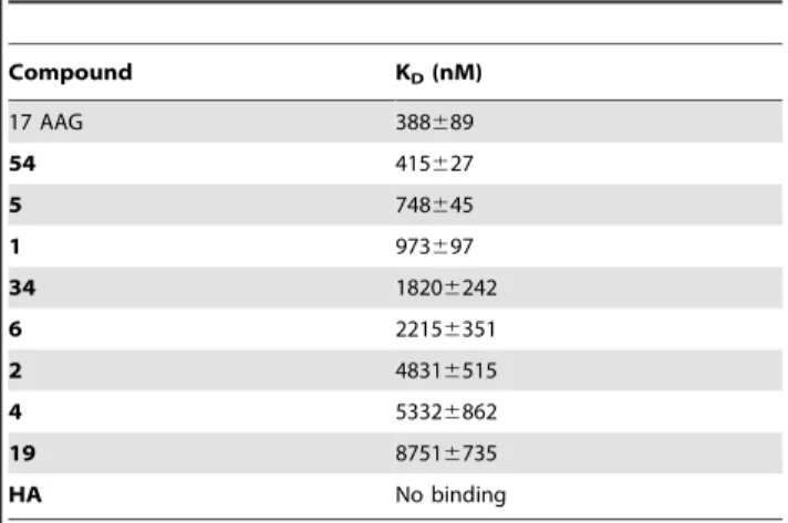
Table 1.Thermodynamic constants measured by SPR for the interaction between tested compounds and immobilized HSP90a.
Compound KD(nM)
17 AAG 388689
54 415627
5 748645
1 973697
34 18206242
6 22156351
2 48316515
4 53326862
19 87516735
HA No binding
Polyphenols, as well as 17-AAG and hardwickiic acid (HA), were dissolved in 100% DMSO to obtain 4 mM solutions, and diluted 1:200 (v/v) in PBS (10 mM NaH2PO4, 150 mM NaCl, pH 7.4) to a final DMSO concentration of 0.5%. Compounds concentration series were prepared as twofold dilutions into running buffer: for each sample, the complete binding study was performed using a six2point concentration series, typically spanning 0.025–1mM, and triplicate aliquots of each compound concentration were dispensed into disposable vials. Binding experiments were performed at 25uC, using a flow rate of 50mL min21
, with 60 s monitoring of association and 300 s monitoring of dissociation. Simple interactions were suitably fitted to a single2site bimolec-ular interaction model (A+B = AB), yielding a singleKD. Sensor-gram elaborations were performed using the BIAevaluation software provided by GE Healthcare.
ATP Hydrolysis Inhibition
The assay was performed using the Discover RX ADP HunterTM Plus Assay kit, following the manufacturer’s instruc-tions. ATPase reactions were carried out for 60 min at 40uC temperature in 100 mM Tris pH 7.4, 100mM ATP and 40 nM Hsp90ain the presence of different concentrations of geraniin or 17-AAG. ADP generation was measured using a Perkin Elmer LS 55 fluorimeter (540 nm excitation and 620 nm emission). Fluo-rescence intensity values measured for Hsp90a without any compound were set at 100% of enzyme activity. The background reaction rate was measured in a reaction lacking enzyme or substrate and subtracted from the experimental rates.
Citrate Synthase Aggregation Assay
Chaperone activity was evaluated by monitoring the therma-l2induced aggregation of citrate synthase (CS) (0.075mM), as reported elsewhere [24], in the absence or presence of a stoichiometric amount of Hsp90a and 0.3mM ATP, and with or without a four2fold molar excess of each testing compound. Aggregation was initiated by unfolding CS incubating the protein in 40 mM HEPES–KOH, pH 7.5 at 43uC. Aggregation was monitored by measuring light scattering at right angles with the Perkin Elmer LS 55 fluorimeter in stirred and thermostatted quartz cells. Both the emission and excitation wavelengths were set at 500 nm, while the band–pass was 2 nm. Kinetics traces reported are the averages of two separate measurements.
Limited Proteolysis
Limited proteolysis experiments were performed on recombi-nant Hsp90a at 37uC, PBS 0.1% DMSO, using trypsin or chymotrypsin as proteolytic agents; 30mL of a 3mM Hsp90a solution were used for each experiment. Binary complex Hsp90a/ geraniin was formed by incubating the protein with a 5:1 molar excess of geraniin at 37uC for 15 min prior to proteolytic enzyme addition. Both Hsp90 and Hsp90/geraniin complex were digested using a 1:100 (w/w) enzyme to substrate ratio. The extent of the reactions was monitored on a time2course basis by sampling the incubation mixture after 5, 15, and 30 min of digestion. Samples were analyzed by MALDITOF/MS using a MALDI micro MX (Waters). Mass data were elaborated using the Masslynx software (Waters). Preferential hydrolysis sites on Hsp90a under different conditions were identified on the basis of the fragments released during enzymatic digestion.
Cell Culture and Treatment
Jurkat and HeLa (epithelial carcinoma) cells, obtained from Cell Bank in GMP-IST (Genova, Italy) were maintained in RPMI 1640
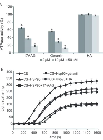
Figure 3. Inhibition of Hsp90a activity.ATPase activity of the chaperone was evaluated in the presence of different concentrations of geraniin, 17-AAG and HA (A). Data are reported as the residual ATPase activity (%) compared to that observed for an untreated sample. Data are the mean of three independent experiments performed in triplicate and were analyzed by t test (HA vs testing compounds): The error bar represents the standard deviation of nine measurements, while * indicates significance at P,0.01. Aggregation kinetics of citrate synthase (CS) at 43uC determined by light scattering (B). The spontaneous aggregation of CS at 43uC (
¤
) and the aggregation of CS at 43uC in the presence of 0.075mM Hsp90aand 0.3mM ATP (m), 0.075mM Hsp90a, 0.3mM ATP and 0.3mM geraniin (&), or 0.075mM Hsp90a, 0.3mM ATP and 0.3mM HA (N
) are shown. Kinetics traces reported are the averages of three separated measurements; the error bar represents the standard deviation of three measurements. doi:10.1371/journal.pone.0074266.g003Figure 4. Schematic representation of the results obtained from limited proteolysis experiments.The preferential cleavage sites detected on recombinant Hsp90aand on the Hsp90a/geraniin complex are in black. The Hsp90aN-terminal domain is highlighted in light grey, the middle domain is boxed and theC-terminal domain is highlighted in grey.
medium or Dulbecco’s modified Eagle medium (DMEM), respectively, supplemented with 10% (v/v) FBS, 2 mML2 gluta-mine and antibiotics (100 U mL21
penicillin, 100mg mL21 streptomycin) purchased from Invitrogen (Carslbad, CA, USA), at 37uC in humidified atmosphere with 5% CO2. To ensure logarithmic growth, cells were subcultured every two days. As control cells, human peripheral blood mononuclear cells (PBMC) were isolated from buffy coats of healthy donors (kindly provided by the Blood Center of the Hospital of Battipaglia, Salerno, Italy) by using standard Ficoll2Hypaque gradients. Freshly isolated PBMC contained 92.863.1% live cells. Proliferation of PBMC was induced by phytohemagglutinin (PHA) (10mg mL21
). Tumor and PBMC cells were treated with different concentrations of geraniin or 17-AAG to evaluate their cytotoxic effects. Stock solutions of geraniin (50 mM in DMSO) and 17-AAG (10 mM in DMSO) were stored a220uC in the dark and diluted just before addition to the sterile culture medium. In all the experiments, the final concentration of DMSO was 0.10% (v/v).
Cell Proliferation and Viability
Jurkat (26104/well) and HeLa (5000/well) cells were seeded in
triplicate in 96 well2plates and incubated for 24 h in the absence
or presence of different concentrations of geraniin (between 0,5mM to 50mM) or 17-AAG (between 0,5 nM to 10mM).
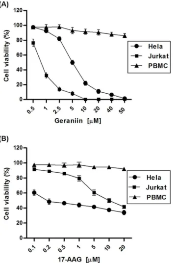
The number of viable cells was determined by using a [3–4,5-dimethyldiazol-2-yl]-2,5-diphenyl tetrazolium bromide (MTT, Sigma-Aldrich) conversion assay, according to the method described by Mosmann [25]. Briefly, following the treatment, 25mL of MTT (5 mg/mL in PBS) was added and the cells were incubated for additional 3 h at 37uC. Thereafter, cells were lysed and suspended with 100mL of buffer containing 50% (v/v) N,N-dimethylformamide, 20% SDS (pH 4.5). The absorbance was Figure 5. Cell viability (%) of cancer cells and normal cells
treated for 24 h with geraniin or 17-AAG.Jurkat, HeLa and PBMC cells were incubated for 24 h with geraniin (A) or 17-AAG (B) used in different concentrations (0.5–50mM) and (0.1–20mM), respectively, and processed for cell proliferation determination by the MTT assay. doi:10.1371/journal.pone.0074266.g005
Figure 6. Effects of geraniin and 17-AAG on cell cycle progression.Percentage of cell cycle stages was analyzed by flow cytometry, (A) PI-stained viable Jurkat cells treated with DMSO, 0.7mM geraniin or 10mM 17-AAG for 24 h. (B) PI-stained viable HeLa cells treated with DMSO, 5mM geraniin or 0.2mM 17-AAG for 24 h, (C) The percentage of hypodiploid cells as treated inA and B. Results are expressed as means6SD of three experiments performed in duplicate (***P,0.001).
measured with a microplate reader (Titertek multiskan MCC7340, LabSystems, Vienna, VA, USA) equipped with a 620 nm filter. Cell population growth inhibition was tested by cytometric counting (trypan blu exclusion). IC50values were calculated from cell viability dose2response curves and defined as the concentra-tion resulting in 50% inhibiconcentra-tion of cell survival, compared to control cells treated with 0.10% DMSO.
Analysis of Cell Cycle and Hypodiploidy by Flow Cytometry
Cell DNA content was measured by propidium iodide (PI) incorporation into permeabilized cells, as described by Nicoletti et al. [26]. Briefly, the cells were harvested after geraniin or 17-AAG treatments were washed with cold PBS and incubated with a PI solution (0.1% sodium citrate, 0.1% Triton X-100 and 50mg mL21of prodium iodide, Sigma-Aldrich, 10mg/ml Rnase A) for 30 min at room temperature. Data from 10.000 events per sample were collected by a FACScalibur flow cytometer (Becton Dickinson, San Jose´, CA) and cellular debris was excluded from analysis by raising the forward scatter threshold. The percentage of cells in the sub G0/G1phase, apoptotic fraction, was quantified using CellQuest software (Becton Dickinson). The distribution of cells in G0/G1, S, G2/M phases was determined using ModFit LT cell cycle analysis software (Becton Dickinson). Results were expressed as a mean 6 SD of three experiments performed in triplicate.
Western Blot Analyses
Treated cells were harvested and disrupted by freeze2thawing in RIPA buffer (50 mM Hepes, 10 mM EDTA, 150 mM NaCl, 1% NP-40, 0.5% sodium deoxycholate, 0.1% SDS, pH 7.4), supplemented with protease inhibitors cocktail (Sigma-Aldrich). Cell debris was removed by centrifugation at 4uC and the supernatant protein concentration was determined, according to the Bio-Rad Protein assay (Biorad Laboratories, CA, USA). Total proteins (30mg) were separated by SDS-PAGE under denatured reducing conditions. Separated proteins were then transferred to nitrocellulose membranes and blocked for 3 h with a solution containing 3% BSA in 50 mM Tris, 200 mM NaCl, 0.1% Tween
20, before incubation at 4uC overnight with the different primary antibodies (diluted 1:1000) against specific proteins. After washing, the membranes were incubated with an appropriate peroxidase-conjugate secondary antibodies at room temperature (1 h). Immunoreactive protein bands were detected by enhanced chemiluminescence reagent (ECL, Rockford, USA), according to the manufacturer’s instructions. Quantitative densitometry anal-yses were performed using a Gel Doc 2000 system (Biorad Laboratories, CA, USA).
Statistical Analysis
All the reported data represent the mean6standard deviation (SD) of at least two independent experiments, performed in triplicate. Data were statistically compared by t-test; in that aim we checked for normal distribution of data and comparable variance among the groups compared. The statistical significance of DNA content of hypodiploid nuclei was examined in the two-way analysis of variance (ANOVA) with Bonferroni post-test analysis.
Results
Screening for Ligand-HSP90 Complex Formation by Surface Plasmon Resonances
Putative interactions of different plant-derived phenolic com-pounds with Hsp90a were evaluated by a SPR-based approach [27]. Natural compounds belonging to different polyphenol classes (38 flavonoids, 12 coumarins and 4 tannins) were assayed for their binding affinity for the chaperone (Figure 1). The geldanamycin derivative 17-AAG, one of the best characterized Hsp90 inhibitors, was used as a positive control, whereas the clerodane diterpene hardwickiic acid (HA) (Figure S1), was selected as a negative control on the basis of our previous observations [28]. In Figure 2 some of the obtained sensorgrams are reported. Eight out of the 54 tested compounds interacted with immobilized Hsp90a, as inferred by the concentration-dependent responses, and by the clearly discernible exponential curves, during the association and dissociation phases. None of the tested coumarins was found to interact with the protein, whereas several of the tested flavonoids and tannins were able to bind to Hsp90a.
To measure the kinetic and thermodynamic parameters for each complex formation, the sensorgrams were fitted to a single2site bimolecular interaction model (A+B = AB); each constant was calculated fitting at least 12 curves, obtained by injecting the different compounds three times at four different concentrations, ranging from 0.025mM to 1mM (Table 1). Among the tested compounds, geraniin showed the highest affinity for the immobilized Hsp90a(Figure 2A). AKDof 415627 nM was measured for the Hsp90a/geraniin complex, comparable to that measured for the Hsp90a/17-AAG complex (388689 nM).
Inhibition of Hsp90a by Geraniin
The elevated affinity towards Hsp90a measured for geraniin, prompted us to investigate further the ability of this ellagitannin to affect different biological activities of the chaperone. Firstly, we evaluated the effects of different concentration of geraniin on the ATPase activity of Hsp90a compared to those produced by 17-AAG or HA, selected as a positive and a negative control, respectively. Geraniin was found to inhibit Hsp90a ATPase activity in a dose2dependent manner, with an inhibitory efficiency similar to that measured for 17-AAG (Figure 3 A).
The next question we addressed was to establish whether geraniin2dependent ATPase inhibition could compromise the chaperone activity of Hsp90a, which was evaluated by monitoring the citrate synthase (CS) thermal induced aggregation in the Figure 7. Effect of geraniin on Hsp90 client protein levels.Equal
presence of Hsp90a, with or without geraniin. Upon incubation at elevated temperatures, CS underwent partial unfolding resulting in a quantitative protein aggregation; the presence of stoichio-metric amounts of Hsp90a changed the aggregation kinetics significantly. When a four2fold molar excess of geraniin was added to the reaction, the curve slope clearly increased, indicating that geraniin, interacting with Hsp90a, affects its affinity towards denatured CS (Figure 3 B). Anti2chaperone activity of the tannin resembled that observed performing the same experiment using a four2fold molar excess of 17-AAG, whereas the addition of the same amount of HA did not perturb the CS+Hsp90acurve.
Study of Hsp90a/geraniin Interaction
In an effort to identify the Hsp90aregion involved in geraniin binding, a limited proteolysis2mass spectrometry2based strategy was used for the structural analysis of the Hsp90a/geraniin complex. This approach is based on the evidence that exposed, weakly structured, and flexible regions of a protein can be recognized by a proteolytic enzyme [29,30]. The proteolytic patterns obtained in the experiments performed on Hsp90aand on the Hsp90a/geraniin complex, using trypsin or chymotrypsin as proteolytic agents, are summarized in Figure 4. Comparison of the differential proteolytic pattern obtained from the digestion of native Hsp90aor of the Hsp90a/geraniin complex confirmed a direct interaction between geraniin and the chaperone. In addition, we observed that peptide bonds following Arg86, Lys237, Lys293, and Tyr380, preferential cleavage sites of the native chaperone in the absence of geraniin, were protected in the complex. Conversely, following interaction with geraniin, peptide bonds located at the level of Lys488 and Lys512 became susceptible to enzymatic hydrolysis. These data suggest that binding of geraniin to Hsp90ainduced significant conformational changes of its three2dimensional structure. Specifically, the observed overall protection of the N2terminal region from proteolysis indicated that this is the protein portion g004mainly involved in interaction with geraniin.
Effect of Geraniin on Cancer Cell Viability
On the basis of the ability of geraniin to bind and inhibit HSP90a activity, we evaluated its potential anti2proliferative or cytotoxic activity in HeLa (epithelial carcinoma) and Jurkat leukemia cancer cell lines. The cancer cell lines and the PBMC (human Peripheral Blood Mononuclear Cells) line were incubated for 24 h with increasing concentrations of geraniin (0.5mM – 50mM) or 17-AAG (0.1mM – 20mM) and cell viability was determined by MTT proliferation assay. Proliferation of Hela and Jurkat cells was inhibited by geraniin treatment in a concentration-dependent manner, with IC50 values of 5.160.2 and 0.7660.1mM, respectively (Figure 5 A). Under the same experimental conditions, IC50 values after 17-AAG treatment were 9.660.15mM in Jurkat and 0.260.3mM in HeLa cell lines (Figure 5 B), in agreement with those reported by Shelton et al. [31] and Bisht et al. [32]. Minimal cytotoxic effects, evaluated by tryptan blue dying, were observed in control PHA-stimulated proliferating PBMC only at high doses of geraniin or 17-AAG (Figure 5).
Induction of Cell Cycle Arrest and Cell Death in Cancer Cell Lines by Geraniin
To establish the mechanism of action underlying the inhibition of cancer cell viability caused by geraniin treatment, the cell cycle progression of cancer cells and PHA-stimulated PBMC were analyzed by flow cytometry.
The Jurkat and HeLa cells were incubated for 24 h with concentrations close to the IC50values of geraniin (0.7 and 5mM, respectively) and 17-AAG (10 and 0.2mM, respectively), The effects of geraniin and 17-AAG-treatments on the distribution of non-apoptotic cells differed significantly. In both tumor cell lines, treatment with geraniin caused a G2/M arrest (Figure 6 A, B), whereas 17-AAG induced a G1and G2/M arrest (Figure 6 A, B), consistent with previous published results [31,33,34]. Moreover, following geraniin treatment the percentage of Jurkat and Hela cells with hypodiploid nuclei was 9.7561.02% and 16.260.7%, respectively, whereas following 17-AGG treatment these values were 6.0560.4% and 11.660.5%, respectively. These results indicate a similar pro-apoptotic effect of the two compounds on both the cell lines. Neither geraniin nor 17-AAG caused any pro-death or cytostatic effects in PHA-stimulated proliferating PBMC, since the levels of hypodiploidy/necrosis in geraniin and 17-AAG treated PBMC were 2.661.3% and 1.860.6%, respectively, very similar to those observed in untreated cells (1.560.8%).
Down2regulation of Hsp90 Client Proteins by Geraniin
Treatment
Geraniin was tested for its effects on the intracellular levels of Hsp70, Hsp90 and Hsp90 client proteins. HeLa and Jurkat cell lines were treated with a geraniin concentration corresponding to their IC50value and at additional lower and higher concentrations (Figure 7). These treatments caused a dose-dependent decrease in the level of the client proteins c-Raf, pAkt and EGFR, whereas the level of Akt was unaffected. Interestingly, the level of Hsp70 expression was enhanced in both tumor cell lines, in a dose-dependent fashion, with 3.560.12-fold increase in Jurkat cells upon 1mM treatment and 2.760.09 fold increase in HeLa cells upon 10mM geraniin treatment.
Discussion
Inhibition of HSP90 has received significant attention in cancer research due to its ability to retard or block tumor growth [6]. In this respect, Hsp90 plays a critical role in the maintenance of multiple oncogenic pathways and is required to maintain the folding, stability and functionally active conformation of many aberrant oncoproteins [7]. In healthy cells, Hsp90 is involved in dynamic, low-affinity interactions with a plethora of proteins during folding and maturation; however, in tumor cells, it assists folding of dysregulated oncoproteins and sustains their aberrant activity [4,5]. Given the diversity of the Hsp90 client proteins involved in critical cellular pathways and processes, inhibition of Hsp90 was predicted to have efficacy in a broad variety of human tumors. However, although several Hsp90 inhibitors have thus far entered into clinical trials, the development of Hsp90 inhibitors has encountered difficulties, including drug solubility and hepatic toxicity [35].
the chaperone activity of the molecule [36]. This mechanism was proved for geraniin, which was able to reduce Hsp90 chaperone activity more efficiently than 17-AAG.
The impaired ATPase and chaperone activities of Hsp90, caused by geraniin binding, induce cytotoxic effects in the tumor cell lines tested, with a large percentage of cells containing hypodiploid DNA. These results are in agreement with those reported in human melanoma cells, where geraniin treatment caused apoptosis, through up-regulation of the Fas ligand expression, the activation of caspase-8, the cleavage of Bid, and the induction of cytochrome c release from mitochondria to the cytosol [21]. Our results also revealed that geraniin induces cell cycle arrest in the G2/M phase in both cancer cell lines, whereas 17-AAG-treated cells accumulated in G1phase of cell cycle.
Moreover, the geraniin-dependent inhibition of Hsp90a chap-erone activity caused a dose-dependent decrease in the level of the oncogenic proteins c-Raf, pAkt and EGFR, further supporting the potential of this compound to interfere with tumor progression. As already reported in other cellular systems, Hsp90 inhibition by geraniin was associated to the up-regulation of Hsp70, as a compensatory mechanism aiding the proper folding and
assem-bling of oncogenic client proteins. Although it is still unclear if the induction of Hsp70 following Hsp90 inhibition attenuates the cytotoxic effects of Hsp90 inhibitors, Hsp70 might also be considered as a pharmacodynamic marker for drug response of HSP90 inhibition [37].
In conclusion, the results presented here, along with the finding that geraniin did not exert any appreciable cytotoxicity in normal cells, encourages further studies on this compound as a promising chemical scaffold for the design of new Hsp90 inhibitors.
Supporting Information
Figure S1 Structure of hardwickiic acid.
(DOCX)
Author Contributions
Conceived and designed the experiments: FDP NDT. Performed the experiments: AV MCV. Analyzed the data: FDP NDT. Contributed reagents/materials/analysis tools: AL NDT. Wrote the paper: FDP NDT AL.
References
1. Pratt WB, Toft DO (2003) Regulation of signaling protein function and trafficking by the hsp90/hsp70-based chaperone machinery. Exp Biol Med (Maywood) 228: 111–133.
2. Zhang H, Burrows F (2004) Targeting multiple signal transduction pathways through inhibition of Hsp90. J Mol Med 82: 488–499. doi: 10.1007/s00109-004-0549-9.
3. Zhang T, Hamza A, Cao X, Wang B, Yu S, et al. (2008) A novel Hsp90 inhibitor to disrupt Hsp90/Cdc37 complex against pancreatic cancer cells. Mol Cancer Ther 7: 162–170.
4. Whitesell L, Lindquist SL (2005) HSP90 and the chaperoning of cancer. Nat Rev Cancer 5: 761–772. doi:10.1038/nrc1716.
5. Holzbeierlein JM, Windsperger A, Vielhauer G (2010) Hsp90: a drug target? Curr Oncol Rep 12: 95–101. doi: 10.1007/s11912-010-0086-3.
6. Wang RE (2011) Targeting heat shock proteins 70/90 and proteasome for cancer therapy. Curr Med Chem 18: 4250–4264.
7. Solit DB, Rosen N (2006) Hsp90: a novel target for cancer therapy. Curr Top Med Chem 6: 1205–1214. doi: 10.2174/156802606777812068.
8. Trepel J, Mollapour M, Giaccone G, Neckers L (2010) Targeting the dynamic Hsp90 complex in cancer. Nat Rev Cancer 10: 537–549. doi: 10.1038/nrc2887. 9. Piper PW, Millson SH (2011) Mechanisms of resistance to Hsp90 inhibitor drugs: a complex mosaic emerges. Pharmaceuticals 4: 1400–1422. doi:10.3390/ ph4111400.
10. Neckers L, Workman P (2012) Hsp90 molecular chaperone inhibitors: are we there yet? Clin Cancer Res 18: 64–76.
11. Kummar S, Gutierrez ME, Gardner ER, Chen X, Figg WD, et al. (2010) Phase I trial of 17-dimethylaminoethylamino-17-demethoxygeldanamycin (17-DMAG), a heat shock protein inhibitor, administered twice weekly in patients with advanced malignancies. Eur J Cancer 46: 340–347. doi: 10.1016/ j.ejca.2009.10.026.
12. Heath EI, Hillman DW, Vaishampayan U, Sheng S, Sarkar F, et al. (2008) A phase II trial of 17-allylamino-17-demethoxygeldanamycin in patients with hormone-refractory metastatic prostate cancer. Clin Cancer Res 14: 7940–7946. doi: 10.1158/1078-0432.CCR-08-0221.
13. Yin Z, Henry EC, Gasiewicz TA (2009) (2)-Epigallocatechin-3-gallate is a novel Hsp90 inhibitor. Biochemistry 48: 336–345. doi:10.1021/bi801637q. 14. Brandt GE, Schmidt MD, Prisinzano TE, Blagg BS (2008) Gedunin, a novel
hsp90 inhibitor: semisynthesis of derivatives and preliminary structure-activity relationships. J Med Chem 51: 6495–6502. doi: 10.1021/jm8007486. 15. Dal Piaz F, Vassallo A, Chini MG, Cordero FM, Cardona F, et al. (2012)
Natural iminosugar (+)-lentiginosine inhibits ATPase and chaperone activity of hsp90. PLoS One 7: e43316. doi: 10.1371/journal.pone.0043316.
16. Oh SH, Woo JK, Yazici YD, Myers JN, Kim WY, et al. (2007) Structural basis for depletion of heat shock protein 90 client proteins by deguelin. J Natl Cancer Inst 99: 949–961. doi: 10.1093/jnci/djm007.
17. Okuda T, YoshidaT, Hatano T (1995) Hydrolyzable tannins and related polyphenols. Fortschr Chem Org Naturst 66: 1–117.
18. Ito H (2011) Metabolites of the ellagitannin geraniin and their antioxidant activities. Planta Med 77: 1110–1115. doi: 10.1055/s-0030-1270749. 19. Clifford MN, Scalbert A (2000) Ellagitannins, occurrence in food, bioavailability
and cancer prevention. J Food Sci Agric 80: 1118–1125.
20. Yang Y, Zhang L, Fan X, Qin C, Liu J (2012) Antiviral effect of geraniin on human enterovirus 71in vitroandin vivo. Bioorg Med Chem Lett 22: 2209– 2211.
21. Lee JC, Tsai CY, Kao JY, Kao MC, Tsai SC, et al. (2008) Geraniin-mediated apoptosis by cleavage of focal adhesion kinase through up-regulation of Fas ligand expression in human melanoma cells. Mol Nutr Food Res 52: 655–663. 22. Dal Piaz F, Malafronte N, Romano A, Gallotta D, Belisario MA, et al. (2012) Structural characterization of tetranortriterpenes from Pseudrocedrela kotschyi and Trichilia emetica and study of their activity towards the chaperone Hsp90. Phytochemistry 75: 78–89. doi: 10.1016/j.phytochem.2011.12.002.
23. Dal Piaz F, Tosco A, Eletto D, Piccinelli AL, Moltedo O, et al. (2010) The identification of a novel natural activator of p300 histone acetyltranferase provides new insights into the modulation mechanism of this enzyme. ChemBioChem 11: 818–827. doi: 10.1002/cbic.200900721.
24. Mosmann T (1983) Rapid colorimetric assay for cellular growth and survival: application to proliferation and cytotoxicity assays. J Immunol Methods 65: 55– 63. doi: 10.1016/0022-1759(83)90303-4.
25. Jakob U, Lilie H, Meyer I, Buchner J (1995) Transient interaction of Hsp90 with early unfolding intermediates of citrate synthase. J Biol Chem 270: 7288–7294. 26. Nicoletti I, Migliorati G, Pagliacci MC, Grignani F, Riccardi C (1991) A rapid and simple method for measuring thymocite apoptosis by propidium iodide staining and flow cytometry. J Immunol Methods 139: 271–279.
27. Cooper MA (2003) Label-free screening of bio-molecular interactions. Anal Bioanal Chem 377: 834–842. doi: 10.1007/s00216-003-2111-y.
28. Faiella L, Dal Piaz F, Bisio A, Tosco A, De Tommasi N (2012) A chemical proteomics approach reveals Hsp27 as a target for proapoptotic clerodane diterpenes. Mol Biosyst 8: 2637–2644. doi: 10.1039/c2mb25171j.
29. Orru` S, Dal Piaz F, Casbarra A, Biasol G, De Francesco R, et al. (1999) Conformational changes in the NS3 protease from hepatitis C virus strain Bk monitored by limited proteolysis and mass spectrometry. Protein Sci 8: 1445– 1454. doi:10.1110/ps.8.7.1445.
30. Giommarelli C, Zuco V, Favini E, Pisano C, Dal Piaz F, et al. (2010) The enhancement of antiproliferative and proapoptotic activity of HDAC inhibitors by curcumin is mediated by Hsp90 inhibition. Cell Mol Life Sci 67: 995–1004. doi: 10.1007/s00018-009-0233-x.
31. Shelton SN, Shawgo ME, Matthews SB, Lu Y, Donnelly AC, et al. (2009) KU135, a novel novobiocin-derived C-terminal inhibitor of the 90-kDa heat shock protein, exerts potent antiproliferative effects in human leukemic cells. Mol Pharmacol 76: 13141322. doi: 10.1124/mol.109.058545.
32. Bisht KS, Bradbury CM, Mattson D, Kaushal A, Sowers A, et al. (2003). Geldanamycin and 17-allylamino-17-demethoxygeldanamycin potentiate the in vitro and in vivo radiation response of cervical tumor cells via the heat shock protein 90-mediated intracellular signaling and cytotoxicity. Cancer Res 63: 8984–8995.
33. Mu¨nster PN, Srethapakdi M, Moasser MM, Rosen N (2001) Inhibition of heat shock protein 90 function by ansamycins causes the morphological and functional differentiation of breast cancer cells. Cancer Res. 61: 2945–2952. 34. Arlander SJ, Eapen AK, Vroman BT, McDonald RJ, Toft DO, Karnitz LM.
(2003) Hsp90 inhibition depletes Chk1 and sensitizes tumor cells to replication stress J Biol Chem 278: 52572–52577.
36. Cunningham CN, Krukenberg KA, Agard DA (2008) Intra- and intermonomer interactions are required to synergistically facilitate ATP hydrolysis in Hsp90. J Biol Chem 283: 21170–21178.