Paracrine Action of Mesenchymal Stem Cells
Revealed by Single Cell Gene Profiling in
Infarcted Murine Hearts
Yan Yao1☯*, Ji Huang1☯, Yongjian Geng2, Haiyan Qian3, Fan Wang4, Xiaohui Liu1, Meisheng Shang1, Shaoping Nie5, Nian Liu1, Xin Du1, Jianzeng Dong1, Changsheng Ma1
1Department of Cardiology, Beijing Anzhen Hospital, Capital Medical University, Beijing Institute of Heart, Lung and Blood Vessel Diseases, Beijing, P. R. China,2Center for Cardiovascular Biology and
Atherosclerosis, Department of Internal Medicine, The University of Texas, Health Science Center at Houston, Medical School, Texas Heart Institute, Houston, TX, United States of America,3Center for Coronary Heart Disease, Department of Cardiology, State Key Laboratory of Cardiovascular Disease, Fuwai Hospital, National Center for Cardiovascular Diseases, Chinese Academy of Medical Sciences and Peking Union Medical College, Beijing, P. R. China,4Department of Biostatistics and Epidemiology, University of Pennsylvania, Philadelphia, PA, United States of America,5Emergency and Critical Care Center, Beijing Anzhen Hospital, Capital Medical University, Beijing Institute of Heart, Lung and Blood Vessel Diseases, Beijing, P. R. China
☯These authors contributed equally to this work. *[email protected]
Abstract
Background
Mesenchymal stem cells (MSCs) have been recently demonstrated as a promising stem cell type to rescue damaged myocardium after acute infarction. One of the most important mechanisms underlying their therapeutic effects is the secretion of paracrine factors. How-ever, the expression profile of paracrine factors of MSCs in infarcted hearts, especially at single cell level, is poorly defined.
Methods and Results
We aimed to depict the transcriptional profile of paracrine factors secreted by MSCsin vivo,
with particular interest in the comparison between normal and infarcted hearts. Bone mar-row mesenchymal stem cells were isolated and injected into mice hearts immediately after infarction surgery. Bioluminescence imaging (BLI) indicated a proportion of cells still alive even up to 10 days post surgery. Paralleled with survived cells, cardiac function was signifi-cantly improved after MSC injection compared to that in PBS-injected mice, indicated by MRI and histology. Despite increased number of vessels in MSC-injected hearts, endotheli-al cells and cardiomyocytes transdifferentiation were not observed in infarcted hearts 5 days after infarction. Furthermore, laser capture microdissection (LCM) followed by high through-put real time PCR was employed in our study, uncovering that the injected MSCs, compared to local cardiomyocytes, displayed elevated levels of secreted factors. To further investigate the regulation of those factors, we performed single cell analysis to dissect the OPEN ACCESS
Citation:Yao Y, Huang J, Geng Y, Qian H, Wang F, Liu X, et al. (2015) Paracrine Action of Mesenchymal Stem Cells Revealed by Single Cell Gene Profiling in Infarcted Murine Hearts. PLoS ONE 10(6): e0129164. doi:10.1371/journal.pone.0129164
Academic Editor:Yiru Guo, University of Louisville, UNITED STATES
Received:October 16, 2014
Accepted:May 5, 2015
Published:June 4, 2015
Copyright:© 2015 Yao et al. This is an open access article distributed under the terms of theCreative Commons Attribution License, which permits unrestricted use, distribution, and reproduction in any medium, provided the original author and source are credited.
Data Availability Statement:All relevant data are within the paper and its Supporting Information files.
Funding:This work was supported by grants from the National Natural Science Foundation of China (No. 81200141, 81070091, 81000130, 81000091), Beijing Novel Program (No. 2011081,
Z131103000413116), Program of International S&T Cooperation Project of China (No. 2013DFB30310) and“973”National Program on Key Basic Research Project of China (No. 2013CB531105).
gene expression profile of MSCs at single cell level in infarcted and normal hearts, respec-tively. Consistent with thein vivoobservation, a similar regulation pattern of those factors
was detected in cultured MSCs under hypoxia.
Conclusions
Our study, for the first time, elucidated gene expression profiles, as well as regulation of paracrine factors, of MSCs at single cell levelin vivo, indicating that paracrine factors from
MSCs account for the improvement of cardiac function after infarction.
Introduction
Mesenchymal stem cell (MSC) is a promising source for the cell-based treatment of acute myo-cardial infarction (AMI). Numerous experimental and clinical studies have displayed their en-couraging therapeutic effects on the improvement of cardiac function [1–3]. However, the mechanisms underlying the benefits of MSC therapy remain debated.
Previously, it was believed that MSCs were able to differentiate into cardiomyocytes, vascu-lar endothelial cells, and vascuvascu-lar smooth muscle cells to repair the damaged myocardium, which is called the trans-differentiation mechanism [4]. However, many investigators have failed to detect persistent engraftment and differentiation of implanted MSCsin vivo, and the
number of newly regenerated cardiomyocytes is too small to explain the significant functional improvement of infarcted hearts [5,6].
Recently, the focus has shifted to paracrine-centric mechanisms of the MSC-mediated pro-tective effects, as it has been shown that various types of cytokines, chemokines, and growth factors released by MSCs play important roles in cardiac remodeling, angiogenesis, apoptosis, and survival [7,8]. Despite previous studies reporting the expression and function of several paracrine factors, we still lack a full-scale profiling of the paracrine factors expressed by MSCs and adjacent cardiomyocytesin vivo, especially in a spatial-temporal manner. So far, few
stud-ies have successfully identified paracrine factors that are beneficial for the MSC-mediated pro-tective effects, due to the technical difficulty to examine the expression and secretion of those paracrine factorsin vivo[9–10].
Single-cell quantitative reverse transcription-polymerase chain reaction (qRT-PCR) is a use-ful approach widely applied in stem cell and cancer research. By use of this technique, we are able to assess the differences at transcriptional level between cell populations taken from in-farcted zones. In the present study, we profiled the expression of twenty-one paracrine factors from MSCs and adjacent cardiomyocytes in infarcted murine hearts, and examined the effect of infarction and hypoxia challenge on their expression patterns bothin vivoandin vitro. We
believe that our studies provide a roadmap to comprehensively understand how MSC exerts its effects on myocardium repair after damage [11].
Materials and Methods
Ethics Statement
Animals
Mice were purchased from the Institute of Laboratory Animal Sciences, Chinese Academy of Medical Sciences (Beijing, China). AMI was performed on 8–10 week old female NOD SCID mice (25~30g). 6–8 week old male transgenic mice (20~25g) of FVB background withβ-actin promoter driving Fluc-eGFP (firefly luciferase- enhanced Green Fluorescent Protein) were used as donors for MSCs isolation.
Isolation, culture and identification of MSCs
MSCs were isolated and cultured as we described previously [12,13]. Briefly, eGFP+/Luc+ trans-genic mice were euthanized, then the tibia and the femur were removed to get the bone mar-row. Mononucleated cells were isolated by ficoll density gradient, washed and resuspended in alpha-MEM (GIBCO, CA, USA) supplemented with 10% fetal bovine serum, streptomycin and penicillin. Afterwards, the obtained cells were cultured at 37°C and 5% CO2. Non-adher-ent cells were removed daily by medium replacemNon-adher-ent. When the cultures reached 80% of con-fluence, cells were recovered by a mixture of trypsin/ EDTA and followed by passages.
The third passage of MSCs were collected, and their purity were subsequently verified by fluorescence-activated cell sorting analysis (FACS). As shown inS1 Fig, those MSCs displayed typical fibroblast-like morphology (S1A Fig), and more than 90% cells expressed specific MSC surface marker CD105 and CD90, while lacking the expression of CD45 and CD34 (S1B Fig). Forin vitroexperiments, cells were exposed to normoxia (20% O2, 5% CO2) or hypoxia (1%
O2, 5% CO2) conditions for 48 hours.
Myocardial infarction model and MSC transplantation
AMI was created in female SCID mice by permanent ligation of left anterior descending coro-nary artery (LAD). The animals were intraperitoneally anesthetized with sodium pentobarbital (50 mg/kg) and mechanically ventilated with room air by using Minivent 845 (Hugo Sachs Electronics, March, Germany). The heart was exposed through a left-sided minithoracotomy, and the left coronary artery was permanently ligated. Infarction was visually confirmed by ob-servation of blanching of the left ventricular myocardium as well as dyskinesis. Immediately after LAD artery ligation, the mice were randomly allocated to receive intramyocardial injec-tions of phosphate-buffered saline (PBS, 20μl) or MSCs (1×10⁶, 20μl) at three sites in the
in-farct border zone.
In vivo
bioluminescence imaging
Bioluminescence imaging analysis was performed at days 1, 4, 7, 10 after cell transplantation to monitor cell survival and engraftment by using IVIS 200 system (Caliper, Hopkinton, MA, USA). The mice were routinely anesthetized and then intraperitoneally injected with 100μl
D-luciferin (200 mg/kg to body weight, dissolved in PBS). 10 minutes after the injection, a series of bioluminescent images were recorded for about 20 minutes. Bioluminescent signals were standardized for exposure time and quantified in units of maximum photons per second per square centimeter per steradian (p/s/cm2/sr). The image with the greatest signal intensity, which represented the viable injected cells in infarcted hearts, was used for quantification anal-ysis at each time point.
Cardiac function assessment and HE staining
using a 7.0T Biospec small animal experimental scanner (Bruker Biospin, Billerica, MA, USA). The electrocardiographic gating was optimized with two cardiogram electrodes attached to each animal’s forelimbs with respiratory motion and body temperature monitors (Small Ani-mal Instruments, Stony Brook, NY, USA). A series of short-axis views were measured using a retrospectively gated, T1-weighted FLASH sequence (TE 3 ms, TR 6 ms, field of view 45 mm × 45 mm, slice thickness 1.0 mm, imaging matrix 128 ×192). Continuously acquired imag-ing data from each slice was reconstructed into 10 cine frames. Planimetry measurements of left ventricular myocardial area were conducted by tracing the epicardial and endocardial bor-ders at end-systole and end-diastole using ParaVision software (Bruker Biospin MRI). Ejection fraction (EF) was calculated as the ratio of (LVEDD–LVESD) to LVEDD.
Mice were euthanized and hearts were harvested at day 11 after surgery. Left ventricle was cut into eight fragments from apex to base, and frozen sections (7 mm thickness, 350 mm apart) were randomly chosen from every fragment. The sections were then subjected to hema-toxylin and eosin staining (HE staining). All the mice were euthanized at the end of the study.
Measurement of angiogenesis
At day 5 after operation, mice were euthanized and the hearts were rapidly excised. Paraffin-embedded tissues were cut in 5μm cross sections through the left ventricle and mounted on
slides. After a brief wash in PBS, heart sections were incubated in a blocking buffer(PBS con-taining 1% bovine serum albumin and 0.1% Triton X-100) at room temperature for 1 hour, then incubated with rabbit anti-CD31 (Abcam, Cambridge, UK) or mouse anti-Actinin 2 (Sigma-Aldrich, MO, USA) primary antibodies at 4°C overnight, followed by secondary anti-bodies against rabbit and mouse IgG (Invitrogen, CA, USA) respectively at 37°C for 1 hour, and subsequently counterstained with DAPI. Angiogenesis analysisin vivowas determined by
the expression of a specific endothelial cell marker, CD31. Capillaries were counted in five ran-domly chosen high-power fields (HPFs) in every chosen section. The results were expressed as capillaries per HPF. The results were expressed as capillaries per HPF. The value was an aver-age of 8 slides per each mouse, total 8 mice per each group.
Laser capture micro-dissection and qRT-PCR analysis
Mice were euthanized and hearts were removed at day 5 after transplantation, embedded in op-timal cutting temperature, and then frozen in liquid nitrogen. Five cyrosections (7μm
thick-ness, 350μm apart) of left ventricle were prepared on polyethylene naphthalate
membrane-coated slides (P.A.L.M. Microlaser Technologies AG, Bernried, Germany). Green fluorescence visualized under laser microscopy was used as an indicator for micro-dissection. eGFP+MSCs, para-eGFP+cells, remote cells and cells from PBS injected hearts were independently captured by a LMD6000 system (Leica, Germany). The dissected tissues were placed on the caps of micro-centrifuge tubes with 50μl lysis enhanced buffer. After dissection, tissues were collected
by centrifugation. Total RNA were extracted from these tissues, and 0.5μg RNA was used in
re-verse transcription–polymerase chain reaction(PCR), followed by real-time PCR (Applied bio-systems, CA, USA) to examine the expression level of paracrine factors. In addition, different zones including remote zone, border zone and infarcted zone were digested into single cells by collagenase type I(1 mg/ml, Sigma), then eGFP+cells were collected to determine the distribu-tion of MSCs in infarcted hearts.
Single cell isolation and qRT-PCR Analysis
(Invitrogen), and then subjected to FACS. MSCs isolated from donor transgenic mice were H2-Kd negative, whereas recipient SCID mice were H2-Kd positive. In addition, dyecycle staining was capable of distinguishing sing cell or fused cell via DNA content, and 2N was con-sidered as a marker of single cell.
Inventoried TaqMan Assays (20×, Applied Biosystems) were pooled and diluted to a final concentration of 0.2× for each of the 24 probes. Individual cells were directly collected into 10μl RT-PreAmp MasterMix (5.0μl CellsDirect 2× Reaction Mix [Invitrogen]; 2.5μl 0.2×
assay pool; 0.5μl RT/Taq enzyme [CellsDirect qRT-PCR kit, Invitrogen]; 2.0μl TE buffer).
The products were analyzed with Universal PCR Master Mix and inventoried TaqMan gene ex-pression assays in 48.48 Dynamic Arrays on a BioMark System (Fluidigm, CA, USA). CT val-ues were calculated from the system’s software (Fluidigm). The resulting values were then normalized to the endogenous controls by subtracting the average of GAPDH expression levels.
Cytokines array
MSCs were cultured under normaxia and hypoxia conditions. Afterwards, supernatants were harvested, and applied for cytokines array (membrane cytokine array, Abcam). Then, the membranes were developed, and quantified by Image J.
LCM and Single-Cell Data Processing
All Ct values of genes were converted into relative expression levels by subtracting the values from the assumed baseline value of 32. The resulting values were then normalized to the endog-enous controls by subtracting the average of GAPDH expression levels.
Statistical Analysis
Data are expressed as mean ± standard deviation (SD). Repeated-measures ANOVA with Bon-ferroni post hoc testing and non-paired Studentttest were used where appropriate. The level
of statistical significance was defined asP<0.05. All data collection and analyses were
per-formed with SPSS 13.0.
Results
Successful engraftment of MSC in infarcted hearts
To assess the functional outcome of MSC transplantation, we first injected firefly luciferase-bearing MSCs into the mouse myocardium after LAD ligation. Since cell engraftment is a key step in stem cell therapy in myocardial infarction, bioluminescence imaging (BLI) was applied to monitor the survival of transplanted MSCs. As shown inS2A Fig, although firefly luciferase signal was decreased after injection, we were still able to detect considerable signal even on day 10 (S2A and S2B Fig). This observation suggested successful engraftment of MSCs in infarcted hearts.
MSC Transplantation improves cardiac function after myocardial
infarction
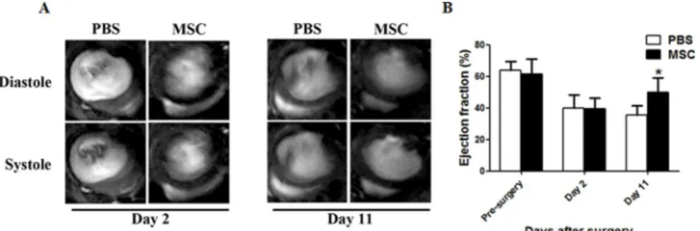
±4.58% vs. 48.62%±7.25%,P<0.01,Fig 1A and 1B). To further confirm the improved outcome
of heart function, we performed HE staining to evaluate the ventricular wall thickness 11 days after myocardial infarction. As shown inS3A and S3B Fig, consistent with a functional im-provement revealed by MRI, MSC transplantation increased ventricular wall thickness by 58.2% compared to PBS injection only (168.6±91.6 vs. 106.6±57.4μm,P<0.01).
None of the MSCs differentiates into endothelial cells or cardiomyocytes
at early stage of infarction
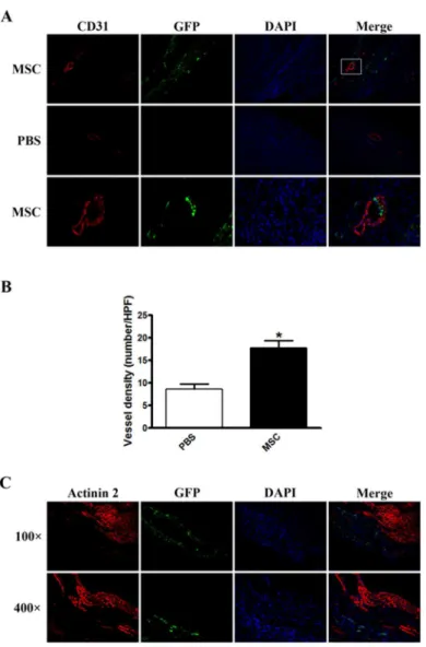
To test whether direct cell transdifferentiation from injected MSCs was responsible for the im-proved cardiac function, we performed immunostaining against CD31, an endothelial cell specif-ic marker, and Actinin 2, a cardiomyocyte specifspecif-ic marker, to determine the transdifferentiation from MSCs to endothelial cells and myocytes, respectively. Compared to the PBS-injected group, MSC injection markedly increased the number of CD31-positive cells, as well as blood vessel density in infarcted hearts 5 days after infarction (17.75±2.6 VS 8.60±1.5/HPF,P<0.01,Fig 2A
and 2B). However, no colocalization between CD31 and GFP was observed (Fig 2A), indicating no transdifferentiation of endothelial cells from MSCs. Similarly, no transdifferentiation of cardiomyocytes from MSCs was present as none of Actinin 2 and GFP double positive cells were observed (Fig 2C). Taken together, these results suggested mechanisms, other than transdifferen-tiation, that accounted for the improved cardiac function.
MSCs express diverse paracrine factors in infarcted hearts
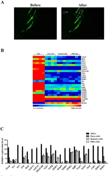
Given no transdifferentiation to endothelial cells and cardiomyocytes from injected MSCs, we hypothesized that paracrine effects play a critical role in the improvement of cardiac function. To test our hypothesis, we first explored the distribution of MSCs in infarcted hearts by FACS. Injected cells were enriched in the infarcted zone after surgery. However, these cells quickly di-minished. On the contrary, the MSCs percentage was dramatically increased in the remote zone on day 5 post-transplantation, while the cell population was sustained in the border zone (S4 Fig). To further determine how different cell population contributed to paracrine effects in infarcted hearts, we carried out laser capture microdissection (LCM) to collect cell clumps from injected MSCs, local cells adjacent to MSCs (Para-cells), local cells in remote zones (Re-mote-cells), and local cells from PBS-injected hearts (PBS-cells), respectively (Fig 3A). The ex-pression profiles of paracrine factors from different cell clumps were tested subsequently. As shown inFig 3B, while IL1 expression levels were comparable among different groups, injected MSCs displayed significantly higher levels of all other factors (VEGF, HGF, IGF1, FGF1, FGF2, Fig 1. Cardiac functional analysis of infarcted hearts with MSCs transplantation.(A) Representative MRI images of hearts at day 2 and 11 in PBS-injected or MSC-transplanted mice. (B) Quantification of ejection fraction (%) at different time points in two groups mice(n = 8,*P<0.01).
AGPT1, AGPT2, PDGF-BB, NGF, CSF1, BMP2, BMP4, IL6, TNF, Itgβ1, TGFβ, MMP2, MMP9, TIMP1, and TIMP2). Interestingly, local cells adjacent to injected MSC exhibited rela-tive higher expression of certain paracrine factors, such as VEGF, HGF, FGF1, CSF1 and BMP4 (Fig 3C). Together, these observations suggested that injected MSCs not only play a pre-dominant role in the expression of paracrine factors, but also affect the paracrine profiles of surrounding cells in infarcted hearts.
The expression profile of paracrine factors is sensitive to the changes of
microenvironment
Given a critical role of the microenvironment on cellular behaviors, and due to heterogeneous nature of MSCs, we performed single cell PCR analysis to evaluate expression levels of Fig 2. Immunostaining for CD31 and Actinin 2 in infarcted hearts with transplanted MSCs.(A) Representative images of immunostaining against CD31 (red fluorescence) in infarcted hearts.
Magnification: 100× in upper and middle panels, 400× in lower panel which is the magnification of the box in upper panel. (B) Statistical analysis of vessel density. The value was an average of 8 slides per each mouse, total 8 mice per each group. (C) Representative images of immunostaining against Actinin 2 (red
fluorescence) in infarcted hearts.
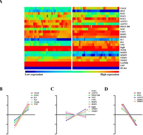
paracrine factors in infarcted versus normal hearts at the single cell level. Probes against H2-Kd, SRY and eGFP were used to ensure the reliability of isolated MSCs (Fig 4A). Despite the existing variability among single MSCs in one condition, the expression profiles of para-crine factors dramatically changed in infarcted hearts (Fig 4A). While the expression of VEGF, FGF2, IL1, IL6, TGFβand TNF significantly increased, HGF, IGF1, AGPT2, MMP2 and MMP9 expression decreased under hypoxic stress. The expression of other factors (FGF1, AGPT1, PDGF-BB, NGF, CSF1, BMP4, BMP2, Itgβ1, TIMP1 and TIMP2), however, remained unchanged (Fig 4B–4D).
To verify the regulatory effects of infarctionin vivo, expression levels of paracrine factors
were also assessed in cultured MSCs under hypoxia conditionin vitro. Similarly, the expression
levels of VEGF, IL1 and TGFβwere significantly increased upon the hypoxia exposure (2.84 ±0.32, 2.68±0.27 and 2.69±0.25 fold vs. normoxia,P<0.01), whereas the expression of AGPT1,
Fig 3. Transcriptomic analysis of gene expression profiles of transplanted MSCs in infarcted hearts.
(A) Representative pictures of frozen slides before and after dissection. Selected cells (white arrow) were indicated by white lines (scale bar: 100μm, magnification 100×). (B) An array displayed overall expression profile of paracrine factors secreted by MSCs, Para-cells, Remote-cells and PBS-cells 5 days after transplantation (n = 8 per group).
HGF and IGF1 was decreased (0.35±0.04, 0.19±0.03 and 0.23±0.03 fold vs. normoxia,P<0.01),
as shown inFig 5A–5F. In addition to mRNA levels, we detected the released paracrine factors using supernatants collected from MSCs under normxia and hypoxia conditions, respectively. As shown inS5A–S5C Fig, EGF, FGF2, TNFα, TGFα, and EGF secretion was dramatically in-creased under hypoxia condition. Interestingly, some of these factors were also upregulated at transcriptional level in our mRNA analysis, suggesting the consistency between mRNA synthe-sis and protein release.
Discussion
In this study, we systemically profiled the expression of twenty-one paracrine factors from transplanted MSCs and evaluated the effect of infarction on their expression pattern in infarct-ed murine heartsin vivoandin situ. Single cell gene profiling revealed that transplanted MSCs
expressed considerable paracrine factorsin vivo. Importantly, we found a significant
heteroge-neity in the expression of these paracrine factors, as they occurred in a site-dependent manner in the infarcted hearts, with the expression levels increasing sequentially from remote zones to proximal zones to MSCs. Furthermore, we showed that infarction shifted the expression pat-tern significantlyin vivo, consistent with the pattern changes caused by hypoxiain vitro.
Over the past decades, MSC transplantation has gained much popularity as a potential ther-apy for AMI and ischemia cardiomyopathy. We and others have demonstrated that MSC treat-ment can improve cardiac perfusion and contractility in infarcted hearts [12–15]. The
Fig 4. Gene expression profiles in transplanted MSCs of normal and infarcted hearts.(A) An array displayed gene expression profile of different paracrine factors from 48 individual MSCs collected from normal and infarcted hearts, respectively (n = 6 each group). (B-D) Relative expression levels of these paracrine factors (n = 6).
mechanism underlying this therapeutic effect has not been clearly defined, with an intense de-bate over“differentiation”versus“paracrine action”. Like reports from other laboratories [16,17], we failed to demonstrate the differentiation of MSCs into mature cardiac and vascular endothelial cells in the acute phase after infarction, despite enhanced blood vessel density in MSC-transplanted mice compared to PBS-injected mice.
Recently, paracrine action has become the dominant theory explaining the protective effects of MSCs, referring to the release of a wide array of factors that exert their effects on surround-ing cells. These factors are involved in the biological processes of angiogenesis, cell prolifera-tion, apoptosis, extracellular matrix remodeling, and inflammation [18,19]. Previous studies have demonstrated MSCs express and secrete a set of paracrine factors, such as FGF2, IGF1, IL1, IL6, TGFβ, TNF, and VEGF, which are increased under hypoxia conditions, while other factors, such as AGPT1 and CSF1, are unchanged (SeeS2 Table.) [20–23]. Although several studies have suggested critical roles of paracrine effects of MSC transplantationin vitro, the
overall paracrine factors expressed by MSCs, especially at the single cell level, remained to be systematically determinedin vivo.
The emergence of single cell analysis technology offers an avenue to discern mechanistic ac-tions of individual cells [24]. By use of single-cell qPCR, we have profiled the expression of twenty-one paracrine factors previously implicated in the pathophysiological processes in in-farcted hearts, and demonstrated that the transplanted MSCs express all of these factorsin vivo. Particularly, the expression of VEGF and IL1 was consistent with previous studies,
sug-gesting the reliability of our analysis [25]. Importantly, we observed that the surrounding cardi-omyocytes of MSCs expressed these paracrine factors, indicating that the protective factors were produced not only by transplanted MSCs but also by adjacent cardiomyocytes. We fur-ther showed that this effect occurred in a site-dependent manner, as the expression of paracrine effects was more pronounced in the cardiomyocytes adjacent to MSCs than those remote to MSCs. The consequence of such paracrine effects is possibly to generate a cascade of reactions within a short time frame to intensively protect the damaged myocardium. Consistent with Fig 5. Gene expression profiles of MSCs cultured under hypoxia.Relative expression levels of VEGF (A), IL1 (B), TGFβ(C), AGPT1 (D), HGF (E) and IGF-1 (F) in cultured MSCs with or without hypoxia condition (n = 6).
this notion, we found that vessel number in MSC-injected hearts was enhanced than PBS-in-jected hearts at 5 days after infarction. Since it is unlikely that angiogenesis occurred in such short time, we assume that the protective effects of paracrine factors underlie the increased ves-sel number in MSC-injected hearts. This may partially explain why the small number of sur-vived MSCs can lead to the considerable beneficial effects observed. The initiation of paracrine effect by transplanted MSCs may thus be a critical event in the protection of the injured heart [25].
Given the high sensitivity of MSCs to local microenvironmental influence, we have exam-ined the responses of MSCs to infarction or hypoxia exposure. The expression profile in in-farcted hearts is significantly different when compared to that in non-inin-farcted hearts.
Interestingly, factors involved in angiogenesis and cell proliferation were preferentially up-reg-ulated, while those associated with extracellular matrix remodeling were down-regulated. Simi-lar changes in paracrine factor expression under hypoxia have been previously reportedin vitro. Ohnishi [23] reported that 135 genes were up-regulated under hypoxia, a large number
of which were involved in cell proliferation, survival and metabolism. Transplantation of hyp-oxic preconditioned MSCs resulted in an increase in angiogenesis, as well as enhanced mor-phologic and functional benefits [26]. Together, these findings suggest that
microenvironmental conditions should be taken into consideration in stem cell therapy. It has been reported that multiple signaling transduction pathways are rapidly activated by MSCs, including PI3K/Akt, Akt/eNOS/Bcl-2, JAK/STAT, ERK and other MAP kinases, there-by promoting survival, proliferation, angiogenesis, anti-apoptosis, extracellular matrix remod-eling and inflammation,etc[27–30]. Inflammation is one of the causes of up-regulation of
VEGF level, a critical factor in cell proliferation, migration, angiogenesis, and cytoprotection [31,32]. IL1 and IL6 can induce VEGF secretion and exert cellular protective function [20,33]. IGF-1 and PDGF have been reported to prevent apoptosis in cardiomyocytes and enhance an-giogenesis [34,35]. Furthermore, some chemokines like AGPT1 and HGF are supposed to in-duce immature stem cells to an injured myocardium and differentiate into large numbers of cardiomyogenic cellsin vivoandin vitroafter infarction [20,36]. Involvement of these
protec-tive factors in MSC paracrine profiles may underlie overall improvement of cardiac function after AMI [37].
Although we have systemically profiled single cell expression of paracrine factors, and as-sessed the regulation patterns in infarction, our study presents several limitations. We have not functionally tested which factors are most important for the protective effects, since the study was not designed to establish a causal relationship. Further studies could be done to clarify the roles of each cytokine and evaluate their molecular mechanisms in MSC-mediated paracrine effects.
In summary, our study, for the first time, has employed laser capture micro-dissection and single cell qRT-PCR to systematically profile the expression of paracrine factors in transplanted MSCs in both infarcted and non-infarcted hearts, providingin vivoevidence that MSCs exert a
paracrine effect on surrounding cardiomyocytes to help improve cardiac function after infarc-tion. These studies will further our understanding on the biological behaviors of MSCsin vivo,
and shed light on the improvement of MSC therapy in myocardial infarction.
Supporting Information
S1 ARRIVE Checklist. The ARRIVE Guidelines Checklist.Animal Research: Reporting In Vivo Experiments.
S1 Fig. Verification of MSCs.(A) Representative images showing typical MSC morphology. (B) FACS to show cultured MSCs displaying MSC specific markers including CD105 and CD90, while lacking the expression of CD45 and CD34.
(TIF)
S2 Fig. Bioluminescence imaging of infarcted hearts with MSCs transplantation.(A) Repre-sentative pictures of bioluminescence imaging, showing the presence of living MSCs more than 10 days. (B) Quantitative analysis the intensity of bioluminescence signals. Values are the mean of 8 mice per each group.
(TIF)
S3 Fig. The ventricular wall thickness with MSC transplantation.(A) Representative images of HE staining 11 days after surgery from two groups. Magnification is 100×. (B) Quantifica-tion on histological secQuantifica-tions from PBS or MSCs injected hearts. Values are means ± SEM. n = 8 per group.P<0.05 versus PBS.
(TIF)
S4 Fig. The distribution of MSCs in infarcted hearts.At day 2 and 5, FACS was conducted to observe the expression of MSCs at different zones, including remote zone, border zone and infarcted zone.
(TIF)
S5 Fig. Cytokines array under normoxia and hypoxia.(A) Blot showing cytokines released from MSCs under normxia and hypoxia conditions. (B) The schematic representing the tested cytokines on the blot. (C) VEGF, FGF2, TNFα, TGFα, and EGF secretion were dramatically in-creased under hypoxia condition.
(TIF)
S1 Table. Primer sequences for RT-PCR. (DOCX)
S2 Table. Comparative analysis of expression levels of factors produced by MSCs. (DOCX)
Acknowledgments
We would like to acknowledge Dr. Yangxin Li (University of Texas Health Science Center and Texas Heart Institute, Houston, USA) for technical assistance and insightful suggestions.
Author Contributions
Conceived and designed the experiments: YY JH YG HQ FW XL MS SN NL XD JD CM. Per-formed the experiments: YY JH XL MS SN NL XD JD CM. Analyzed the data: YY JH YG HQ FW. Contributed reagents/materials/analysis tools: YY JH XL MS SN NL XD JD CM. Wrote the paper: YY JH XL MS SN NL.
References
1. Schuleri KH, Feigenbaum GS, Centola M, Weiss ES, Zimmet JM, Turney J, et al. Autologous mesen-chymal stem cells produce reverse remodelling in chronic ischaemic cardiomyopathy. Eur Heart J. 2009; 30:2722–2732. doi:10.1093/eurheartj/ehp265PMID:19586959
2. van der Spoel TI, Jansen of Lorkeers SJ, Agostoni P, van Belle E, Gyöngyösi M, Sluijter JP, et al. Human relevance of pre-clinical studies in stem cell therapy: systematic review and meta-analysis of large animal models of ischaemic heart disease. Cardiovasc Res. 2011; 91:649–658. doi:10.1093/cvr/
3. Williams AR, Trachtenberg B, Velazquez DL, McNiece I, Altman P, Rouy D, et al. Intramyocardial stem cell injection in patients with ischemic cardiomyopathy: functional recovery and reverse remodeling. Circ Res. 2011; 108:792–796. doi:10.1161/CIRCRESAHA.111.242610PMID:21415390
4. Pittenger MF, Martin BJ. Mesenchymal stem cells and their potential as cardiac therapeutics. Circ Res. 2004; 95:9–20. PMID:15242981
5. Nygren JM, Jovinge S, Breitbach M, Sawen P, Roll W, Hescheler J, et al. Bone marrow-derived he-matopoietic cells generate cardiomyocytes at a low frequency through cell fusion, but not transdifferen-tiation. Nat Med. 2004; 10:494–501. PMID:15107841
6. Aicher A, Brenner W, Zuhayra M, Badorff C, Massoudi S, Assmus B, et al. Assessment of the tissue dis-tribution of transplanted human endothelial progenitor cells by radioactive labeling. Circulation. 2003; 107:2134–2139. PMID:12695305
7. Gnecchi M, Zhang Z, Ni A, Dzau VJ. Paracrine mechanisms in adult stem cell signaling and therapy. Circ Res. 2008; 103:1204–1219. doi:10.1161/CIRCRESAHA.108.176826PMID:19028920 8. Burchfield JS, Iwasaki M, Koyanagi M, Urbich C, Rosenthal N, Zeiher AM, et al. Interleukin-10 from
transplanted bone marrow mononuclear cells contributes to cardiac protection after myocardial infarc-tion. Circ Res. 2008; 103:203–211. doi:10.1161/CIRCRESAHA.108.178475PMID:18566343 9. Wang X, Zhao T, Huang W, Wang T, Qian J, Xu M, et al. Hsp20- engineered mesenchymal stem cells
are resistant to oxidative stress via enhanced activation of Akt and increased secretion of growth fac-tors. Stem Cells. 2009; 27:3021–3031. doi:10.1002/stem.230PMID:19816949
10. Xu M, Uemura R, Dai Y, Wang Y, Pasha Z, Ashraf M. In vitro and in vivo effects of bone marrow stem cells on cardiac structure and function. J Mol Cell Cardiol. 2007; 42:441–448. PMID:17187821 11. Narsinh KH, Sun N, Sanchez-Freire V, Lee AS, Almeida P, Hu S, et al. Single cell transcriptional
profil-ing reveals heterogeneity of human induced pluripotent stem cells. J Clin Invest. 2011; 121:1217–
1221. doi:10.1172/JCI44635PMID:21317531
12. Yang YJ, Qian HY, Huang J, Geng YJ, Gao RL, Dou KF, et al. Atorvastatin treatment improves survival and effects of implanted mesenchymal stem cells in post-infarct swine hearts. Eur Heart J. 2008; 29:1578–1590. doi:10.1093/eurheartj/ehn167PMID:18456710
13. Yang YJ, Qian HY, Huang J, Li JJ, Gao RL, Dou KF, et al. Combined therapy with simvastatin and bone marrow–derived mesenchymal stem cells increases benefits in infarcted swine hearts. Arterioscler
Thromb Vasc Biol. 2009; 29:2076–2082. doi:10.1161/ATVBAHA.109.189662PMID:19762786 14. Trachtenberg B, Velazquez DL, Williams AR, McNiece I, Fishman J, Nguyen K, et al. Rationale and
de-sign of the transendocardial injection of autologous human cells (bone marrow or mesenchymal) in chronic ischemic left ventricular dysfunction and heart failure secondary to myocardial infarction (TAC-HFT) trial: a randomized, double-blind, placebo controlled study of safety and efficacy. Am Heart J. 2011; 161: 487–493. doi:10.1016/j.ahj.2010.11.024PMID:21392602
15. Hare JM, Traverse JH, Henry TD, Dib N, Strumpf RK, Schulman SP, et al. A randomized, double-blind, placebo-controlled, dose-escalation study of intravenous adult human mesenchymal stem cells (pro-chymal) after acute myocardial infarction. J Am Coll Cardiol. 2009; 54:2277–2286. doi:10.1016/j.jacc.
2009.06.055PMID:19958962
16. Nygren JM, Jovinge S, Breitbach M, Sawen P, Roll W, Hescheler J, et al. Bone marrow-derived he-matopoietic cells generate cardiomyocytes at a low frequency through cell fusion, but not transdifferen-tiation. Nat Med. 2004; 10:494–501. PMID:15107841
17. Aicher A, Brenner W, Zuhayra M, Badorff C, Massoudi S, Assmus B, et al. Assessment of the tissue dis-tribution of transplanted human endothelial progenitor cells by radioactive labeling. Circulation. 2003; 107:2134–2139. PMID:12695305
18. Uemura R, Xu M, Ahmad N, Ashraf M. Bone marrow stem cells prevent left ventricular remodeling of is-chemic heart through paracrine signaling. Circ Res. 2006; 98:1414–1421. PMID:16690882
19. Jeevanantham V, Butler M, Saad A, Abdel-Latif A, Zuba-Surma EK, Dawn B. Adult bone marrow cell therapy improves survival and induces long-term improvement in cardiac parameters. Circulation. 2012; 126:551–568. doi:10.1161/CIRCULATIONAHA.111.086074PMID:22730444
20. Kinnaird T, Stabile E, Burnett MS, Shou M, Lee CW, Barr S, et al. Local delivery of marrow-derived stro-mal cells augments collateral perfusion through paracrine mechanisms. Circulation. 2004; 109:1543–
1549. PMID:15023891
21. Gnecchi M, He H, Noiseux N, Liang OD, Zhang L, Morello F, et al. Evidence supporting paracrine hy-pothesis for Akt-modified mesenchymal stem cell-mediated cardiac protection and functional improve-ment. FASEB J. 2006; 20:661–669. PMID:16581974
23. Ohnishi S, Yasuda T, Kitamura S, Nagaya N. Effect of hypoxia on gene expression of bone marrow-de-rived mesenchymal stem cells and mononuclear cells. Stem Cells. 2007; 25:1166–1177. PMID:
17289933
24. Rubakhin SS, Romanova EV, Nemes P, Sweedler JV. Profiling metabolites and peptides in single cells. Nat Methods. 2011; 8:S20–29. doi:10.1038/nmeth.1549PMID:21451513
25. Imanishi Y, Saito A, Komoda H, Kitagawa-Sakakida S, Miyagawa S, Kondoh H, et al. Allogenic mesen-chymal stem cell transplantation has a therapeutic effect in acute myocardial infarction in rats. J Mol Cell Cardiol. 2008; 44:662–671. doi:10.1016/j.yjmcc.2007.11.001PMID:18343403
26. Hu X, Yu SP, Fraser JL, Lu Z, Ogle ME, Wang JA, et al. Transplantation of hypoxia- preconditioned mesenchymal stem cells improves infarcted heart function via enhanced survival of implanted cells and angiogenesis. J Thorac Cardiovasc Surg. 2008; 135:799–808. doi:10.1016/j.jtcvs.2007.07.071PMID:
18374759
27. Shabbir A, Zisa D, Lin H, Mastri M, Roloff G, Suzuki G, et al. Activation of host tissue trophic factors through JAK-STAT3 signaling: a mechanism of mesenchymal stem cells- mediated cardiac repair. Am J Physiol Heart Circ Physiol. 2010; 299: H1428–1438. doi:10.1152/ajpheart.00488.2010PMID:
20852053
28. Haider HKh, Jiang S, Idris NM, Ashraf M. IGF-1-overexpressing mesenchymal stem cells accelerate bone marrow stem cell mobilization via paracrine activation of SDF-1alpha/CXCR4 signaling to pro-mote myocardial repair. Circ Res. 2008; 103:1300–1308. doi:10.1161/CIRCRESAHA.108.186742
PMID:18948617
29. Fang J, Chen L, Fan L, Wu L, Chen X, Li W, et al. Enhanced therapeutic effects of mesenchymal stem cells on myocardial infarction by ischemic postconditioning through paracrine mechanisms in rats. J Mol Cell Cardiol. 2011; 51: 839–847. doi:10.1016/j.yjmcc.2011.06.013PMID:21763697
30. Cantoni S, Cavallini C, Bianchi F, Bonavita F, Vaccari V, Olivi E, et al. Rosuvastatin elicits KDR-depen-dent vasculogenic response of human placental stem cells through PI3K/AKT pathway. Pharmacol Res. 2012; 65:275–284. doi:10.1016/j.phrs.2011.12.004PMID:22207243
31. Williams AR, Hare JM. Mesenchymal stem cell: biology, pathophysiology, translational findings, and therapeutic implications forcardiac disease. Circ Res. 2011; 109:923–940. doi:10.1161/
CIRCRESAHA.111.243147PMID:21960725
32. Zisa D, Shabbir A, Mastri M, Taylor T, Aleksic I, McDaniel M, et al. Intramuscular VEGF activates an SDF1-dependent progenitor cell cascade and an SDF1-independent muscleparacrine cascade for car-diac repair. Am J Physiol Heart Circ Physiol. 2011; 301:H2422–2432. doi:10.1152/ajpheart.00343.
2011PMID:21963833
33. Haynesworth SE, Baber MA, Caplan AI. Cytokine expression by human marrow-derived mesenchymal progenitor cells in vitro: effects of dexamethasone and IL-1 alpha. J Cell Physiol. 1996; 166:585–592.
PMID:8600162
34. Padin-Iruegas ME, Misao Y, Davis ME, Segers VF, Esposito G, Tokunou T, et al. Cardiac progenitor cells and biotinylated insulin-like growth factor-1 nanofibers improve endogenous and exogenous myo-cardial regeneration after infarction. Circulation. 2009; 120:876–887. doi:10.1161/
CIRCULATIONAHA.109.852285PMID:19704095
35. Windmolders S, De Boeck A, Koninckx R, Daniëls A, De Wever O, Bracke M, et al. Mesenchymal stem cell secreted platelet derived growth factor exerts a pro-migratory effect on resident cardiac atrial ap-pendage stem cells. J Mol Cell Cardiol. 2014; 66:177–188. doi:10.1016/j.yjmcc.2013.11.016PMID:
24326234
36. Deuse T, Peter C, Fedak PW, Doyle T, Reichenspurner H, Zimmermann WH, et al. Hepatocyte growth factor or vascular endothelial growth factor gene transfer maximizes mesenchymal stem cell-based myocardial salvage after acute myocardial infarction. Circulation. 2009; 120:S247–S254. doi:10.1161/
CIRCULATIONAHA.108.843680PMID:19752375
37. Mirotsou M, Jayawardena TM, Schmeckpeper J, Gnecchi M, Dzau VJ. Paracrine mechanisms of stem cell reparative and regenerative actions in the heart. J Mol Cell Cardiol. 2011; 50:280–289. doi:10.