Abstract
Mesenchymal progenitor cells play a vital role in bone regenerative medicine and tissue engineering strategies. To be clinically useful osteoprogenitors should be readily available with the potential to form bone matrix. While mesenchymal stromal cells from bone marrow have shown promise for tissue engineering, they are obtained in small numbers and there is risk of donor site morbidity. Osteogenic progenitor cells derived from dermal tissue may provide a more abundant and easily expandable source of cells. Bone turnover in vivo is regulated by mechanical
forces, particularly oscillatory luid shear stresses (FSS),
and in vitro osteogenic progenitors have been shown to be regulated by mechanical stimuli. The aim of this study was
to assess what effect osteogenic media and FSS, generated
by a simple rocking platform, had on cell behaviour and
matrix production in human progenitor dermal ibroblasts (HDFs) and the embryonic stem cell-derived mesenchymal
progenitor cell line (hES-MP).
Osteogenic media stimulated alkaline phosphatase
activity (ALP) and calcium deposition in HDFs. The addition of FSS further enhanced ALP activity and
mineralised matrix deposition in both progenitor cells cultured in osteogenic media. Both types of progenitor cell
subjected to FSS showed increases in collagen secretion
and apparent collagen organisation as imaged by second harmonic generation.
Keywords: Mesenchymal stem cells; dermal ibroblasts;
luid shear stress; second harmonic generation; osteogenesis;
matrix production; collagen.
*Address for correspondence: Gwendolen Reilly
Department Materials Science and Engineering Kroto Research Institute
University of Shefield
Broad Lane
Shefield , S3 7HQ, UK.
Telephone Number: +44 (0)114 222 5986
FAX Number: +44 (0) 114 222 5945
E-mail: g.reilly@shef.ac.uk
Introduction
Mesenchymal progenitor cells play a vital role in bone regenerative medicine and tissue engineering strategies, and to be clinically useful they should be readily available with the potential to undergo osteogenesis. Mesenchymal stem or stromal cells (MSCs) harvested from bone marrow have shown great potential as an autologous bone cell source with self-renewing and multipotent properties capable of in vitro differentiation along the osteogenic lineage (Jaiswal et al., 1997; Mauney et al., 2004; Pittenger et al., 1999). However, bone marrow extraction carries the risk of donor site morbidity and only a small number of MSCs are obtained from bone marrow, which
are dificult to expand to suficient numbers in vitro. This has led researchers to search for alternative multipotent cell reservoirs. Progenitor cells with similar phenotypic characteristics and differentiation capabilities have been obtained from a variety of other adult tissues including adipose (De Ugarte et al., 2003; Zuk et al., 2001), tendon (Rui et al., 2011), and skeletal muscle (Asakura et al., 2001; Bosch et al., 2000), as well as foetal tissues such as umbilical cord blood (Erices et al., 2000; Goodwin et al., 2001) and amniotic luid (Soncini et al., 2007).
Another recently identiied tissue that might harbour a
suitable cell source for bone repair is the dermis of skin.
Dermal ibroblasts were initially thought to be terminally
differentiated, but it has been reported that dermal
ibroblasts may be more plastic than irst thought and
are able to switch their lineage preference (Rutherford et al., 2002; Sommar et al., 2009) while numerous studies report that multipotent progenitor cells reside in the dermal tissue of rodents and humans (Bartsch et al., 2005; Chen et al., 2007; Toma et al., 2001; Xue and Li, 2011; Young et al., 2001). Chen et al. (2007) established single cell
clones from dermal foreskin ibroblasts and found that
around 30 % upregulated alkaline phosphatase (ALP) and osteocalcin (OCN) mRNA along with strong staining of deposited calcium when cultured in osteogenic media. Others have observed osteogenic differentiation from a population of skin cells (Buranasinsup et al., 2006; Lorenz et al., 2008). Lorenz et al. (2008) observed an upregulation in OCN and osteonectin (ON) mRNA in cells derived from human juvenile foreskin when cultured in osteogenic media, while Buranasinsup et al. (2006) observed positive ALP and mineral staining in cells derived from middle-age human skin biopsies. This suggests that dermal tissue has the potential to be an easily accessible source of cells suitable for use in bone tissue engineering.
MSC behaviour and function can be controlled by biochemical stimuli such as growth factors, cytokines and signalling events (Augello and De Bari, 2010) as well as
MATRIX PRODUCTION AND COLLAGEN STRUCTURE ARE ENHANCED IN TWO
TYPES OF OSTEOGENIC PROGENITOR CELLS BY A
SIMPLE FLUID SHEAR STRESS STIMULUS
R. M. Delaine-Smith1, S. MacNeil1 and G. C. Reilly1*
interstitial bone space as a result of repetitive loading and unloading of the bone. It is also thought that oscillatory
FSS would be experienced by cells in the bone marrow cavity. The magnitude of FSS in mature bone have been
predicted to be in the range of 0.8-3 Pa (Weinbaum et al.,
1994) and although the magnitude of the FSS in bone
marrow are not known, they are predicted to be much less due to the higher porosity and lower stiffness of the marrow (Gurkan and Akkus, 2008). Recent in vitro
stimulation of osteoprogenitors using FSS have shown
that osteogenic differentiation can be induced or enhanced on 2D substrates as well as 3D scaffolds, see recent reviews Delaine-Smith and Reilly (2011) and McCoy and O’Brien (2010). At present there are no studies that we know of that have looked at what effect the combination
of osteogenic supplements and oscillatory FSS have on dermal ibroblasts.
While it is has been established that mechanical forces
can inluence MSC differentiation, it is still not clear what
the best conditions are to achieve this. This is mainly due to the number of parameters that are relevant – the magnitude of the force, the number of cycles, the length of stimulation and the number of rest periods. Therefore, a simple system that is capable of mechanically stimulating large numbers of samples and testing a wide variety of parameters, which also allows for easy monitoring of cell differentiation and matrix production, would be ideal. It has been suggested that a rocking ‘see-saw’ system holding
culture wells containing media is able to create FSS suitable
for stimulating cells (Zhou et al., 2010; Tucker et al., 2011 ). This simple system is an easily accessible device that has many advantages over other more commonly used apparatus including smaller amounts of medium per sample, cheap and easy operation, no special chambers required and high throughput. Therefore, this system seems
ideal for testing a wide range of luid low regimes and how they inluence the osteogenic differentiation of progenitor
cells in a rapid and straightforward manner. The simplicity of the apparatus also allows for fast and easy monitoring of the production of the major bone matrix protein, collagen, by conventional methodologies and, as we assess in this study, by non-invasive monitoring using second harmonic generation (SHG). SHG is a multiphoton-based technique, which allows the imaging of non-centrosymmetric molecules such as collagen (Bayan et al., 2009). The collagen molecule is excited by two near-infrared incident photons, which come together to produce a visible photon with exactly half the wavelength and twice the energy. This photon can be detected at half the wavelength of that used
the production and organisation of cell secreted tissue engineered collagen using SHG.
Methods Cell culture
Three cell types were used in this study: primary human
dermal ibroblasts (HDFs) isolated from dermal tissue
taken from one consenting patient undergoing surgery (procedures were approved by the National Health Service Research ethics committee); the human embryonic cell-derived mesenchymal progenitor cell line hES-MP 002.5 (hES-MP) (Cellartis, Gothenburg, Sweden); and the late stage osteoblast/early stage osteocyte mouse cell line MLO-A5 kindly donated by Professor Lynda Bonewald (University of Missouri, Kansas City, MO, USA) under a Material Transfer Agreement with the University of Texas.
HDFs were expanded in basal media, which consisted of Dulbecco’s modiied Eagle’s medium (DMEM) (Biosera,
Ringmer, UK) supplemented with 10 % foetal calf serum
(FCS), 2 mM L-glutamine and 100 mg/mL penicillin and
streptomycin (P/S). MLO-A5 and hES-MP cells were
cultured in basal α-media, which consisted of Minimum Essential Alpha Medium (α-MEM) (Lonza, Verviers, Belgium) containing 10 % FCS, 2 mM L-glutamine and
100 mg/mL P/S and seeded onto gelatine coated T-75
lasks. All cells were incubated at 37 °C in the presence
of 5 % CO2 and fresh media changes were made every
2-3 d. For each experiment, hES-MPs were used between passages 3-7 and cultured in either basal α-media with
50 mg/mL ascorbic acid-2-phosphate (AA) and 5 mM
β-glycerophosphate (βGP) (non-Dex containing media (NM)), or basal α-media with 50 mg/mL AA, 5 mM βGP
and 100 nM dexamethasome (Dex) (osteogenic media
(OM)). HDFs were used between passages 2-3 and were cultured either in the presence of ibroblastic media (FM)
containing basal media with 50 µg/mL AA, or in OM. MLO-A5 cells were cultured in NM and used between passages 25-30. All reagents were obtained from Sigma-Aldrich (Gillingham, UK) unless otherwise stated.
Application of luid shear stress
For experiments, all cells were seeded onto gelatine-coated
standard 6-well plates at a density of 10,000 cells per well in their respective basal media. Media with supplements were added 24 h after attachment. Cells of each media type were either cultured under static conditions (no forces) or
subjected to luid shear stresses (FSS) (also referred to as
min for 1 h per day, for 5 days per week. The rocking platform had a maximum tilt angle of 6 degrees and each well contained 2 mL of media. Bouts of rocking were performed outside of an incubator and static controls were also placed outside of the incubator for the same period
of time. Media was changed every 2-3 days. The FSS
generated were calculated for three separate points in space along the well bottom using a lubrication-based model previously described by Zhou et al. (2010) for a circular
well. Values of FSS were obtained using the equation for
calculating the characteristic shear stress (Eq. 1), where µ is the luid viscosity (10-3 Pa s), θ
max is the maximal lip angle, δ is the ratio of the luid depth to the well length, and T is the time for one cycle. Briely, the model assumes
that luid movement is mainly driven by gravity, and that the luid free surface remains horizontal. Secondly, the centrifugal forces acting on the luid are neglected due to
the low angular acceleration and velocity.
(Eq. 1)
Cellular morphology
Cellular morphology was visualised at day 7 using
fluorescence microscopy. DAPI
(4′,6-diamidino-2-phenylindole dihydrochloride) (1 µg/mL) and phalloidin TRITC (phalloidin-tetramethylrhodamine B isothiocyanate) (1 µg/mL) (Sigma) staining were used for the cell nucleus and the cell actin-cytoskeleton, respectively, and images were captured using an Image Express™ fluorescent microscope (Axon Instruments, Wokingham, UK) using the built-in x20 objective and preset DAPI and Rhodamine
ilters. Images from DAPI and phalloidin TRITC channels were combined using ImageJ software.
Alkaline phosphatase activity and total DNA measurement
Total DNA was measured using a luorescent
Quant-iT™ PicoGreen® dsDNA reagent assay kit (Invitrogen,
Paisley, UK) per the manufacturer’s instructions. Briely,
cells were lysed in a carbonate buffer solution and freeze-thawed three times before a known volume of cell lysate was added to the provided Tris-buffered EDTA solution.
The Quant-iT™ PicoGreen® reagent was then added,
which binds to double-stranded DNA in solution, and
fluorescence intensity was recorded using a FLx800 microplate luorescence reader (BioTek, Potten, UK) using
485 nm excitation and 520 nm emission. Total DNA was converted to ng DNA/sample from a standard curve. ALP activity in the cells was assessed using a colorimetric assay; a known quantity of cell lysate was added to a p-nitrophenol phosphate substrate (Sigma) and the subsequent conversion to p-nitrophenyl was measured by recording the rate of colour change from colourless to yellow at 405 nm. ALP activity was calculated as nmol of substrate converted per minute using a standard curve and then normalised to total DNA.
Collagen and calcium staining
Total cellular collagen production was quantiied at days
7, 14 and 21 by staining the deposited collagen using a 0.1 % Picrosirius red solution (Sigma) for 1 h on a platform shaker. The unbound Picrosirius red solution was washed
away by three washes with deionised water and the resulting stain was removed with methanol:0.2 M NaOH (1:1) for 10 min on a platform shaker. The absorbance of the resulting solution was then measured at 490 nm on a 96-well plate reader. Calcium deposition by the cells was visualised at day 21 by staining with a 1 % Alizarin red solution for 15 min (Sigma). Excess Alizarin solution was
removed by washing with deionised water ive times and then deposited calcium was quantiied by removing the
Alizarin stain with 5 % v/v perchloric acid for 10 min and reading the absorbance of the resulting solution at 405 nm.
Second harmonic generation (SHG)
Deposited collagen fibres were visualised from SHG
images obtained using a Zeiss Axioskop 2 FS MOT
(Carl Zeiss MiroImaging, Jena, Germany) laser-scanning confocal microscope equipped with a tuneable Chameleon Ti:sapphire multiphoton laser (Coherent, Santa Clara, CA, USA). Excitation of the samples was performed at 940 nm and SHG emission was collected in the backwards
scattering direction and iltered through a primary dichroic (HFT KP650) before entering a descanned LSM 510 Meta
detector (Carl Zeiss MicroImaging) set with a narrow
10 nm bandpass ilter centred around 469 nm with the
pinhole set to maximum. Samples of all conditions were imaged at days 7, 14 and 21 of culture using a 40x
NA 1.3 Plan Neoluar oil immersion objective (Carl Zeiss
MicroImaging) focused on the central region of each sample with a power of 20 mW.
Statistics
All rocker experiments were performed two or three times with triplicate samples for each condition (n = 6 or
9). For collagen visualisation using SHG, one sample of
each condition was imaged at each time point (n = 2-3), with images being obtained from the centre region approximately 10 µm into the sample. Cells of the same type and cultured using the same media conditions were
compared for signiicant differences between statically
cultured and rocked groups using an unpaired Student’s t-test. All graphs are mean ± SD and signiicant differences are marked for p < 0.05 and p < 0.01.
Results
Fluid shear stress proiles
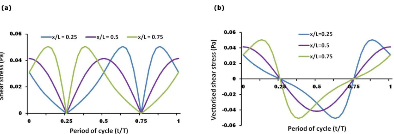
The FSS at the base of the culture wells were calculated
at 3 separate points in space (x/L = 0.25, 0.5, 0.75, where x is the distance from the edge of the well and L is the diameter of the well) along the middle of the well parallel
to the luid movement during one rocking cycle. The FSS
varied in a spatiotemporal fashion and were oscillatory in
nature (Fig. 1a-b). Under the conditions used, the shear
stress at the centre of the well (x/L = 0.5) was found to vary in a sinusoidal manner peaking at 0.041 Pa, while at the other two locations (x/L = 0.25 or 0.75) the stress peaked at 0.051 Pa and deviated from a typical sinusoidal
wave. The stress proiles at locations x/L = 0.25 and 0.75
were identical except for a phase difference of 180 degrees.
The effect of FSS on matrix formation by MLO-A5 cells
The ALP activity of MLO-A5 cells peaked in both static and rocked groups at day 7 and remained constant up to
days 14 and 21 (Fig. 2a). Cells exposed to rocking appeared
to have 15 % lower ALP activity across all three time
points but this was not statistically signiicant. Collagen
production measured at day 14 was not affected by rocking, but by day 21 samples exposed to rocking had 25 % more
secreted collagen than static controls (Fig. 2b). Calcium
deposition assayed at day 21 was 2-fold higher when cells
were subjected to rocking (Fig. 2c). At day 21 collagen
and calcium staining in the rocked groups appeared more
uniform across the culture dish, whereas statically cultured
groups showed patchy staining (Fig. 2d).
The effect of FSS on the morphology of progenitor cells
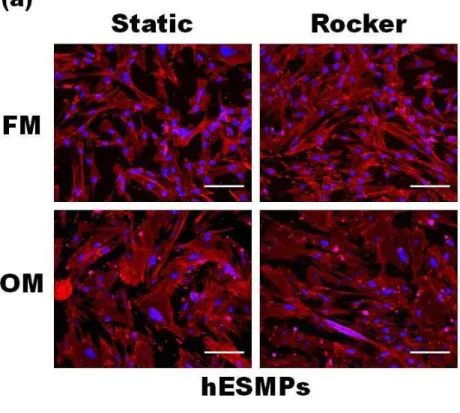
hES-MP cells cultured under static conditions in non-Dex
containing media (NM) had a ibroblastic, spindle-shaped
morphology, whereas hES-MPs cultured in osteogenic media (OM) were larger and more cuboidal in shape,
indicative of an osteoblastic cell (Fig. 3a). HDFs cultured in ibroblastic media (FM) showed a typical ibroblastic
morphology, however when cultured in OM they showed a
more cuboidal morphology (Fig. 3b) similar to the hES-MP
Fig. 1. (a) Calculated shear stress proiles for one cycle experienced at the base of a 6-well plate for three different locations, x/L = 0.25, 0.5, or 0.75, where x is the distance from the edge of the well and L is the diameter of the well. (b) The oscillatory nature of the luid low-induced shear stresses indicated by positive and negative stress.
cells and generally appeared larger than those cultured in
FM. Cellular alignment did not appear to be inluenced by luid low induced by rocking in any areas of the culture
well for either cell type.
The effect of FSS on total DNA and ALP activity of progenitor cells
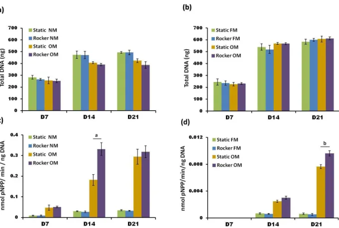
Total DNA content, an indicator of total cell number, increased for both cell types in all cell groups between days
7 and 14 and then remained constant up to day 21 (Fig.
4a-b). There were no statistically signiicant differences
in total DNA between cells that were rocked or cultured under static conditions but it was noticed that hES-MP cells cultured in OM did have 20 % less DNA at days 14 and 21 compared with cells cultured in NM. Normalised ALP activity increased in both cell types for all cell
groups up to day 21 (Fig. 4c-d), however no ALP activity was detectable in any HDF culture group at day 7. ALP
activity in hES-MPs was an order of magnitude higher in OM cultured cells compared to those cultured in NM. Fig. 3. Fluorescent DAPI staining
of cell nucleus (blue) and phalloidin T R I T C s t a i n i n g o f c e l l a c t i n cytoskeleton (red). (a) hES-MP cells cultured in either non-Dex containing media (NM) or osteogenic media (OM) under static or rocked conditions; or (b) HDFs cultured in either ibroblastic
media (FM) or OM under static or
rocked conditions. Cells cultured in
FM or NM show a more ibroblastic
Fig. 4. The effect of FSS on total DNA content of hES-MPs (a) and HDFs (b) and ALP activity (plotted normalised to total DNA) for hES-MPs (c) and HDFs (d) measured at days 7, 14 and 21. FSS did not affect total DNA for any
cell groups, but did cause statistically signiicant higher normalised ALP activity in Dex-treated cell groups. ALP activity in non-Dex-treated groups was minimal and FSS did not enhance this. Note the y axis range for HDFs is
smaller than that for hES-MPs due to lower ALP activity. All bar graphs are mean ± SD (n = 9) and signiicant differences between static and rocked cells are a = p < 0.01 and b = p < 0.05.
When OM was combined with rocking, ALP activity in hES-MPs was 2-fold higher at day 14 compared with their static counterparts. It appears that the rocking accelerated the upregulation of ALP activity, as by day 21 the static controls were as high as the rocked samples. However, there was no effect seen when NM was combined with
rocking. HDFs cultured in FM did not produce enough
detectable ALP above baseline values at any time point; when cultured in OM they began producing detectable levels of ALP at day 14, and by 21 these levels had increased 4 times compared to day 14. When OM cultured
HDFs were subjected to FSS, ALP activity increased
by 20 % over static counterparts at day 21, which was
statistically signiicant.
The effect of FSS on total collagen and calcium production by progenitor cells
Total collagen production quantiied by Picrosirius red
staining showed that cells cultured with Dex had produced less collagen at all time points, compared to those cultured
without Dex (Fig. 5a-b). For hES-MPs cultured in either
media group, the application of rocking caused the total amount of collagen deposited by day 21 to be 20 % higher (p < 0.05) (Fig. 5a). For HDFs subjected to rocking,
signiicantly more total collagen (p < 0.01) was seen at
days 14 and 21 for both media groups (Fig. 5b). Calcium
deposition assayed at day 21 was 3-fold higher in Dex treated hES-MPs subjected to rocking compared to static
counterparts and this staining was more uniform across the culture dish while static cells showed patchy staining
(Fig. 5c). In comparison, HDFs cultured in OM showed a
relatively small amount of calcium staining at day 21 but
rocking signiicantly increased the amount deposited by 50 % (Fig. 5d). The Alizarin stain was seen to concentrate
more around the centre of the wells and became fainter towards the outside of the well. Both hES-MP cells and
HDFs cultured without Dex did not produce any calcium
as visualised by the absence of Alizarin red stain.
Assessment of collagen production by second harmonic generation
The effect of FSS on collagen deposition and maturation was monitored in both HDFs and hESMP cells using SHG at days 7, 14 and 21 (Fig. 6a-b). Signal intensity
SHG intensity in hES-MP cells (Fig. 6a) than in HDFs (Fig.
6b) when compared with static counterparts. While rocking did not appear to induce a preferred direction of collagen orientation with either cell type in any media groups, rocking did appear to improve collagen organisation at day 14 and even more clearly at day 21. Statically cultured
groups showed short and disorganised collagen ibres, whereas FSS groups had thicker and longer bundles of ibres and this effect was more evident in groups cultured
without Dex.
Discussion
Our aims were to undertake research towards progressing tissue engineering of bone towards the clinic – examining a convenient possible source of osteogenic progenitor cells, assessing a simple methodology to apply mechanical stimulation to these cells, and monitoring collagen production and orientation using the minimally invasive technique of SHG.
This study was the irst, to our knowledge, to use a
simple platform rocking method to directly stimulate
progenitor cells using oscillatory FSS for enhancing osteogenic differentiation. This is also the irst study we know of to subject dermal ibroblasts and the hES-MP cell line to oscillatory FSS for the purpose of stimulating the
production of a mineralised collagenous matrix. Although it has been demonstrated that osteoprogenitor MSCs
respond to a variety of mechanical stimuli in a range of 2D and 3D bioreactor conditions (Delaine-Smith and Reilly, 2011), here we present the interesting result that
dermal ibroblasts cultured in osteogenic media produce a
mineralised collagenous matrix that is further enhanced by
oscillatory FSS. The reasons for selecting HDFs for use in
this study is that isolating progenitor cells from the dermis would have many advantages over other osteoprogenitor sources in that any donor will have large quantities of easily accessible skin and operations to remove it are simple and less painful than procedures to remove bone marrow.
HDFs also have a high proliferative potential and can be
expanded into large numbers in vitro.
The FSS calculated in this study were much lower than
those estimated to occur within mature bone (0.8-3 Pa (Weinbaum et al., 1994)), peaking at 0.041 Pa in the well
centre, and are also much lower than the FSS generally used
by others for mechanically stimulating osteoblastic cells, particularly in 2D (McCoy and O’Brien, 2010). However, the mature osteoblast MLO-A5 cell line responded to the shear forces with a noticeable increase in collagen and calcium production at day 21. Collagen production and organisation was improved in both the hES-MP cells and
the HDFs when subjected to these FSS. When cultured
in combination with osteogenic media, both cell types upregulated ALP activity and calcium production. Previous studies have also shown that hMSCs cultured in osteogenic supplements are mechanosensitive to relatively small shear Fig. 5. Total collagen production at days 7, 14 and 21 as quantiied by Picrosirius red staining for hES-MPs (a) and
HDFs (b). The application of FSS caused a statistically signiicant increase in collagen deposition for all cells at day 21, and cells treated with Dex produced less collagen than those without Dex. Calcium deposition was visualised at day 21 by Alizarin red staining for hES-MPs (c) and HDFs (d) and the application of FSS caused an increase in
Fig. 6. Second harmonic generation (SHG) images of deposited collagen produced by hES-MPs (a) and HDFs (b) at days 7, 14 and 21. Increases in collagen deposition and organisation are indicated by an increase in SHG intensity
and area coverage. A more organised collagen matrix can be observed in cells subjected to FSS, indicated by the appearance of thicker collagen bundles and more deined ibres. Images with a dark appearance did not produce
forces (0.036 Pa) applied for only short periods of time (Kreke et al., 2005). In a recent study, a T-75 lask rocking system was used to stimulate osteoblasts and osteocytes to condition media for MSCs (Hoey et al., 2011), and
although they did not calculate the FSSs present, it is likely
they would have also been relatively low. It is unclear whether tissue engineers should be attempting to replicate the mature bone environment or rather a developmental or fracture-healing environment where bone cells differentiate in vivo. Immature, developing bone tissue resembles a healing wound and not a mature tissue and so the forces experienced are likely to be different from those of a fully developed tissue, although little is known about what these forces are (Willie et al., 2010).
The shear stress proiles and the peak shear force
varied for different locations within the well plate, but the resulting calcium staining for MLO-A5 cells and hES-MPs showed a rather uniform pattern across the well. This indicates that either the range of forces being experienced by the cells have a similar effect on their differentiation or that the cells are communicating with each other, such as via gap junctions (Donahue, 2000; Taylor et al., 2007).
Another contributing factor could be that the luid low is
inducing chemotransport (Donahue et al., 2003) and so biochemical factors regulating bone cell metabolism, such as prostaglandin E2 (Genetos et al., 2005), are released into the media by the cells and moved around due to mass transport. While we did not test the mechanisms
by which luid low enhances osteogenic differentiation,
the data presented combined with other studies suggests that osteoblastic differentiation may be guided by soluble factors that accumulate in the media from a combination of externally applied chemical stimulants and direct mechanical stress on the cells (Hoey et al., 2011).
MLO-A5s are a late stage osteoblast/early stage osteocyte murine cell line that mineralise rapidly
(8-10 d) even in the absence of βGP (Kato et al., 2001) and they have been shown to respond to bouts of mechanical conditioning with enhanced matrix production (Morris et al., 2010; Sittichokechaiwut et al., 2010). MLO-A5s express high levels of ALP activity due to their advanced
stage of maturity and FSS were not seen to have a signiicant effect on these levels at the selected time points,
but calcium production was increased 2-fold. ALP activity is present in the early stages of osteogenesis and also plays a part in the initial stages of mineralisation via its enzymatic
hydrolysis activity (Yadav et al., 2011).
This study showed that hES-MPs and HDFs cultured in osteogenic media had signiicantly higher ALP activity than
those cultured in non-Dex containing media and this level continued to rise up to day 21. Some authors report ALP activity as a biphasic process, rising to a peak level before gradually decreasing again (Bancroft et al., 2002; Datta et al., 2006), but this was not seen here. FSS increased ALP activity in both sets of progenitor cells when cultured in osteogenic media and both cells subsequently increased their calcium deposition. ALP activity in the hES-MPs was
at least ten-fold higher than that in HDFs. The hES-MPs
are a relatively homogeneous population of cells derived from a single source of embryonic stem cells already
characterised as mesenchymal lineage speciic and able
to undergo osteogenesis in induction media (Karlsson et al., 2009). However, HDFs are a much more variable cell population from a mature adult donor and it is likely that only a sub-population of the cells can undergo osteogenic differentiation, or that the cells have varying levels of differentiation potential (Chen et al., 2007).
Cells derived from dermal tissue have previously been reported to show osteogenic differentiation potential (Bartsch et al., 2005; Chen et al., 2007) and this study
showed that HDFs produced ALP and deposited calcium
when cultured in osteogenic supplements. Previous studies
have tended to culture HDFs under static conditions but this study showed that the application of oscillatory FSS
could further enhance this osteogenic differentiation. In a previous study by Sommar et al. (2009), HDFs were cultured in a macroporous gelatine construct in the
presence of osteogenic media and subjected to FSS in a rotating spinner lask. They noticed the formation of
bone-like tissue, with further enhancements in the amount of
deposited mineral in constructs cultured in spinner lasks.
This observation, along with the present study, suggests
that FSS can enhance mineralised matrix in HDFs cultured in osteogenic media. This revelation that HDFs can be
induced towards an osteogenic phenotype using osteogenic
supplements and FSS highlights their potential use in the repair of bone. A limitation of our study was that only HDFs
from one patient were used and as with other progenitor populations, there will be cell variability resulting from variations in the source of tissue, such as tissue type and location within the body, donor characteristics, or in vitro passaging conditions.
Fibroblasts were only used to passage 3 because after
this they did not consistently make calcium at day 21, although they did continue to up-regulate ALP activity to similar levels (data not shown). The majority of studies
have used dermal ibroblast populations taken from foetal
or juvenile skin (Lavoie et al., 2009; Xue and Li, 2011), with the authors reporting loss of osteogenic potential or decreased potential at higher passage numbers. Some have reported that dermal progenitor populations display a delayed differentiation potential, often taking longer to mineralise than other MSC populations, anywhere between 4-8 weeks (Buranasinsup et al., 2006; Jaager and Neuman, 2011; Lorenz et al., 2008). However, there are a number of studies that have shown osteogenic differentiation to occur
in cells from mature and aged dermis (Xue and Li, 2011; Young et al., 2001). This loss of differentiation potential and donor variation could be a potential limitation with the future use of these cells for autologous bone repair, and so it is clear that more studies from a larger number of donors are required to assess their bone forming potential.
Monitoring matrix development by progenitor cells is very important for a successful tissue construct to
be developed. Collagen type 1 ibres are the primary
is visualised very clearly from the SHG images (Fig. 6) and this is the irst study that we know of to show the true
extent of this effect of Dex on collagen production using SHG. It has been reported that MSCs treated with Dex in vitro show a reduction in collagen production (Leboy et al., 1991; Ogston et al., 2002) and large concentrations of Dex used to treat patients for various conditions can cause bone loss or impairment of bone formation leading to osteoporosis (Scutt et al., 1996). Cells subjected to FSS also appeared to be more organised into thicker and longer
bundles of ibres when imaged using SHG; information
that could not be obtained from Picrosirius red staining. This enhanced collagen organisation suggests that cells
subjected to these FSS would produce tissues with stronger
tensile properties.
The process of converting mechanical stimulation into a biochemical response, mechanotransduction, is thought to occur through a number of mechanically-sensitive mechanisms including the cytoskeleton and integrins, ion channels, the glycocalyx and the primary cilia (Jacobs et al., 2010; Morris et al., 2010; Reilly et al., 2003; Weinbaum et al., 2007). Through these mechanisms,
the application of FSS initiates a number of signalling
events, including the synthesis and release of nitric oxide and prostaglandins (Klein-Nulend et al., 2005), a calcium signalling response and phosphorylation of the
mitogen-activated protein (MAP) kinase ERK (You et al., 2001). During osteogenic differentiation, the actin cytoskeleton in hMSCs remodels resulting in a morphological switch
from a ibroblastic fusiform shape to a square shape which
is more osteoblast-like. This was observed to happen with the hES-MP cells cultured in osteogenic media and a very
similar morphological switch was observed when HDFs were cultured in osteogenic media. When subjected to low,
both cells appeared to be more elongated in either media condition. This is thought to be due to a stiffening of the cell cytoskeleton, and it has been seen that stiffer cells tend to become more mechano-responsive perhaps due to the
forces being transmitted more eficiently (Yourek et al., 2010). Previous studies have shown that actin cytoskeletal tension is required for the activation of mechanosensors or signalling mechanisms involved in the regulation of intracellular processes and protein expression resulting
from FSS (Arnsdorf et al., 2009). Also a number of studies have shown that remodelling of the cell cytoskeleton can induce changes in the organisation and distribution of deposited collagen (Brammer et al., 2009; Koepsell et al., 2011).
have on progenitor cells is now a major research focus for musculoskeletal tissue engineers and their potential to aid healing and direct differentiation are being realised.
The simple system employed here created FSS that
enhanced osteogenic differentiation in mature bone cells and bone progenitor cells in the presence of osteogenic supplements. Using SHG, we saw an enhanced production and organisation of the major bone matrix protein collagen
caused by FSS. This system has many advantages in that
it is simple to use, can be used with many experimental samples, and could be easily scaled up for large defects. This system could be used for a number of tissue engineering strategies, such as pre-treating cells before injection into a scaffold or directly into a tissue defect or for stimulating cells cultured on thin scaffold sheets to be layered to form a 3D implantable tissue.
Acknowledgments
We gratefully acknowledge funding from the Engineering and Physical Sciences Research Council. Dr Steven
Matcher, University of Shefield kindly provided advice
on the SHG technique. Imaging work was performed at the
Kroto Research Institute Confocal Imaging Facility, using
the LSM510 Meta confocal microscope, thanks to Nicola Green for facility maintenance and technical assistance.
We wish to conirm that there are no known conlicts of
interest associated with this publication and there has been
no signiicant inancial support for this work that could have inluenced its outcome.
References
Arnsdorf EJ, Tummala P, Kwon RY, Jacobs CR (2009)
Mechanically induced osteogenic differentiation - the role of RhoA, ROCKII and cytoskeletal dynamics. J Cell Sci 122: 546-553.
Asakura A, Komaki M, Rudnicki MA (2001) Muscle satellite cells are multipotential stem cells that exhibit myogenic, osteogenic, and adipogenic differentiation. Differentiation 68: 245-253.
Augello A, De Bari C (2010) The regulation of differentiation in mesenchymal stem cells. Hum Gene Ther 21: 1226-1238.
Augst A, Marolt D, Freed LE, Vepari C, Meinel L, Farley M, Fajardo R, Patel N, Gray M, Kaplan DL,
grown using human mesenchymal stem cells, silk scaffolds and bioreactors. J R Soc Interface 5: 929-939.
Bancroft GN, Sikavitsas VI, van den Dolder J, Shefield TL, Ambrose CG, Jansen JA, Mikos AG (2002) Fluid low
increases mineralized matrix deposition in 3D perfusion culture of marrow stromal osteoblasts in a dose-dependent manner. Proc Natl Acad Sci USA 99: 12600-12605.
Bartsch G, Yoo JJ, De Coppi P, Siddiqui MM, Schuch G, Pohl HG, Fuhr J, Perin L, Soker S, Atala A (2005)
Propagation, expansion, and multilineage differentiation of human somatic stem cells from dermal progenitors. Stem Cell Dev 14: 337-348.
Bassey EJ, Ramsdale SJ (1994) Increase in femoral bone-density in young-women following high-impact excercise. Osteoporosis Int 4: 72-75.
Bayan C, Jonathan ML, Miller E, Kaplan D,
Georgakoudi I (2009) Fully automated, quantitative, noninvasive assessment of collagen iber content and
organiaztion in thick collagen gels. J Appl Phys 105: 102042
Bosch P, Musgrave DS, Lee JY, Cummins J, Shuler F, Ghivizzani SC, Evans C, Robbins PD, Huard J (2000)
Osteoprogenitor cells within skeletal muscle. J Orthop Res 18: 933-944.
Brammer KS, Oh S, Cobb CJ, Bjursten LM, van der Heyde H, Jin S (2009) Improved bone-forming functionality on diameter-controlled TiO(2) nanotube surface. Acta Biomater 8: 3215-3223.
Buranasinsup S, Sila-asna M, Bunyaratvej N, Bunyaratvej A (2006) In vitro osteogenesis from human skin-derived precursor cells. Dev Growth Differ 48: 263-269.
Chen FG, Zhang WJ, Bi D, Liu W, Wei X, Chen FF, Zhu L, Cui L, Cao Y (2007) Clonal analysis of nestin(-) vimentin(+) multipotent ibroblasts isolated from human
dermis. J Cell Sci 120: 2875-2883.
Chen YJ, Huang CH, Lee IC, Lee YT, Chen MH, Young TH (2008) Effects of cyclic mechanical stretching
on the mRNA expression of tendon/ligament-related and
osteoblast-speciic genes in human mesenchymal stem
cells. Connect Tissue Res 49: 7-14.
Dalby MJ, Gadegaard N, Tare R, Andar A, Riehle MO, Herzyk P, Wilkinson CDW, Oreffo ROC (2007) The control of human mesenchymal cell differentiation using nanoscale symmetry and disorder. Nat Mater 6: 997-1003.
Datta N, Pham QP, Sharma U, Sikavitsas VI, Jansen
JA, Mikos AG (2006) In vitro generated extracellular
matrix and luid shear stress synergistically enhance 3D
osteoblastic differentiation. Proc Natl Acad Sci USA 103: 2388-2493.
De Ugarte DA, Morizono K, Elbarbary A, Alfonso Z, Zuk PA, Zhu M, Dragoo JL, Ashjian P, Thomas B, Benhaim
P, Chen I, Fraser J, Hedrick MH (2003) Comparison of
multi-lineage cells from human adipose tissue and bone marrow. Cells Tissues Organs 174: 101-109.
Delaine-Smith RM, Reilly GC (2011) The effects of mechanical loading on mesenchymal stem cell differentiation and matrix production. Vitam Horm 87: 417-480.
Donahue HJ (2000) Gap junctions and biophysical regulation of bone cell differentiation. Bone 26: 417-422.
Donahue TLH, Haut TR, Yellowley CE, Donahue
HJ, Jacobs CR (2003) Mechanosensitivity of bone cells to oscillating fluid flow induced shear stress may be modulated by chemotransport. J Biomech 36: 1363-1371.
Erices A, Conget P, Minguell JJ (2000) Mesenchymal progenitor cells in human umbilical cord blood. Brit J Haematol 109: 235-242.
Genetos DC, Geist DJ, Liu DW, Donahue HJ, Duncan
RL (2005) Fluid shear-induced ATP secretion mediates
prostaglandin release in MC3T3-E1 osteoblasts. J Bone Miner Res 20: 41-49.
Goodwin HS, Bicknese AR, Chien SN, Bogucki BD,
Oliver DA, Quinn CO, Wall DA (2001) Multilineage
differentiation activity by cells isolated from umbilical cord blood: Expression of bone, fat, and neural markers. Biol Blood Marrow Transplant 7: 581-588.
Gurkan UA, Akkus O (2008) The mechanical environment of bone marrow: a review. Ann Biomed Eng 36: 1978-1991.
Hoey DA, Kelly DJ, Jacobs CR (2011) A role for the primary cilium in paracrine signaling between mechanically stimulated osteocytes and mesenchymal stem cells. Biochem Biophys Res Commun 412: 182-187.
Jaager K, Neuman T (2011) Human dermal ibroblasts
exhibit delayed adipogenic differentiation compared with mesenchymal stem cells. Stem Cell Dev, 20: 1327-1336.
Jacobs CR, Temiyasathit S, Castillo AB (2010) Osteocyte mechanobiology and pericellular mechanics. Ann Rev Biomed Eng 12: 369-400.
Jaiswal N, Haynesworth SE, Caplan AL, Bruder SP
(1997) Osteogenic differentiation of puriied,
culture-expanded human mesenchymal stem cells in vitro. J Cell Biochem 64: 295-312.
Janmey PA, McCulloch CA (2007) Cell mechanics: Integrating cell responses to mechanical stimuli. Ann Rev Biomed Eng 9: 1-34.
Karlsson C, Emanuelsson K, Wessberg F, Kajic K,
Axell MZ, Eriksson PS, Lindahl A, Hyllner J, Strehl R (2009) Human embryonic stem cell-derived mesenchymal progenitors. Potential in regenerative medicine. Stem Cell Res 3: 39-50.
Kato Y, Boskey K, Spevak L, Dallas M, Hori M, Bonewald LF (2001) Establishment of an osteoid
preosteocyte-like cell MLO-A5 that spontaneously mineralizes in culture. J Bone Miner Res 16: 1622-1633.
Klein-Nulend J, Bacabac RG, Mullender MG (2005) Mechanobiology of bone tissue. Pathol Biol 53: 576-580.
Koepsell L, Remund T, Bao J, Neuield D, Fong H, Deng Y (2011) Tissue engineering of annulus ibrosus using electrospun ibrous scaffolds with aligned polycaprolactone ibers. J Biomed Mater Res Part A 4: 564-575.
Kreke MR, Huckle WR, Goldstein AS (2005) Fluid
flow stimulates expression of osteopontin and bone sialoprotein by bone marrow stromal cells in a temporally dependent manner. Bone 36: 1047-1055.
Lavoie JF, Biernaskie JA, Chen Y, Bagli D, Alman B, Kaplan DR, Miller FD (2009) Skin-derived precursors
differentiate into skeletogenic cell types and contribute to bone repair. Stem Cell Dev 18: 893-905.
partially demineralized bone scaffolds in vitro. Tissue Eng 10: 81-92.
McCoy RJ, O’Brien FJ (2010) Inluence of shear stress
in perfusion bioreactor cultures for the development of three-dimensional bone tissue constructs: A review. Tissue Eng Part B Rev 16: 587-601.
Morris HL, Reed CI, Haycock JW, Reilly GC (2010) Mechanisms of fluid-flow-induced matrix production in bone tissue engineering. Proc Inst Mech Eng H, 224: 1509-1521.
Ogston N, Harrison AJ, Cheung HFJ, Ashton BA,
Hampson G (2002) Dexamethasone and retinoic acid differentially regulate growth and differentiation in an immortalised human clonal bone marrow stromal cell line with osteoblastic characteristics. Steroids 67: 895-906.
Pittenger MF, Mackay AM, Beck SC, Jaiswal RK,
Douglas R, Mosca JD, Moorman MA, Simonetti DW, Craig S, Marshak DR (1999) Multilineage potential of adult human mesenchymal stem cells. Science 284: 143-147.
Reilly GC, Engler AJ (2010) Intrinsic extracellular matrix properties regulate stem cell differentiation. J Biomech 43: 55-62.
Reilly GC, Haut TR, Yellowley CE, Donahue HJ, Jacobs CR (2003) Fluid low induced PGE(2) release by
bone cells is reduced by glycocalyx degradation whereas calcium signals are not. Biorheology 40: 591-603.
Rui YF, Lui PPY, Ni M, Chan LS, Lee YW, Chan KM
(2011) Mechanical loading increased BMP-2 expression which promoted osteogenic differentiation of tendon-derived stem cells. J Orthop Res 29: 390-396.
Rutherford RB, Moalli M, Franceschi RT, Wang D, Gu
K, Krebsbach PH (2002) Bone morphogenetic
protein-transduced human ibroblasts convert to osteoblasts and
form bone in vivo. Tissue Eng 8: 441-452.
Scutt A, Bertram P, Brautigam M (1996) The role of glucocorticoids and prostaglandin E(2) in the recruitment of bone marrow mesenchymal cells to the osteoblastic
lineage: Positive and negative effects. Calciied Tissue Int
59: 154-162.
Sharp LA, Lee YW, Goldstein AS (2009) Effect of low-frequency pulsatile low on expression of osteoblastic
genes by bone marrow stromal cells. Ann Biomed Eng 37: 445-453.
Sittichokechaiwut A, Edwards JH, Scutt AM, Reilly GC (2010) Short bouts of mechanical loading are as effective as dexamethasone at inducing matrix production by human bone marrow mesenchymal stem cells. Eur Cells Mater 20: 45-57.
strain on bone morphogenetic protein (BMP-2) mRNA expression. Tissue Eng 12: 3459-3465.
Taylor AF, Saunders MM, Shingle DL, Cimbala JM,
Zhou Z, Donahue HJ (2007) Mechanically stimulated osteocytes regulate osteoblastic activity via gap junctions. Am J PhysiolCell Physiol 292: C545-C552.
Toma JG, Akhavan M, Fernandes KJL, Barnabe-Heider F, Sadikot A, Kaplan DR, Miller FD (2001) Isolation of
multipotent adult stem cells from the dermis of mammalian skin. Nat Cell Biol 3: 778-784.
Tucker RP, Franklin S, Okech W, Chen D, Ventikos Y,
Thompson MS (2011) Validated in vitro cyclic shear stress alters human tenocyte ECM synthesis. Histol Histopathol 26 (suppl 1): 27.P6.
Weinbaum S, Cowin SC, Zeng Y (1994) A model for
the excitation of osteocytes by mechanical loading-induced
bone luid shear stress. J Biomech 27: 339-360.
Weinbaum S, Tarbell JM, Damiano ER (2007) The structure and function of the endothelial glycocalyx layer. Ann Rev Biomed Eng 9: 121-167.
Willie BM, Petersen A, Schmidt-Bleek K, Cipitria
A, Mehta M, Strube P, Lienau J, Wildemann B, Fratzl
P, Duda G (2010) Designing biomimetic scaffolds for bone regeneration: why aim for a copy of mature tissue properties if nature uses a different approach? Soft Matter 6: 4976-4987.
Xue S, Li L (2011) Upregulation of collagen type 1
in aged murine dermis after transplantation of dermal multipotent cells. Clin Exp Dermatol 36: 775-781.
Yadav MC, Simao AMS, Narisawa S, Huesa C, McKee MD, Farquharson C, Millan JL (2011) Loss of skeletal
mineralization by the simultaneous ablation of PHOSPHO1
and alkaline phosphatase function: A uniied model of the mechanisms of initiation of skeletal calciication. J Bone
Miner Res 26: 286-297.
You J, Reilly GC, Zhen XC, Yellowley CE, Chen Q, Donahue HJ, Jacobs CR (2001) Osteopontin gene regulation by oscillatory luid low via intracellular calcium
mobilization and activation of mitogen-activated protein kinase in MC3T3-E1 osteoblasts. J Biol Chem 276: 13365-13371.
Young HE, Steele TA, Bray RA, Hudson J, Floyd JA,
Yourek G, McCormick SM, Mao JJ, Reilly GC (2010)
Shear stress induces osteogenic differentiation of human mesenchymal stem cells. Regen Med, 5:713-724.
Zhou XZ, Liu DW, You LD, Wang LY (2010) Quantifying luid shear stress in a rocking culture dish. J
Biomech 43: 1598-1602.
Zuk PA, Zhu M, Mizuno H, Huang J, Futrell JW,
Katz AJ, Benhaim P, Lorenz HP, Hedrick MH (2001) Multilineage cells from human adipose tissue: Implications for cell-based therapies. Tissue Eng 7: 211-228.
Discussion with Reviewers
Reviewer I: Why use ESC derived MSC and not bone
marrow/adipose derived? Are the authors satisied that
these are a good representation and what surface markers are they positive for?
Authors: The human embryonic stem cell-derived mesenchymal progenitor (hES-MP) cell line are free of undifferentiated hESC markers (Oct-4, TRA 1-60, TRA 1-81, SSEA-3, SSEA-4) but express the MSC markers (CD105, CD166, CD10, CD13) along with vimentin and desmin. hES-MP cells are readily available (Cellartis) and easy to culture and represent a relatively homogenous MSC cell line that can be passaged up to 20 times without
any signiicant loss of proliferative capacity. They show
similar differentiation potential to bone marrow-derived MSCs (Karlsson et al., 2009) but perform with a higher consistency. We used this cell line as model for MSCs where there would be less variability than with primary human MSCs, and suggest they are a good cell type to use for optimisation of mechanical stimulation parameters to subsequently be used for primary adult MSCs.
Reviewer I: Do the authors believe HDFs to have a good future in bone tissue engineering?
Authors: It is not currently known whether the cells derived from a skin biopsy which undergo osteogenesis
are the HDFs themselves or progenitor cells residing amongst the HDFs. There are also relatively few studies looking at the osteogenic potential of HDFs compared with
those of bone marrow or adipose-derived MSCs. Some of these studies suggest that they are a potential abundant source of autologous cells for bone tissue engineering but that more in vivo studies are needed to see how well they can incorporate into bone tissue. Certainly, autologous keratinocytes have proved very valuable for treating patients with extensive skin loss due to burns, wounds or chronic ulcers (MacNeil, 2007), and more recently
dermal ibroblasts have been shown to have within them
a population of cells which appear to be pre-angiogenic (Krajewska et al., 2011). The latter study suggests that
the HDF cultures contain progenitor cells for several
differentiation pathways.
Reviewer I: How speciic is SHG to collagen detection and what else might be visualised (if anything)? Do the
authors see this as a simple technique that could be picked up in different labs with the right kit?
Authors: Only molecules lacking a centre of symmetry
can exhibit SHG, and this is ampliied with increasing
organisation of the structure. Collagen is a very strong emitter of SHG and is often very well organised, e.g. in tendon, so tissues or tissue-engineered constructs containing collagen can produce SHG. It is unsure whether other tissue components produce SHG, but if they do then
it is not expected to be signiicant. When using multiphoton
lasers to produce SHG, there is also the possibility of unwanted two-photon fluorescence from other tissue components spilling into the SHG emission window,
however with the correct ilters this can be minimised. We
have carried out wavelength dependant studies and found that the selection of 940 nm as the excitation source not only increases SHG intensity at a given power compared with other excitation wavelengths (800-1000 nm) but also
removes unwanted luorescence from other components.
The equipment required to visualise SHG is a confocal microscope and a multiphoton laser capable of excitation at wavelengths anywhere between 700 to 1060 nm. Using the right imaging conditions, any user familiar with confocal microscopy should be able to perform SHG easily, and as the sample requires no processing it is a very quick method for imaging collagen.
Reviewer II: With respect to the impact on the patient, how much skin would need to be removed to produce a
clinically relevant number of low passage HDFs?
Authors: A small 3 mm skin biopsy is suficient to extract
and expand dermal ibroblasts. After 2-3 weeks of culture,
tens of millions of cells will be available by passage 3.
Reviewer IV: The rocking system only allows for
oscillatory low, not for continuous low. This may be a disadvantage for looking at different low modes. Please
elaborate.
Authors: Yes this is true. However, in mature bone (and many other tissues in the body, such as the bone marrow),
luid low is oscillatory in nature and so we are interested
in replicating this stimulus. It is true that one could not
compare, for instance, unidirectional to oscillatory low in this system. For that one could use the parallel plate low
chamber system. However, the rocker system has many advantages in that it has simple operation, a large number of samples can be stimulated at once, and it requires small volumes of media.
Additional References
MacNeil S (2007) Progress and opportunities in tissue engineering of skin. Nature 445: 874-880.


