Ambient Temperature and Cerebrovascular
Hemodynamics in the Elderly
Wen-Chi Pan1, Melissa N. Eliot1, Petros Koutrakis2, Brent A. Coull3, Farzaneh A. Sorond4, Gregory A. Wellenius1*
1Department of Epidemiology, Brown University School of Public Health, Providence, RI, United States of America,2Department of Environmental Health, Harvard School of Public Health, Boston, MA, United States of America,3Department of Biostatistics, Harvard School of Public Health, Boston, MA, United States of America,4Department of Neurology, Brigham and Women’s Hospital and Institute for Aging Research, Hebrew SeniorLife, Boston, MA, United States of America
Abstract
Background and Purpose
Some prior studies have linked ambient temperature with risk of cerebrovascular events. If causal, the pathophysiologic mechanisms underlying this putative association remain unknown. Temperature-related changes in cerebral vascular function may play a role, but this hypothesis has not been previously evaluated.
Methods
We evaluated the association between ambient temperature and cerebral vascular function among 432 participants65 years old from the MOBILIZE Boston Study with data on cere-brovascular blood flow, cerecere-brovascular resistance, and cerecere-brovascular reactivity in the middle cerebral artery. We used linear regression models to assess the association of mean ambient temperature in the previous 1 to 28 days with cerebrovascular hemodynam-ics adjusting for potential confounding factors.
Results
A 10°C increase in the 21-day moving average of ambient temperature was associated with a 10.1% (95% confidence interval [CI], 2.2%, 17.3%) lower blood flow velocity, a 9.0% (95% CI, 0.7%, 18.0%) higher cerebrovascular resistance, and a 15.3% (95%CI, 2.7%, 26.4%) lower cerebral vasoreactivity. Further adjustment for ozone and fine particulate matter (PM2.5) did not materially alter the results. However, we found statistically significant
interac-tions between ambient temperature and PM2.5such that the association between
tempera-ture and blood flow velocity was attenuated at higher levels of PM2.5.
Conclusions
In this elderly population, we found that ambient temperature was negatively associated with cerebral blood flow velocity and cerebrovascular vasoreactivity and positively OPEN ACCESS
Citation:Pan W-C, Eliot MN, Koutrakis P, Coull BA, Sorond FA, Wellenius GA (2015) Ambient Temperature and Cerebrovascular Hemodynamics in the Elderly. PLoS ONE 10(8): e0134034. doi:10.1371/ journal.pone.0134034
Editor:James M. Brophy, McGill University Health Center / Royal Victoria, CANADA
Received:February 15, 2015
Accepted:July 5, 2015
Published:August 10, 2015
Copyright:© 2015 Pan et al. This is an open access article distributed under the terms of theCreative Commons Attribution License, which permits unrestricted use, distribution, and reproduction in any medium, provided the original author and source are credited.
Data Availability Statement:The authors confirm that all third-party data underlying the findings are fully available without restriction. The data are available upon request to the MOBILIZE Boston Study Data Repository Principal Investigator at the Hebrew SeniorLife Institute for Aging Research. Requests for the data may be sent to Robert R. McLean, DSc, MPH ([email protected]).
associated with cerebrovascular resistance. Changes in vascular function may partly underlie the observed associations between ambient temperature and risk of cerebrovas-cular events.
Introduction
Ambient temperature has been associated with increased risk of cerebrovascular events and cerebrovascular death [1–9], although results of prior studies have been inconsistent. For example, risk of ischemic stroke has been both positively [3–5] and negatively [6–9] associated with ambient temperature. In the peripheral circulation, higher ambient temperatures have been associated with reduced flow-mediated dilation in the brachial artery [10,11], a marker of vascular reactivity. However, the effects of ambient temperature on the cerebral circulation have not been previously studied.
The potential effects of ambient temperature on cerebrovascular hemodynamics must be considered in the context of ambient air pollution, which has been repeatedly linked to changes in peripheral vascular function [12–15]. We have previously reported an association between ambient fine particulate matter air pollution (PM2.5) and resting cerebrovascular flow and
resistance [16]. While that analysis adjusted for potential confounding by ambient tempera-ture, we did not consider the potential interactions between temperature and PM2.5or the
potential associations with O3.
Understanding the relationships between ambient temperature and cerebrovascular func-tion may yield insights into the mechanisms of weather-related cerebrovascular events and inform future prevention or treatment strategies. Accordingly, we evaluated the association between ambient temperature and cerebrovascular flow, resistance, and vasoreactivity in a pro-spective cohort of community-dwelling elderly in the Boston metropolitan area. A secondary goal was to evaluate the joint effects on cerebral hemodynamics of temperature with either PM2.5or O3.
Materials and Methods
Study Design
We evaluated the association between short-term changes in ambient temperature and markers of cerebrovascular hemodynamics among 423 participants in the MOBILIZE Boston study, a prospective, community-based cohort study [17]. Briefly, between 2005 and 2008 we recruited
765 non-institutionalized men and women aged≧65 years who were able to communicate in
English, resided within 5 miles (8.0 km) from the study clinic, and were able to walk 20 feet (6.1 m) without personal assistance. Individuals with a Mini-Mental State Examination score <18 were not eligible to participate. On enrollment, subjects participated in an in-home
inter-view followed within 4 weeks by a clinic examination. We assessed participant characteristics, medical history, medication inventory, smoking history, blood pressure, height, and weight, as previously described [18]. A second in-home interview and clinic examination (follow-up visit) were performed a median of 16.5 months after the baseline visit. All participants provided writ-ten informed consent upon enrollment. The study was approved by the Institutional Review Boards at Hebrew Senior Life and Brown University.
Participants were classified as normotensive if blood pressure was<140/90mmHg and there was no history of hypertension or receiving antihypertensive medications; controlled hypertensive if blood pressure was<140/90 mmHg and there was a history of hypertension or
Disorders and Stroke, and Environmental Health Sciences (NIEHS), NIH, and grant RD83479801 (PK) from the US Environmental Protection Agency (US EPA). The contents of this report are solely the responsibility of the authors and do not necessarily represent the official views of the sponsoring institutions.
receiving antihypertensive medication; and uncontrolled hypertensive if blood pressure was
140/90mmHg. A blood sample was collected during the clinic visit and participants were
classified as having diabetes mellitus if they reported a past diagnosis of diabetes, they reported
using any diabetes medications, were found to have a hemoglobin A1c6.5%, or had a
ran-dom glucose measurement200 mg/dl. Height and weight were measured during the clinic
visit according to a standard protocol and body mass index calculated.
Cerebrovascular Hemodynamics
At each clinic examination we evaluated participants’cerebrovascular hemodynamics at rest and during provocative stimulation, as previously described [17]. Briefly, we used transcranial Doppler ultrasound (TCD) to continuously measure cerebral blood flow velocity in the middle cerebral artery (MCA) while participants sat in a chair. A 2-MHz TCD probe (MultiDop X4, DWL-Transcranial Doppler Systems Inc., Sterling, VA) was placed over the right or left tempo-ral bone with the best signal and held in place during recordings using a Velcro headband. TCD data could not be obtained in some participants because of the absence of a suitable acoustic window to insonate the MCA. We measured arterial blood pressure using a Finometer photoplethysmographic system (Finapres Medical Systems, Arnhem, the Netherlands) placed on a finger and held at heart level with a sling. The envelope of the velocity waveform was digi-tized at 500 Hz, displayed simultaneously with the blood pressure, ECG, and end-tidal CO2
sig-nals; and stored for later offline analysis. Cerebrovascular resistance was calculated as the ratio of mean arterial pressure to blood flow velocity [19].
After a 5-minute resting period, we assessed cerebral vasoreactivity by asking participants to breathe room air normally for 2 minutes, inspire a gas mixture of 8% CO2, 21% O2, and balance
N2for 2 minutes, and then mildly hyperventilate to an end-tidal CO2of25 mm Hg for 2
minutes. Cerebral vasoreactivity was calculated as the slope of the linear regression of mean
MCA blood flow velocity versus end-tidal CO2during the maneuver.
Meteorological and Air Pollution Data
We obtained hourly ambient temperature and other meteorological data from the National Weather Service station at Boston’s Logan Airport. PM2.5was measured continuously at the
Boston/Harvard ambient monitoring station, as previously described [20]. This monitor was <10 km of the clinic site and<20 km from participants’residential address. Hourly O3 mea-surements were obtained from the Massachusetts Department of Environmental Protection’s Greater Boston monitoring sites and averaged. For each participant we estimated average ambient temperature, dew point temperature, PM2.5, and O3levels in the 1, 2, 3, 5, 7, 14, 21,
and 28 days prior to the clinical visit when cerebrovascular hemodynamics were measured.
Statistical Methods
Of the 765 participants in the MOBILIZE Boston Study, we excluded 76 participants with a his-tory of stroke, 1 participant with a clinical visit on a weekend (making it difficult to statistically adjust for potential day of week effects), and 265 subjects due to the absence of a suitable acous-tic window to insonate the MCA, leaving 423 paracous-ticipants for this analysis. Among these 423 participants, TCD data on cerebrovascular hemodynamics were available at two visits in 258 (61%) participants and at one visit in 165 participants.
were all natural log-transformed and results are expressed as the percent difference (and 95% confidence interval [CI]) in each outcome per 10°C change in ambient temperature, 10μg/m3
change in PM2.5, or 10 ppb change in O3. All models were adjusted for age (natural cubic spline
with 3 degrees of freedom), sex, race (whiteversusothers), smoking status (neverversusever),
hypertension status (normotension, controlled hypertension,versusuncontrolled
hyperten-sion), diabetes mellitus, body mass index (natural cubic spline with 3 degrees of freedom), visit number (baselineversusfollow-up visit), day of week (indicator variables), seasonal pattern
(sine and cosine of calendar time with period of 1 year), and long-term temporal trends (cen-tered time as linear and quadratic functions). Where indicated, we used natural cubic splines with 3 degrees of freedom, resulting in a function with 2 internal knots placed at the upper and lower tertiles of the distribution of the relevant variable. In sensitivity analyses we further adjusted for dew point temperature, PM2.5, or O3in separate models. The exposure-response
function of each outcome with temperature, PM2.5, and O3was initially modeled as a linear
function and, subsequently, as a natural cubic spline with 3 degrees of freedom.
In a set of secondary analyses, we evaluated potential interactions between ambient
temper-ature and PM2.5and between ambient temperature and O3by assuming linear
exposure-response functions and including interaction terms in our main models. In additional sensitiv-ity analyses, we verified these results using a low rank tensor product smoothing which allows more flexible exposure-response functions for the main effects and 2-way interactions [21]. Analyses were performed using R statistical software (version 3.1.0). A 2-sidedP-value of
<0.05 was considered statistically significant.
Results
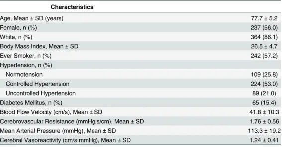
At baseline, the 423 participants with at least one assessment of cerebrovascular hemodynamics were predominantly female (56.0%) and white (86.1%), with a mean age of 77.7 (standard devi-ation [SD], 5.2) years (Table 1). Over the course of the study period, the mean ambient temper-ature was 11.0°C (SD: 9.2°C), mean levels of PM2.5were 9.0μg/m3(SD: 5.3μg/m3), and mean
O3levels were 22.9 ppb (SD: 10.7 ppb). Average pollutant levels over the 1 to 28 days prior
to each clinical assessment are shown in Table A inS1 Tables. The intraclass correlation
Table 1. Characteristics of 423 Participants from the MOBILIZE Boston Study.
Characteristics
Age, Mean±SD (years) 77.7±5.2
Female, n (%) 237 (56.0)
White, n (%) 364 (86.1)
Body Mass Index, Mean±SD 26.5±4.7
Ever Smoker, n (%) 242 (57.2)
Hypertension, n (%)
Normotension 109 (25.8)
Controlled Hypertension 224 (53.0)
Uncontrolled Hypertension 89 (21.0)
Diabetes Mellitus, n (%) 65 (15.4)
Blood Flow Velocity (cm/s), Mean±SD 41.8±10.3
Cerebrovascular Resistance (mmHg.s/cm), Mean±SD 1.76±0.56
Mean Arterial Pressure (mmHg), Mean±SD 113.3±19.2
Cerebral Vasoreactivity (cm/s.mmHg), Mean±SD 1.24±0.41
Abbreviations: SD, standard deviation.
coefficients for resting cerebrovascular resistance, mean arterial pressure, and blood flow veloc-ity ranged from 0.81 to 0.84, indicating high within-person reproducibilveloc-ity of these measures over time.
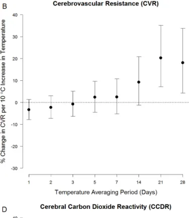
Measures of cerebral hemodynamics were associated with ambient temperature averaged over longer periods (Fig 1). For example, a 10°C increase in ambient temperature averaged over the prior 21 days (i.e.: the 21-day moving average) was associated with a 10.1% (95% CI: 2.2%, 17.3%) decrease in resting blood flow velocity, a 20.3% (95% CI: 7.1%, 35.1%) increase in resting cerebrovascular resistance, a 9.0% (95% CI: 0.7%, 18.0%) increase in resting mean arte-rial pressure, and a 15.3% (95%CI: 2.7%, 26.4%) decrease in cerebral vasoreactivity (Table B in
S1 Tables, Main Model). We used natural cubic splines and confirmed that the
exposure-response functions underlying these observed associations were approximately linear (Fig 2
andS1 Fig). Ambient temperature was not associated with any outcome at shorter moving
averages.
In sensitivity analyses, we assessed the robustness of the relationship between ambient tem-perature and cerebrovascular hemodynamics by further adjusting for PM2.5, O3, or dew point
temperature in separate models (Table B inS1 Tables). Adjustment for PM2.5modestly
attenu-ated the results although the overall pattern of the results remained unchanged. Adjustment for O3had little or no impact on the results. Adjustment for dew point temperature had an
inconsistent effect on the results, but substantially increased the width of the confidence inter-vals suggesting strong colinearity between ambient temperature and dew point (Spearman’s rank correlation coefficients 0.93 to 0.98).
We evaluated the potential interaction between temperature and PM2.5on cerebrovascular
hemodynamics and found that PM2.5modified the relationship between temperature and
rest-ing blood flow velocity, reachrest-ing statistical significance for the 5-, 14-, 21-, and 28-day movrest-ing averages (Table 2). Specifically, the association of ambient temperature with blood flow velocity was significantly attenuated at higher PM2.5concentrations andvice versa. For example, a 10°C
increase in the 21-day moving average of ambient temperature was associated with a 9.0% lower resting blood flow velocity at the 25thpercentile of PM2.5(6.76μg/m3), but a 6.4% lower
resting blood flow velocity at the 75thpercentile of PM2.5(9.85μg/m3) (Pfor interaction = 0.02).
We found no statistically significant interactions between ambient temperature and PM2.5on
other outcomes. We confirmed these findings using a more flexible 2-way interaction model that showed a similar pattern of results (data not shown). We found no evidence of interaction
between ambient temperature and O3with any measure of cerebrovascular function (Table C
inS1 Tables).
Discussion
In this cohort of elderly participants, we evaluated the association between cerebral hemody-namics and mean ambient temperature in the prior 1 to 28 days and found that, at longer aver-aging times, higher ambient temperatures were associated with higher mean arterial pressure, higher resting cerebrovascular resistance, lower resting blood flow velocity, and lower cerebro-vascular reactivity in response to changing end-tidal CO2levels. Additionally, at longer
averag-ing times we found evidence of an interaction between ambient temperature and PM2.5in
association with blood flow velocity.
Fig 1. Association between ambient temperature and (A.) resting blood flow velocity (BFV), (B.) resting cerebrovascular resistance (CVR), (C.) resting mean arterial pressure (MAP), (D.) and cerebral varoreactivity (Cerebral VR).They-axis denotes the % change (and 95% confidence interval) in
each outcome per 10°C increase in temperature, adjusted for age, sex, race, smoking, hypertension, diabetes, BMI, visit number, day of week, season, and long-term time trends. The x-axis denotes the averaging period for ambient temperature (in days) prior to the TCD assessment.
Fig 2. Exposure-response functions between 21-day average ambient temperature and (A.) resting blood flow velocity, (B.) resting cerebrovascular resistance, (C.) resting mean arterial pressure, and (D.) cerebral varoreactivity modeled using natural cubic splines with 3 degrees of freedom.Dashed lines represent 95% confidence intervals.
vascular reactivity [22], suggesting elevated body temperature may perturb cerebrovascular regulation and potentially lead to increased risk of stroke [23].
More is known about the effects of ambient temperature on the peripheral circulation. Nawrot et al [10]. found that higher ambient temperatures in the prior 1 to 21 days were associ-ated with reduced brachial artery flow-mediassoci-ated vasodilatation, indicative of reduced endothe-lial function. Similarly, in the Framingham Heart Study, Widlanskyet al [11]. found that
higher same-day temperatures were associated with reduced hyperemic flow velocity, a marker of peripheral microvascular vasodilator function. These results may be considered analogous to and qualitatively consistent with our findings of reduced cerebrovascular reactivity in the MCA territory in association with ambient temperature, albeit over different time scales. On the other hand, Widlanskyet al. [11] found no association between same-day temperature and
either resting brachial artery diameter or flow-mediated dilation. In a repeated-measures study among patients with type 2 diabetes, Zanobettiet al. found that same-day temperature was
pos-itively associated with brachial artery diameter but not with either flow-mediated dilation or nitroglycerin-mediated dilation [24]. These studies differ from ours, in part, in the characteris-tics of participants and time periods considered. Importantly, only the study by Nawrot et al. [10] considered the effects of averages of temperature longer than 5 days on markers of endo-thelial function.
We have previously reported that in this cohort PM2.5is associated with higher resting
cere-brovascular resistance and lower resting blood flow velocity at longer moving averages [16]. In the current study we found evidence of an interaction between PM2.5and ambient temperature
for blood flow velocity, but not other outcomes. The physiologic basis for this interaction is unclear, but since the observed associations with temperature and PM2.5are in the same
direc-tion (i.e.: to decrease blood flow velocity), it is plausible that as blood flow velocity decreases other compensatory mechanisms are activated to preserve cerebral blood flow. Nonetheless, if our findings on cerebral circulation can be extrapolated to the peripheral circulation, our results suggest that there may be important interactions between ambient temperature and at
Table 2. Joint effects of ambient temperature and PM2.5on blood flow velocity.
Moving Average (Days prior to clinic visit)
P Interactiona
% Difference (95% CI) in Blood Flow Velocity per 10°C Increase in Ambient
Temperature
% Difference (95% CI) in Blood Flow Velocity per 10μg/m3Increase in PM2.5
PM2.5at 25th percentileb
PM2.5at 75th percentileb
Temperature at 25th percentilec
Temperature at 75th percentilec
1 0.08 0.1(-4.5, 4.8) 1.4 (-3.1, 6.3) -4.2 (-10.0, 3.2) 0.3(-8.7, 4.6)
2 0.22 -0.9 (-6.1, 4.6) 0.2 (-5.0, 5.8) -5.2 (-11.9, 3.1) -1.7 (-10.9, 4.3) 3 0.10 -1.7 (-7.4, 4.4) -0.1 (-6.0, 6.0) -7.3 (-14.0, 1.9) -2.2 (-12.6, 3.5) 5 0.05 -4.6 (-11.3, 2.7) -2.7 (-9.5, 4.7) -8.9 (-16.0, 2.3) -1.4 (-14.3, 4.3) 7 0.11 -3.5 (-11.2, 4.9) -1.8 (-9.6, 6.7) -10.9 (-18.9, 0.7) -4.6 (-17.6, 2.4) 14 0.02 -7.8 (-17.0, 2.5) -4.9 (-14.4, 5.7) -15.7 (-23.6, -1.2) -3.5 (-21.2, 1.9) 21 0.02 -9.0 (-19.4, 2.7) -6.4 (-17.0, 5.7) -19.6 (-27.5, -5.0) -7.5 (-25.4, -2.2) 28 <0.01 -7.6 (-18.7, 5.0) -4.2 (-15.7, 8.9) -22.1 (-29.2, -6.4) -6.5 (-26.6, -3.0)
Abbreviations: PM2.5,fine particulate matter.
aFrom models including covariates for temperature, PM
2.5, the cross product of temperature and PM2.5, and adjusting for potential confounding factors. bThe 25thpercentile of PM
2.5at ranged from 5.19 to 6.88μg/m3, and the 75thpercentile ranged from 9.93 to 10.2μg/m3. cThe 25thpercentile of ambient temperature ranged from 2.81 to 3.38°C, and the 75thpercentile ranged from 18.1 to 18.7°C.
least some ambient air pollutants in eliciting endothelial dysfunction, but these interactions might only be observed at longer averaging times.
Arterial blood pressure has been shown to be negatively associated with same-day ambient temperature [25–29]. We similarly observed a modest negative association between mean arte-rial pressure and ambient temperature averaged over the prior 1–3 days, although these results did not reach statistical significance. However, at longer averaging periods, we found that ambient temperature was positively and significantly associated with mean arterial pressure. Few prior studies have considered comparably longer averaging periods. However, previous authors report that nighttime temperature is positively associated with blood pressure, poten-tially via reduced sleep quality or duration [30,31], suggesting that temperature may affect blood pressure via different physiologic mechanisms depending on both the time frame and the context of exposure. Our findings that the association between temperature and arterial blood pressure may be in opposite directions depending on the time frame considered raises potentially interesting questions about the differential effects of temperature on the risk of car-diovascular events. Indeed, time-series studies have often found that excess heat is associated with increased risk of cardiovascular events over the next 1–3 days while excess cold increases the risk of cardiovascular events over the next month or so [32,33]. Additional studies con-firming or refuting our observations are clearly needed.
Impaired vasoreactivity assessed by TCD is an established predictor of subsequent stroke, especially among patients with high grade carotid artery disease [34]. If higher temperatures do indeed increase the risk of cererbrovascular events (although, as mentioned above, the evi-dence for this remains equivocal), our results implicate impaired vasoreactivity as one potential mechanism.
We found no association between O3and any measure of cerebrovascular hemodynamics.
This finding is consistent with previous experimental and observational studies failing to find associations between recent O3levels and peripheral vascular function [29,35,36].
Addition-ally, our findings indicated that the association between ambient temperature and blood flow velocity was modified by PM2.5levels. Although we are not aware of any studies examining
how air pollutants interact with temperature on cerebrovascular function, several observational studies suggest the interplay of meteorological variables and air pollutants on peripheral vascu-lar function. For example, Hampelet al. [37] found a significant interaction between PM2.5and
ambient temperature on blood pressure among pregnant women. Further experimental studies may be needed to uncover the biological mechanism underlying the potentially complex inter-actions between pollutants and meteorological variables in eliciting vascular responses.
individuals. On the other hand, our study has several strengths including a novel hypothesis, a relatively large sample size, a well-characterized study population, and a detailed assessment of cerebrovascular hemodynamics.
Conclusions
In conclusion, in this cohort of elderly participants, we found that ambient temperature was associated with lower cerebral blood flow velocity, higher cerebrovascular resistance, higher mean arterial blood pressure, and lower cerebral vasoreactivity. The association between ambi-ent temperature and blood flow velocity was attenuated at high levels of PM2.5, but unaffected
by ambient O3levels. These findings build upon and extend the growing literature on
periph-eral vascular effects of temperature and air pollution and provide insights into the potential mechanisms of weather-related cardiovascular morbidity and mortality.
Supporting Information
S1 Fig. Exposure-response functions between ambient temperature and markers of cerebral hemodynamics among 423 participants in the MOBILIZE Boston Study.Natural cubic splines with 3 degrees of freedom were applied to model the association between ambient tem-perature and each outcome (blood flow velocity, cerebrovascular resistance, mean arterial pres-sure, and cerebral vasoreactivity) averaging temperature over different periods (1-, 7-, 14-, 21-, or 28-day) prior to the clinic visit. The dashed lines denote the 95% confidence intervals. The carpet plots along thex-axis denote the density of temperature values. All models were adjusted
for age, sex, race, smoking status, hypertension status, diabetes, body mass index, visit number, day of week, season, and long-term temporal trends. The concentration-response plot of 1-day moving average was similar to the 2- and 3-day plots, and the 7-day moving average plot was similar approximate to the 5-day plot.
(TIFF)
S1 Tables. Supplemental tables.
(DOCX)
Author Contributions
Conceived and designed the experiments: GAW W-CP FAS. Performed the experiments: FAS. Analyzed the data: W-CP MNE GAW. Contributed reagents/materials/analysis tools: BAC PK FAS. Wrote the paper: W-CP MNE PK FAS GAW.
References
1. Pan WH, Li LA, Tsai MJ. Temperature extremes and mortality from coronary heart disease and cerebral infarction in elderly Chinese. Lancet. 1995; 345(8946):353–5. PMID:7845116.
2. Michelozzi P, Accetta G, De Sario M, D'Ippoliti D, Marino C, Baccini M, et al. High temperature and hos-pitalizations for cardiovascular and respiratory causes in 12 European cities. Am J Respir Crit Care Med. 2009; 179(5):383–9. doi:10.1164/rccm.200802-217OCPMID:19060232.
3. Basu R, Pearson D, Malig B, Broadwin R, Green R. The effect of high ambient temperature on emer-gency room visits. Epidemiology. 2012; 23(6):813–20. Epub 2012/09/26. doi:10.1097/EDE. 0b013e31826b7f97PMID:23007039.
4. Ostro B, Rauch S, Green R, Malig B, Basu R. The effects of temperature and use of air conditioning on hospitalizations. American journal of epidemiology. 2010; 172(9):1053–61. Epub 2010/09/11. doi:10. 1093/aje/kwq231PMID:20829270.
6. Hong YC, Rha JH, Lee JT, Ha EH, Kwon HJ, Kim H. Ischemic stroke associated with decrease in tem-perature. Epidemiology. 2003; 14(4):473–8. PMID:12843774.
7. Magalhaes R, Silva MC, Correia M, Bailey T. Are stroke occurrence and outcome related to weather parameters? Results from a population-based study in northern portugal. Cerebrovascular diseases. 2011; 32(6):542–51. doi:10.1159/000331473PMID:22104569.
8. Goggins WB, Woo J, Ho S, Chan EY, Chau PH. Weather, season, and daily stroke admissions in Hong Kong. International journal of biometeorology. 2012; 56(5):865–72. doi:10.1007/s00484-011-0491-9 PMID:21915799.
9. Mostofsky E, Wilker EH, Schwartz J, Zanobetti A, Gold DR, Wellenius GA, et al. Short-term changes in ambient temperature and risk of ischemic stroke. Cerebrovascular diseases extra. 2014; 4(1):9–18. Epub 2014/02/28. doi:10.1159/000357352PMID:24575110; PubMed Central PMCID: PMC3934677.
10. Nawrot TS, Staessen JA, Fagard RH, Van Bortel LM, Struijker-Boudier HA. Endothelial function and outdoor temperature. European journal of epidemiology. 2005; 20(5):407–10. PMID:16080588.
11. Widlansky ME, Vita JA, Keyes MJ, Larson MG, Hamburg NM, Levy D, et al. Relation of season and temperature to endothelium-dependent flow-mediated vasodilation in subjects without clinical evidence of cardiovascular disease (from the Framingham Heart Study). The American journal of cardiology. 2007; 100(3):518–23. PMID:17659939; PubMed Central PMCID: PMC1994775.
12. Brook RD, Brook JR, Urch B, Vincent R, Rajagopalan S, Silverman F. Inhalation of fine particulate air pollution and ozone causes acute arterial vasoconstriction in healthy adults. Circulation. 2002; 105 (13):1534–6. PMID:11927516.
13. O'Neill MS, Veves A, Zanobetti A, Sarnat JA, Gold DR, Economides PA, et al. Diabetes enhances vul-nerability to particulate air pollution-associated impairment in vascular reactivity and endothelial func-tion. Circulafunc-tion. 2005; 111(22):2913–20. PMID:15927967.
14. Briet M, Collin C, Laurent S, Tan A, Azizi M, Agharazii M, et al. Endothelial function and chronic expo-sure to air pollution in normal male subjects. Hypertension. 2007; 50(5):970–6. PMID:17875820.
15. Brook RD, Urch B, Dvonch JT, Bard RL, Speck M, Keeler G, et al. Insights into the mechanisms and mediators of the effects of air pollution exposure on blood pressure and vascular function in healthy humans. Hypertension. 2009; 54(3):659–67. Epub 2009/07/22. doi:10.1161/HYPERTENSIONAHA. 109.130237PMID:19620518; PubMed Central PMCID: PMC3706996.
16. Wellenius GA, Boyle LD, Wilker EH, Sorond FA, Coull BA, Koutrakis P, et al. Ambient fine particulate matter alters cerebral hemodynamics in the elderly. Stroke. 2013; 44(6):1532–6. Epub 2013/05/28. doi: 10.1161/STROKEAHA.111.000395PMID:23709640; PubMed Central PMCID: PMC3722046.
17. Leveille SG, Kiel DP, Jones RN, Roman A, Hannan MT, Sorond FA, et al. The MOBILIZE Boston Study: design and methods of a prospective cohort study of novel risk factors for falls in an older popu-lation. BMC geriatrics. 2008; 8:16. PMID:18638389. doi:10.1186/1471-2318-8-16
18. Wellenius GA, Wilhelm-Benartzi CS, Wilker EH, Coull BA, Suh HH, Koutrakis P, et al. Ambient particu-late matter and the response to orthostatic challenge in the elderly: the Maintenance of Balance, Inde-pendent Living, Intellect, and Zest in the Elderly (MOBILIZE) of Boston study. Hypertension. 2012; 59 (3):558–63. doi:10.1161/HYPERTENSIONAHA.111.180778PMID:22275528; PubMed Central PMCID: PMC3286019.
19. Sorond FA, Galica A, Serrador JM, Kiely DK, Iloputaife I, Cupples LA, et al. Cerebrovascular hemody-namics, gait, and falls in an elderly population: MOBILIZE Boston Study. Neurology. 2010; 74 (20):1627–33. Epub 2010/05/19. doi: 74/20/1627 [pii] doi:10.1212/WNL.0b013e3181df0982PMID: 20479362; PubMed Central PMCID: PMC2875129.
20. Kang CM, Koutrakis P, Suh HH. Hourly Measurements of Fine Particulate Sulfate and Carbon Aerosols at the Harvard-U.S. Environmental Protection Agency Supersite in Boston. Journal of the Air & Waste Management Association. 2010; 60(11):1327–34. WOS:000283971800005.
21. Wood SN. Low-rank scale-invariant tensor product smooths for generalized additive mixed models. Biometrics. 2006; 62(4):1025–36. doi:10.1111/j.1541-0420.2006.00574.xPMID:17156276.
22. Lavinio A, Timofeev I, Nortje J, Outtrim J, Smielewski P, Gupta A, et al. Cerebrovascular reactivity dur-ing hypothermia and rewarmdur-ing. British journal of anaesthesia. 2007; 99(2):237–44. doi:10.1093/bja/ aem118PMID:17510046.
23. Silvestrini M, Vernieri F, Pasqualetti P, Matteis M, Passarelli F, Troisi E, et al. Impaired cerebral vasor-eactivity and risk of stroke in patients with asymptomatic carotid artery stenosis. Jama. 2000; 283 (16):2122–7. PMID:10791504.
25. Madsen C, Nafstad P. Associations between environmental exposure and blood pressure among par-ticipants in the Oslo Health Study (HUBRO). European journal of epidemiology. 2006; 21(7):485–91. PMID:16858621.
26. Alperovitch A, Lacombe JM, Hanon O, Dartigues JF, Ritchie K, Ducimetiere P, et al. Relationship between blood pressure and outdoor temperature in a large sample of elderly individuals: the Three-City study. Archives of internal medicine. 2009; 169(1):75–80. doi:10.1001/archinternmed.2008.512 PMID:19139327.
27. Halonen JI, Zanobetti A, Sparrow D, Vokonas PS, Schwartz J. Relationship between outdoor tempera-ture and blood pressure. Occupational and environmental medicine. 2011; 68(4):296–301. doi:10. 1136/oem.2010.056507WOS:000288164700011. PMID:20864465
28. Modesti PA, Morabito M, Massetti L, Rapi S, Orlandini S, Mancia G, et al. Seasonal Blood Pressure Changes An Independent Relationship With Temperature and Daylight Hours. Hypertension. 2013; 61 (4):908–14. doi:10.1161/Hypertensionaha.111.00315WOS:000316112800037. PMID:23381792
29. Hoffmann B, Luttmann-Gibson H, Cohen A, Zanobetti A, de Souza C, Foley C, et al. Opposing effects of particle pollution, ozone, and ambient temperature on arterial blood pressure. Environ Health Per-spect. 2012; 120(2):241–6. doi:10.1289/ehp.1103647PMID:22020729; PubMed Central PMCID: PMC3279434.
30. Brook RD, Shin HH, Bard RL, Burnett RT, Vette A, Croghan C, et al. Can personal exposures to higher nighttime and early-morning temperatures increase blood pressure? Journal of clinical hypertension. 2011; 13(12):881–8. doi:10.1111/j.1751-7176.2011.00545.xPMID:22142347; PubMed Central PMCID: PMC3234119.
31. Modesti PA, Morabito M, Massetti L, Rapi S, Orlandini S, Mancia G, et al. Seasonal blood pressure changes: an independent relationship with temperature and daylight hours. Hypertension. 2013; 61 (4):908–14. doi:10.1161/HYPERTENSIONAHA.111.00315PMID:23381792.
32. Nordio F, Zanobetti A, Colicino E, Kloog I, Schwartz J. Changing patterns of the temperature-mortality association by time and location in the US, and implications for climate change. Environment interna-tional. 2015; 81:80–6. Epub 2015/05/13. doi:10.1016/j.envint.2015.04.009PMID:25965185.
33. Anderson GB, Bell ML. Heat waves in the United States: mortality risk during heat waves and effect modification by heat wave characteristics in 43 U.S. communities. Environmental health perspectives. 2011; 119(2):210–8. Epub 2010/11/19. doi:10.1289/ehp.1002313PMID:21084239; PubMed Central PMCID: PMC3040608.
34. Reinhard M, Schwarzer G, Briel M, Altamura C, Palazzo P, King A, et al. Cerebrovascular reactivity pre-dicts stroke in high-grade carotid artery disease. Neurology. 2014; 83(16):1424–31. Epub 2014/09/14. doi:10.1212/WNL.0000000000000888PMID:25217057; PubMed Central PMCID: PMC4206163.
35. Fakhri AA, Ilic LM, Wellenius GA, Urch B, Silverman F, Gold DR, et al. Autonomic effects of controlled fine particulate exposure in young healthy adults: effect modification by ozone. Environ Health Per-spect. 2009; 117(8):1287–92. Epub 2009/08/13. doi:10.1289/ehp.0900541PMID:19672410; PubMed Central PMCID: PMC2721874.
36. Barath S, Langrish JP, Lundback M, Bosson JA, Goudie C, Newby DE, et al. Short-term exposure to ozone does not impair vascular function or affect heart rate variability in healthy young men. Toxicologi-cal sciences: an official journal of the Society of Toxicology. 2013; 135(2):292–9. doi:10.1093/toxsci/ kft157PMID:23872581; PubMed Central PMCID: PMC3807622.