Changes in red blood cell osmotic
fragility induced by total plasma and
plasma fractions obtained from rats
bearing progressive and regressive
variants of the Walker 256 tumor
1Laboratório de Pesquisas Bioquímicas, Centro de Assistência Integral à Saúde da
Mulher, and 2Departamento de Bioquímica, Instituto de Biologia,
Universidade Estadual de Campinas, Campinas, SP, Brasil
3Departamento de Ciências da Saúde, Colegiado de Medicina,
Universidade Estadual de Santa Cruz, Ilhéus, BA, Brasil T.C. Cavalcanti1,
C.C. Gregorini2,
F. Guimarães1,
O. Rettori3 and
A.N. Vieira-Matos3
Abstract
Two variants (A and B) of the widely employed Walker 256 rat tumor cells are known. When inoculated sc, the A variant produces solid, invasive, highly metastasizing tumors that cause severe systemic effects and death. We have obtained a regressive variant (AR) whose sc growth is slower, resulting in 70-80% regression followed by development of immunity against A and AR variants. Simultaneously with the beginning of tumor regression, a temporary anemia devel-oped (~8 days duration), accompanied by marked splenomegaly (~300%) and changes in red blood cell osmotic fragility, with mean corpuscular fragility increasing from 4.1 to 6.5 g/l NaCl. The possibil-ity was raised that plasma factors associated with the immune response induced these changes. In the present study, we identify and compare the osmotic fragility increasing activity of plasma fractions obtained from A and AR tumor bearers at different stages of tumor develop-ment. The results showed that by day 4 compounds precipitating in 60% (NH4)2SO4 and able to increase red blood cell osmotic fragility appeared in the plasma of A and AR tumor bearers. Later, these compounds disappeared from the plasma of A tumor bearers but slightly increased in the plasma of AR tumor bearers. Furthermore, by day 10, compounds precipitating between 60 and 80% (NH4)2SO4 and with similar effects appeared only in plasma of AR tumor bearers. The salt solubility, production kinetics and hemolytic activity of these compounds resemble those of the immunoglobulins. This, together with their preferential increase in rats bearing the AR variant, suggest their association with an immune response against this tumor.
Correspondence
T.C. Cavalcanti
Laboratório de Pesquisas Bioquímicas CAISM/UNICAMP
Caixa Postal 6150 13083-970 Campinas, SP Brasil
E-mail: [email protected]
Research supported by CAPES and CAISM/UNICAMP. C.C. Gregorini was the recipient of a CAPES/CNPq fellowship. Publication supported by FAPESP.
Received August 21, 2002 Accepted April 22, 2003
Key words
•Plasma fractions
•Temporary anemia
•RBC osmotic fragility
•Tumor regressive variant
•Cancer immunity
Introduction
The Walker 256 (W256) tumor cells have been extensively used in studies of cancer pathophysiology (1-4). Two variants of this tumor have been reported, namely A and B (3). The A variant has been widely em-ployed. It is known that tumor variants can be obtained by repeated passages through different media, a phenomenon that can be reversed by successive passages through the original medium (5). In our laboratory, after 60 successive intraperitoneal (ip) passages, a regressive variant (AR) of the W256 A tumor was obtained. The subcutaneous (sc) unifocal growth of the AR tumor variant is slower when compared to the A variant, and in 70-80% of the individuals tumor regres-sion occurs followed by development of im-munity against both the A and AR variants (Rettori O, Vieira-Matos AN, Guimarães F and Cavalcanti TC, unpublished data). Simi-lar examples of the development of immu-nogenic variants of several tumors can be found in the literature (6).
Previous studies of the host’s response to the AR variant have shown that 8 to 12 days after unifocal inoculation and preceding overt tumor regression, a temporary anemia al-ways develops, followed by an increase in red blood cell (RBC) osmotic fragility, a marked hypertrophy of the spleen (7) and presence in plasma of a substance able to increase the osmotic fragility of healthy RBC (Rettori O, Vieira-Matos AN, Guimarães F and Cavalcanti TC, unpublished data). Sple-nectomy always greatly augmented the lev-els of anemia, with a further increase in RBC osmotic fragility. The hemolytic mechanism of the anemia and its early and temporary manifestation (both suggesting the involve-ment of polyclonal immunoglobulins), the apparent role of the spleen in modulating its evolution and the fact that it appeared only in the rats bearing the AR tumor variant raised the possibility that factors appearing in the plasma, associated with the immune response,
could be responsible for the increase in RBC osmotic fragility observed in rats bearing the AR tumor variant, and that splenectomized tumor bearers could be a convenient source of these factors.
Considerable difficulty in experimental cancer studies, which usually employ uni-focal tumor inoculations, is related to the fact that the initiation of the systemic effects of cancer in individuals of the same group is a random phenomenon. It may start any time between 6 and 47 days or more after tumor inoculation, with deaths occurring between days 14 and 58 or more (7). By employing multifocal simultaneous sc inoculations (2, 4 or more inoculations/rat), the beginning of the systemic effects of cancer is rapid and highly synchronized among individuals of the same group, beginning 3-5 days after tumor cell inoculation and leading to death within 10-13 days (7-9). Therefore, studies employing multifocal inoculations yield rapid and highly reproducible results.
The purpose of the present study was to partially characterize the putative plasma factors responsible for the increase in RBC osmotic fragility in AR tumor-bearing rats. For this purpose, we monitored the time course of appearance of increasing RBC osmotic fragility activity in fractions ob-tained from plasma of splenectomized rats receiving multifocal inoculations of the A and AR variants of W256 tumor cells.
Material and Methods
Experimental design
re-ceived the same amount of Ringer-lactate containing the appropriate tumor cells. Be-fore tumor inoculation, 0.7 ml blood was collected from the suborbital plexus into a syringe containing 0.05 ml heparin (5000 IU/ml). A similar blood sample was also obtained on day 4 after the inoculations. On day 10, all animals were sacrificed under anesthesia and a large blood sample was collected following thoracotomy, iv heparin injection and heart section. Hemoglobin lev-els and the osmotic fragility of fresh RBC were also determined in all blood samples. On days 0, 4 and 10 after the inoculations, pools of plasma from 6 animals were ob-tained. One aliquot of each pool was frozen (-18ºC) for further testing of its effect on healthy RBC osmotic fragility, and the re-mainder was used in ammonium sulfate pre-cipitation studies. Finally, the effect of the plasma and its fractions on RBC osmotic fragility was tested employing fresh RBC from normal donors as described below.
Tumor and animals
The W256 A tumor line was provided by Dr. Maria C. Cintra Gomes of the Departa-mento de Fisiologia, Instituto de Biologia, UNICAMP. The line originally came from the National Cancer Institute Bank, Cam-bridge, MA, USA. This tumor and its AR variant are stored under liquid N2, and
main-tained by ip or sc passage in rats.
Eight-week-old male Wistar rats were used in this study. The animals were housed (5 per cage) at controlled room temperature (21º ± 2ºC) on a 12-h light-dark cycle. Ani-mals were allowed free access to standard rat chow (Labina/Purina, Campinas, SP, Brazil) and water. The rats were splenectomized and then randomly divided into 3 groups: a) controls (N = 6), b) W256 A variant tumor bearers (A, N = 6), and c) W256 AR variant tumor bearers (AR, N = 6). Each tumor-bearing rat received multifocal simultaneous sc inoculations at four dorsal sites, spaced at
least 1 cm apart, with 5 × 10⁶ tumor cells (A
or AR) suspended in 250 µl Ringer-lactate, in each site. The controls received identical inoculations of Ringer-lactate (placebo). Plasma pools (6 animals/pool) were used in the ammonium sulfate fractionation studies. Tumor cells with 98% viability assessed by Trypan blue were obtained from the ascitic fluid of donor rats. General United Kingdom Coordinating Committee on Cancer Research guidelines for animal welfare were followed (10).
Ammonium sulfate fractionation of plasma proteins
Ammonium sulfate was slowly added to the pool of total plasma (2.0-5.0 ml, diluted 1/10 in PBS) immediately after collection from control and tumor-bearing rats under constant stirring to a final concentration of 60%, w/v (11). Stirring was maintained for another 60 min and the solution was then centrifuged at 375 g for 30 min. The super-natant and precipitates were named S60 and P60, respectively. The ammonium sulfate concentration of the S60 fractions was in-creased to 80% to obtain a P80 fraction by centrifugation as described earlier. The P60 and P80 fractions were resuspended in 2.0 to 5.0 ml of 10 mM PBS, pH 7.4. Finally, all fractions were extensively dialyzed against PBS for 48 h with six buffer changes. Samples were diluted in PBS or concentrated with a 10-kDa molecular membrane (Spectrum, type C, POR
, Houston, TX, USA) to reconsti-tute the original volumes. All operations were carried out on ice at ± 4ºC. The protein content of the different fractions was meas-ured by the method of Lowry et al. (12).
Determination of hemoglobin and RBC osmotic fragility
cor-puscular fragility was employed for data anal-ysis of RBC osmotic fragility defined as the NaCl concentration (g/l) causing 50% RBC lysis (13).
RBC incubation tests
RBC from normal rats were used to test the osmotic fragility changes induced by the putative factors present in the plasma and its fractions from tumor bearers. After the cen-trifugation of blood, 150 µl of normal packed RBC was resuspended (final hematocrit: ~20%) and incubated at 37ºC for 30, 120 and 240 min in 0.6 ml of the following solutions: a) plasma from splenectomized controls; b) plasma from A tumor bearers; c) plasma from AR tumor bearers; d) fractions S60 control, A or AR; e) fractions P60 control, A or AR; f) fractions P80 control, A or AR; g) autologous plasma from RBC donors and last change dialysis buffer as controls. Be-fore incubation, in order to achieve a glucose concentration of ~150 mg/100 ml, 3.5 µl of 25% glucose was added to the solutions lacking glucose (dialysis buffer and dialyzed fractions).
Autopsy
Autopsies were performed in all tumor-bearing rats. Special attention was paid to the eventual presence of metastases, signifi-cant tissue invasion and bleeding.
Statistical analysis
The results are reported as means ± SEM and means(diff) ± SEM(diff) when analyzing
paired data. Statistical significance was de-termined by ANOVA followed by the post hoc Dunnett test (P < 0.05), and by the paired Student t-test.
Results
Development of anemia
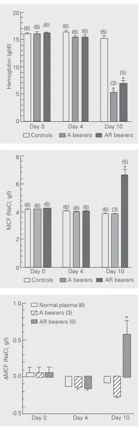
Although there were no changes on day 4, ten days after tumor inoculation (11-12 days after splenectomy), hemoglobin levels dropped markedly in tumor bearers, from 15.2 ± 0.5 to
Figure 1. Development of ane-mia by splenectomized rats in-oculated multifocally with A and AR Walker 256 (W256) tumor cells. Tumor cells (5 x 10⁶) were
simultaneously inoculated sc in four dorsal sites at least 1 cm apart. Data are reported as means ± SEM. The number of rats in each group is given in parentheses. *P < 0.01 com-pared to control on day 10 (paired Student t-test).
Figure 2. Changes of osmotic fragility of fresh RBC from sple-nectomized rats multifocally in-oculated with A and AR Walker 256 (W256) tumor cells. Data are reported as means ± SEM. The number of rats in each group is given in parentheses. MCF = mean corpuscular fragility. *P < 0.01 compared to control on day 10 (paired Student t-test).
Figure 3. Changes in osmotic fragility induced in normal donor RBC by incubation (30 min at 37ºC) with plasma obtained from controls, Walker 256 (W256) A tumor bearers and W256 AR tu-mor bearers at different inter-vals after tumor inoculation. Data are reported as means ± SEM. The number of experi-ments is given in parentheses. MCF = mean corpuscular fragil-ity. *P < 0.05 compared to con-trol on day 10 (ANOVA followed by the post hoc Dunnett test).
Day 0 Day 4 Day 10 Controls A bearers AR bearers (6) (6) (6) (6) (6) (6)
(6) (3) (5)
*
MCF (NaCl, g/l)
8
6
4
2
0
Day 0 Day 4 Day 10 1.0
0.5
0.0
-0.5
123 123 123 123 123 123 123
123 123 123
123
Normal plasma (6) 12
12
A bearers (3) AR bearers (5)
∆
MCF (NaCl, g/l)
*
Hemoglobin (g/dl)
20
15
10
5
0
(6) (6) (6) (6)
(6) (6) (6)
(3) (5)
* *
5.2 ± 0.4 g/100 ml in A tumor bearers and from 15.2 ± 0.5 to 6.7 ± 0.8 g/100 ml in AR tumor bearers. In control splenectomized rats, no significant changes were observed in hemo-globin levels (Figure 1).
Changes in RBC osmotic fragility induced by tumor growth
There was no significant change in the fresh RBC osmotic fragility of A tumor bear-ers in spite of the pronounced degree of anemia, while in AR tumor bearers a marked 58% rise in RBC osmotic fragility was ob-served on day 10 after inoculation (Figure 2). The increase in RBC osmotic fragility in these splenectomized animals usually started at about day 8 and reached a maximum (with intravascular hemolysis) around day 10, when the animals were sacrificed.
Changes in RBC osmotic fragility induced by unfractioned plasma and plasma fractions from A and AR tumor bearers
Only plasma samples obtained on day 10 from AR tumor bearers were able, after incu-bation at 37ºC, to induce a significant in-crease in osmotic fragility of fresh RBC from normal donor rats (Figure 3). A full effect of increase in osmotic fragility was already observed after 30 min of incubation at 37ºC and maintained from then on up to 240 min (data not shown).
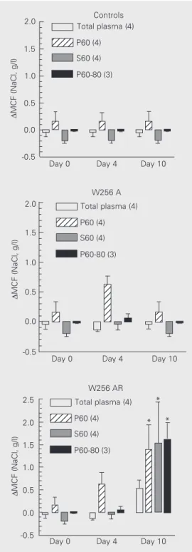
Regarding the plasma fractions, four days after inoculation, P60 obtained from A and AR plasmas induced an increase in RBC osmotic fragility of fresh RBC from normal donor rats. On day 10, this activity disap-peared in the plasma of the A tumor bearers, but remained in the plasma of the AR tumor-bearing rats (Figure 4). In addition, an im-portant activity increasing RBC osmotic fra-gility appeared on day 10 in the S60 fraction from AR tumor bearers. This new activity precipitated completely at 80% ammonium sulfate saturation; no RBC osmotic fragility
1234 1234
Total plasma (4) P60 (4) S60 (4) P60-80 (3) 12 12 12 12 12 12 12 12 12 12 12 12 12 12 12 12 12 12 12 12 2.5 1.0 0.5 -0.5 ∆
MCF (NaCl, g/l)
1.5 0.0 2.0 W256 AR 12 12 123 123 123 123 123 123 123 123 123 123
Day 0 Day 4 Day 10 12345
12345
Total plasma (4) P60 (4) S60 (4) P60-80 (3) 2.0 1.0 0.5 -0.5 ∆
MCF (NaCl, g/l)
1.5 0.0 W256 A 2.0 1.0 0.5 -0.5 ∆
MCF (NaCl, g/l)
1.5
0.0
Day 0 Day 4 Day 10 12345 12345 12345 12 12 12 12 12 12 Total plasma (4) P60 (4) S60 (4) P60-80 (3)
Controls
Day 0 Day 4 Day 10
increasing activity could be detected in the S80 fraction (Figure 4). Full effects were already detectable after a 30-min incubation and therefore the results obtained after 120-and 240-min incubation have not been in-cluded.
Figure 4. Changes in osmotic fragility induced in normal do-nor RBC by incubation with to-tal plasma and fractions from controls and from Walker 256 (W256) A and W256 AR tumor bearers obtained at different times after tumor inoculation. Data are reported as means ± SEM. The number of experi-ments is given in parentheses. P60: precipitate from 60% am-monium sulfate saturation; S60: supernatant from 60% ammo-nium sulfate saturation; P60-80: precipitate from 60-80% ammo-nium sulfate saturation. MCF, mean corpuscular fragility. *P < 0.05 compared to total plasma (ANOVA followed by the
post hoc Dunnett test).
* *
Autopsy
No evidence of metastasis was found in any of the experimental animals, nor signifi-cant tissue invasion or bleeding that could have masked the present observations.
Discussion
Cancer anemia
Cancer anemia is classified as an anemia of chronic diseases, whose pathogenic mech-anism has been difficult to establish. Bone marrow production of RBC is usually slightly increased but insufficient to compensate for a moderately increased RBC destruction (14-17). At present, there are no clear explana-tions for either phenomenon, i.e., bone mar-row relative insufficiency and increased RBC destruction. It has been suggested that the activation of the immune and inflammatory systems through the increasing levels of pro-inflammatory cytokines such as interleukins 1 and 6, TNF-α and INF-δ would induce iron
retention by the reticuloendothelial system, gastrointestinal tract and liver, therefore ex-erting an inhibitory effect on erythroid pre-cursors (18-20). The erythropoietin produc-tion and bone marrow response to it are decreased in cancer anemia (4,18,20,21) and treatment with recombinant erythropoietin has been reported to be only of temporary help (14,22). Macro- and/or microscopic al-terations of bone marrow have long been ruled out as factors involved in cancer ane-mia in humans (23) and rats (4). In the terminal stage of cancer, RBC are rapidly destroyed, even transfused ones, at a rate of up to five times the physiological rate, with-out an increase in RBC osmotic fragility (8). This terminal anemia is not temporary but continues to increase with a very high corre-lation (r2 = 0.86) with the other severe
sys-temic effects involved in terminal general failure (8). Development of hemolytic ane-mia associated with increased RBC osmotic
fragility is not frequent in cancer and has been associated with a) autoimmune anti-bodies, b) microangiopathic alterations, and c) chemotherapy (24,25).
The present study emphasizes the com-plexity of the mechanisms involved in ane-mia induced by cancer. We had previously reported that in intact rats bearing the pro-gressive A variant of the W256 cancer the rapid terminal destruction of RBC was asso-ciated with a decrease or no change in RBC osmotic fragility (8). In contrast, rats inocu-lated with the AR tumor variant systemati-cally developed a temporary anemia associ-ated with a marked increase in RBC osmotic fragility at the beginning of tumor regres-sion. This anemia progressed and rapidly led to lethal intravascular hemolysis only in sple-nectomized rats, a model used in order to amplify the effect we wanted to study in the present investigation. Chemotherapy was not employed in the present study and the char-acteristic fragments of RBC present in mi-croangiopathic anemia were not found in the morphological studies performed on blood. The hemolytic nature of the anemia, its early and temporary manifestation in intact AR tumor bearers and the apparent role of the spleen in modulating it raise the possibility that immunoglobulins associated with an in-itial polyclonal immune response to the tu-mor could be involved in its mechanism, perhaps signaling the elaboration of a further step toward the development of an adaptive host immune response, whose success, how-ever, is still not guaranteed.
Development of anemia and changes in RBC osmotic fragility induced by tumor growth
rats, whose RBC are destroyed at about five times the physiological rate around the 10th day, as previously reported (8). Since sple-nectomized rats were used in the present experiments, this marked degree of anemia indicates that the spleen may not be the only site of RBC destruction in rats bearing the W256 tumor.
Ten days after inoculation a marked in-crease in osmotic fragility was detected only in RBC obtained from the animals receiving the AR variant of the W256 tumor cells (Figure 2). The presence of this activity only in plasma from rats bearing the immuno-genic AR variant is consistent with the idea discussed previously that this hemolytic ac-tivity could reflect some kind of immunore-sponse, maybe immunoglobulin production, against the W256 tumor. Contrary to the just temporary increase in RBC osmotic fragility observed in the intact AR tumor bearers, the increase in RBC osmotic fragility in the sple-nectomized AR tumor bearers progressed and fatally evolved to intravascular hemoly-sis and ultimately death, had the animals not been sacrificed in advance. These observa-tions confirmed preliminary experiments in-dicating an exacerbation of the increase in RBC osmotic fragility in splenectomized W256 AR tumor bearers, and supported the use of splenectomized animals in the experi-ments that followed this study in order to isolate the putative substance that increases RBC osmotic fragility. In non-tumor-bear-ing rats, as can be seen in Figures 1 and 2, splenectomy by itself induced no significant degree of anemia or alteration of RBC os-motic fragility up to day 10 postinoculation (days 11-12 postsplenectomy; see Material and Methods).
Another interesting observation shown in Figures 1 and 2 is that the anemia induced by the W256 tumor cells was independent of the changes in RBC osmotic fragility, since the degree of anemia found in the splenecto-mized A tumor bearers, whose RBC showed no significant changes in osmotic fragility,
was even higher than that observed in the AR tumor bearers, whose RBC osmotic fra-gility were markedly increased. In a previous study, intact rats inoculated with the W256 A tumor cells presented a decrease in RBC osmotic fragility associated with tumor growth (8). The lack of significant changes in RBC osmotic fragility in A tumor bearers observed in these experiments may be re-lated to the fact that splenectomized animals were used in the latter experiments. These observations indicate the complexity of the mechanisms involved in anemia induced by cancer.
Changes in RBC osmotic fragility induced by plasma from W256 A and AR tumor bearers
Consistent with the increase in RBC os-motic fragility observed in animals 10 days after inoculation with the AR tumor variant, only the plasma from AR tumor bearers was able to increase the RBC osmotic fragility of normal non-tumor-bearing donors under in-cubation at 37ºC (Figure 3). These tions confirmed our preliminary observa-tions and indicate that factors present in AR plasma may be responsible for the increase in RBC osmotic fragility observed in the animals bearing this regressive tumor vari-ant.
Changes in RBC osmotic fragility induced by plasma fractions from A and AR tumor bearers
activity disappeared on day 10 from the plasma of the W256 A tumor bearers, while persisting in the plasma of the animals bear-ing the AR tumor variant.
These results suggest that, during W256 tumor growth, compounds with a high mo-lecular weight, which are able to increase RBC osmotic fragility in A and AR tumor bearers, were produced. These molecules could be part of an initial immune response against the tumor, which would be sustained only by the animals bearing the AR tumor variant. Furthermore, only the AR bearers were able to produce other additional mol-ecules of smaller molecular weight with simi-lar activities, suggesting the initiation of a possible adaptive immune response against the W256 cancer. The biochemical nature of these molecules is not clear, but their pro-duction kinetics, salt solubility characteris-tics and hemolytic activity resemble those of
the immunoglobulin superfamily involved in immune responses.
In conclusion, changes in osmotic fragil-ity of RBC occur during growth of the W256 tumor, with early and temporary increase in osmotic fragility being characteristic of the immunogenic AR variant. Products associ-ated with this activity can be isolassoci-ated from plasma of tumor bearers, whose chemical nature and pathophysiological role deserve further studies.
Acknowledgments
The authors are indebted to Dr. Maria C. Cintra Gomes from the Departamento de Fisiologia, Instituto de Biologia, UNICAMP, for providing the original Walker 256 A tumor line and Mr. Amilton Garcia for ani-mal care.
References
1. Toal JN, Millar FK, Brooks RH & White J (1960). Sodium retention by rats bearing the Walker carcinosarcoma 256. American Journal of Physiology, 200: 175-181.
2. Morrison SB (1971). Water intake and exchange and hydration of rats during growth of Walker 256 carcinoma. Journal of the National Cancer Institute, 46: 825-830.
3. Guaitani A, Recchia M, Carli M, Rochetti M, Bart4sek I & Garatini S (1982). Walker carcinoma 256: a model for studies on tumor-in-duced anorexia and cachexia. Oncology, 39: 173-178.
4. Zucker S, Lysik RM & Di Stefano J (1977). Pathogenesis of anemia in rats with Walker 256 carcinosarcoma. Journal of Clinical Medi-cine, 90: 502-511.
5. Tannock IF (1983). Biology of tumor growth. Hospital Practice, 18: 81-93.
6. Martin F, Caignard A, Jeannin JF, Leclerc A & Martin MS (1983). Selection by trypsin of two sub-lines of rat colon cancer cells forming progressive or regressive tumors. International Journal of Cancer, 32: 623-627.
7. Rettori O, Vieira-Matos AN & Tahin QS (1995). Variability and dis-continuity of the pathognomonic systemic effects caused by Walker 256 tumor progression rats. Tumori, 81: 370-377.
8. Vido AA, Cavalcanti TC, Guimarães F, Vieira-Matos AN & Rettori O (2000). The hemolytic component of cancer anemia: effects of osmotic and metabolic stress on the erythrocytes of rats bearing multifocal inoculations of the Walker 256 tumor. Brazilian Journal of Medical and Biological Research, 33: 815-822.
9. Rettori O, Vieira-Matos AN & Gontijo JAR (2000). Re-assessment of the renal hydrosaline dysfunction in rats bearing the Walker tumor.
Renal Failure, 22: 769-784.
10. United Kingdom Coordinating Committee on Cancer Research (UKCCR) (1988). Guidelines for the welfare of animals in experimen-tal neoplasia. Laboratory Animals, 22: 195-201.
11. Green AA & Hughes WL (1955). Protein fractionation on the basis of solubility in aqueous solutions of salts and organic solvents. Meth-ods in Enzymology, 1: 67-90.
12. Lowry OH, Rosebrough NJ, Farr AL & Randall RJ (1951). Protein measurement with the Folin phenol reagent. Journal of Biological Chemistry, 193: 265-275.
13. Dacie JV, Lewis SM & Gordon SEC (1984). Practical Hematology. 6th edn. Churchill Livingstone, New York.
14. Spivak JL (1994). Cancer-related anemia: its causes and characteris-tics. Seminars in Oncology, 21: 3-8.
15. Erslev A (1995). Anemia of chronic disease. In: Beutler E, Lichtman MA, Coller BS & Kipps TJ (Editors), Williams Hematology. McGraw Hill Inc., New York, 518-524.
16. Ludwig H & Fritz E (1998). Anemia in cancer patients. Seminars in Oncology, 25: 2-6.
17. Cantoni S & Morra E (2001). Anemia of cancer: an overview. Tumori, 87: 54-57.
18. Jelkmann W (1998). Proinflammatory cytokines lowering erythro-poietin production. Journal of Interferon and Cytokine Research, 18: 555-558.
19. Andrews NC (1999). Disorders of iron metabolism. New England Journal of Medicine, 341: 1986-1995.
factor-alpha and erythropoietin serum levels in B-cell chronic lymphatic leukemia patients with anemia. Acta Haematologica, 108: 84-89. 21. Erslev AJ (2000). Erythropoietin and anemia of cancer. European
Journal of Haematology, 64: 353-358.
22. Abels R (1993). Erythropoietin for anaemia in cancer patients. Euro-pean Journal of Cancer, 22A: S2-S8.
23. Berlin IN (1974). Anemia of cancer. Annals of the New York Acade-my of Sciences, 230: 209-211.
24. Rytting M, Worth L & Jaffe N (1996). Hemolytic disorders associ-ated with anemia. Hematology/Oncology Clinics of North America, 10: 365-376.