The he m o lytic co m po ne nt o f cance r
ane m ia: e ffe cts o f o sm o tic and
m e tabo lic stre ss o n the e rythro cyte s
o f rats be aring m ultifo cal ino culatio ns
o f the Walke r 2 5 6 tum o r
1Departamento de Bioquímica, Instituto de Biologia, and
2Laboratório de Pesquisas Bioquímicas, Centro de Atenção Integral à Saúde
da Mulher, Universidade Estadual de Campinas, Campinas, SP, Brasil A.A. Vido1, T.C. Cavalcanti2,
F. Guimarães2,
A.N. Vieira-Matos2
and O . Rettori2
Abstract
Cancer anemia is classified as an anemia of chronic diseases, although it is sometimes the first symptom of cancer. Cancer anemia includes a hemolytic component, important in the terminal stage when even transfused cells are rapidly destroyed. The presence of a chronic component and the terminal complications of the illness limit studies of the hemolytic component. A multifocal model of tumor growth was used here to simulate the terminal metastatic dissemination stage (several simultaneous inoculations of Walker 256 cells). The hemolyt-ic component of anemia began 3-4 days after inoculation in 100% of the rats and progressed rapidly thereafter: Hb levels dropped from 14.9 ± 0.02 to 8.7 ± 0.06 from days 7 to 11 (~5 times the physiologically normal rate in rats) in the absence of bleeding. The development of anemia was correlated (r2
= 0.86) with the development of other systemic effects such as anorexia. There was a significant decrease in the osmotic fragility of circulating erythrocytes: the NaCl concentra-tion causing 50% lysis was reduced from 4.52 ± 0.06 to 4.10 ± 0.01 (P<0.01) on day 7, indicating a reduction in erythrocyte volume. However, with mild metabolic stress (4-h incubation at 37oC), the erythrocytes showed a greater increase in osmotic fragility than the controls, suggesting marked alteration of erythrocyte homeostasis. These effects may be due to primary plasma membrane alterations (transport and/or permeability) and/or may be secondary to metabolic changes. This multifocal model is adequate for studying the hemolytic component of cancer anemia since it is rapid, highly reproducible and causes minimal animal suffering.
Co rre spo nde nce
O . Rettori
Laboratório de Pesquisas Bioquímicas, CAISM, UNICAMP Caixa Postal 6151
13081-970 Campinas, SP Brasil
Fax: + 55-19-788-9383 E-mail: rettori@ caism.unicamp.br A.A. Vido was the recipient of a CAPES fellowship. Publication supported by FAPESP.
Received September 24, 1999 Accepted March 10, 2000
Ke y wo rds
·Hemolytic component ·Cancer anemia ·O smotic fragility ·Systemic effects ·Walker 256 tumor
Intro ductio n
Cancer anemia is usually classified as an anemia of chronic diseases (ACD), but is sometimes the first symptom to appear and therefore not necessarily a chronic
manifes-tation of cancer (1-3). Cancer anemia in-cludes a hemolytic component which mark-edly increases in the final stage of the illness, when even transfused red blood cells (RBC) are rapidly destroyed (4,5).
component are not well understood, and their study is difficult because of the simulta-neous presence of a chronic component, and because of ethical difficulties, treatments and other complications, particularly in the terminal stages.
In experimental studies such as those
previously performed with the Walker256
tumor inoculated at a single subcutaneous (sc) site, the development of anemia showed the general characteristics of ACD (5,6). However, if instead of the usual unifocal tumor growth model, the studies are initiated using multifocal simultaneous tumor inocu-lations (simulating the metastatic dissemina-tion of the final stage), the systemic effects of cancer, including anemia, appear rapidly and synchronously in all animals (7,8). The development of anemia under these condi-tions is similar to that found in terminal cancer patients, in which the rate of RBC destruction is very high (4,9). This multifo-cal model using the Walker 256 tumor there-fore seems to be particularly suited for the study of the hemolytic component of cancer anemia.
In the present study, we examined the time-course of the changes occurring in he-moglobin levels (Hb) and RBC osmotic fra-gility (OF) and correlated them with other systemic effects of cancer such as anorexia. The OF was studied in fresh whole blood samples and in the same RBC after
meta-bolic stress (in vitro incubation at 37o
C).
Mate rial and Me tho ds
Tum o r and anim als
The Walker 256 A tumor line was kindly provided by Dr. Maria C. Cintra Gomes, Department of Physiology, IB/ UNICAMP. The line originally came from the National Cancer Institute Bank, Cam-bridge, MA, USA. The tumor is currently
kept in the laboratory under liquid N2 and is
maintained through intraperitoneal or sc
pas-sages in rats.
Thirty-five 9-week-old male Wistar rats were used. The animals were housed at
con-trolled temperature (21o
C), on a 12-h light-dark cycle and fed a commercial rat diet (Labina/Purina, Campinas, SP, Brazil). The rats were randomly divided into experimen-tal (tumor) and pair-fed control groups of 20 and 15 animals, respectively. The tumor bear-ers were allowed free access to food, while the pair-fed controls were allowed to eat only the amount ingested the day before by the tumor bearers. Both had free access to water. Tumor-bearing rats received four dor-sal sc inoculations of 5 x 10⁶
tumor cells each in 0.25 ml of Ringer-lactate spaced at least 1 cm apart. Healthy tumor cells with 98% viability (assessed by Trypan blue) were obtained from the ascitic fluid of a donor rat. Control rats received four identical inocula-tions of vehicle only.
The general UKCCR guidelines for ani-mal welfare were followed (10). One of the authors was responsible for the daily clinical evaluation of the animals. Somatic and vis-ceral pain was explored by palpation and gentle compression of the limbs and axillary, inguinal, tumoral, peritoneal and thoracic regions. No animal needed to be sacrificed before the end of the experiment.
Expe rime ntal de sign
He m o gram
Hemograms were performed using a Cell-dyn 1600 apparatus and the results were corrected for the relative volume of heparin present in each blood sample.
O smo tic fragility and RBC incubatio n te sts
Red blood cell OF was determined as de-scribed by Dacie et al. (11). Packed RBC obtained from tumor bearers and controls (0.2 ml, followed by a 5-min centrifugation at 978 g) at sacrifice were incubated for 4 h at 37o
C after resuspension in different media: a) RL (1.0 ml of Ringer-lactate), b) RL + G (1.0 ml of Ringer-lactate + 2 mg of glucose), c) PLa (1.0 ml of autologous plasma) and d) PLa + G (1.0 ml of autologous plasma + 1 mg of glucose). After incubation, the OF was again deter-mined. To facilitate data analysis the median corpuscular fragility (MCF) was often used. MCF has been defined as the NaCl concentra-tion (g/l) causing 50% of RBC lysis (11).
Auto psy
Autopsies were performed on all tumor-bearing rats. The tumors were dissected and weighed, and special attention was paid to the eventual presence of metastases, the in-vasion of important tissues and bleeding.
Statistical analysis
The results are reported as means ± SEM. The statistical significance of the changes in fresh blood was determined by the Student
paired t-test. The significance of the metabolic
stress studies was tested by ANOVA and by the paired Student t-test (12).
Re sults
Tum o r de ve lo pm e nt
Tumors grew at all inoculated sites (4/
rat) and were palpable within 3 days. Their mean weights were 2.8 ± 0.2 g and 10.3 ± 0.6 g on days 7 and 11, respectively.
D e ve lo pm e nt o f ane m ia
Figure 1 shows the time-course of the changes in Hb and food intake during the experiment. The Hb levels decreased rapidly (P<0.001) in tumor bearers, dropping from 14.9 ± 0.2 to 8.7 ± 0.6 g/dl in the last 4 days
(1.55 g dl-1 day-1, about 5 times the normal
rate of RBC destruction in rats). The de-crease in Hb levels was correlated with the decrease in food intake: on days 4, 7 and 11 Hb levels decreased 2.3 ± 2.1, 16.7 ± 1.1 and 48.3 ± 3.3%, while the respective average
food intake decreased 27, 34 and 72% (r2 =
0.86, P<0.001).
O smo tic fragility change s in fre sh RBC
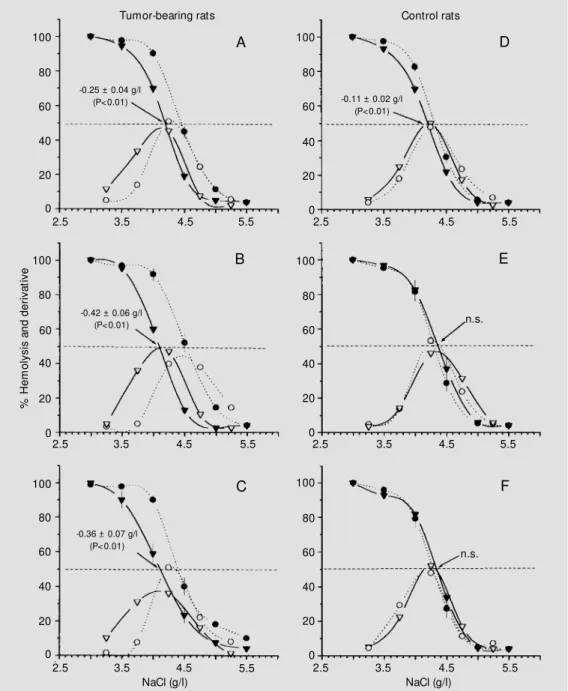
Figure 2 shows the OF curves of fresh RBC from tumor bearers and pair-fed con-trols, 4, 7 and 11 days after tumor inocula-tion. A significant shift to the left was ob-served in all tumor bearers (Figure 2A, B and C) which, expressed in MCF, corresponded to -0.25 ± 0.04, -0.42 ± 0.06 and -0.36 ± 0.07 g/l, respectively (P<0.01 in all instances). In the control rats there was a slight shift to the left on day 4 but no changes on days 7 and 11
Figure 1 - Changes in the hemo-globin levels and daily food take of rats w ith multifocal in-oculations of the Walker 256 tu-mor and of their pair-fed con-trols. The pair-fed control data w ere shifted backw ards one day in order to cancel the method-ological delay in the protocol (see M at erial and M et hods). * Tum or-bearing and pair-f ed control animals.
H
b
(
g
/d
l)
20
F
o
o
d
i
n
ta
k
e
(
g
/d
a
y
)
60
15
10
5
0
50
40
30
20
10
0 Hb Control
Hb Tumor
P<<0.001
Food intake*
0 10 20 30 40
(Figure 2D, E and F).
Consistent with the changes in OF the hemogram showed a decrease in median RBC corpuscular volume, expressed as fl, with values of 53.2 (range 53-54), 51.6 (range 51-52) and 50.0 (range 49-52) for tumor bearers on days 4, 7 and 11 after tumor
inoculation, respectively (r2 = 0.77, P<0.001).
In the control group, the values were 51.6 (range 51-53), 51.8 (range 50-53) and 50.2
(range 49-52), respectively (r2
= 0.21, not significant).
Another change observed on day 11 was the decrease in the slope of the RBC OF curves (Figure 2C). The maximal derivative value, which corresponds to the maximal percent variation in RBC lysis, induced by a change of 0.5 g/l in NaCl concentration dropped from 51.7 ± 1.8% on day zero to 43.7 ± 2.1% per 0.5 g/l on day 11 (-8.0 ±
%
H
e
m
o
ly
s
is
a
n
d
d
e
ri
v
a
ti
v
e
100
80
60
40
20
0
100
80
60
40
20
0
100
80
60
40
20
0
100
80
60
40
20
0
100
80
60
40
20
0 100
80
60
40
20
0
2.5 3.5 4.5 5.5 2.5 3.5 4.5 5.5
2.5 3.5 4.5 5.5
2.5 3.5 4.5 5.5
2.5 3.5 4.5 5.5
NaCl (g/l)
2.5 3.5 4.5 5.5
NaCl (g/l)
-0.25 ± 0.04 g/l (P<0.01)
-0.42 ± 0.06 g/l (P<0.01)
-0.36 ± 0.07 g/l (P<0.01)
A D
B E
C F
n.s.
Tumor-bearing rats Control rats
Figure 2 - Osm ot ic f ragilit y curves for fresh RBC from rats m ult if ocally inoculat ed w it h Walker 256tumor (left) and for the pair-fed controls (right) sacri-ficed on days 4 (A and D), 7 (B and E) and 11 (C and F) after inoculation. Filled symbols: % hemolysis. Open symbols: de-rivative values (difference be-tw een be-tw o consecutive points on the hemolysis curve). Broken lines and circles: mean values on day zero. Solid lines and in-verted triangles: mean values on the day of sacrifice. n.s.: Nonsig-nificant.
-0.11 ± 0.02 g/l (P<0.01)
2.1% per 0.5 g/l, P<0.01). Under the same conditions the changes in the control group were not significant, i.e., 52.0 ± 2.4 to 48.0 ± 1.0% per 0.5 g/l (-4.0 ± 2.2% per 0.5 g/l).
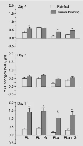
O sm o tic fragility afte r in vitro incubatio n
The sensitivity of RBC from tumor-bear-ing rats and their pair-fed controls to the metabolic stress induced by incubation at
37o
C for 4 h was tested using Ringer-lactate buffer as incubation medium and autologous plasma with or without glucose addition. This treatment caused only a small increase in the MCF of RBC from the pair-fed con-trols, whereas RBC from tumor bearers were more sensitive to the stress, particularly 11 days after tumor inoculation when the in-crease in MCF was marked and the differ-ence, compared with the controls, was sig-nificant for all incubation media tested (Fig-ure 3).
Auto psy
There were no signs of bleeding or mac-roscopic visceral metastases. Only some en-larged regional lymph nodes were observed, particularly in the retroperitoneum and around the thymus, but in no instance were neighboring tissues invaded.
D iscussio n
All rats rapidly and synchronously devel-oped anemia and anorexia, the systemic ef-fects characteristic of the final stage of can-cer, after multifocal tumor inoculation. The effects began 3-4 days after inoculation and progressed rapidly thereafter, and would have resulted in death 8-9 days later (7,8) if the rats had not been sacrificed at fixed times according to the experimental protocol. The effects developed in the absence of compli-cations such as bleeding, invasions or visible metastases, as confirmed at autopsy. The early onset, rapid course and magnitude of
the decrease in Hb levels in the tumor bear-ers indicated an acute form of anemia which, in the absence of bleeding, could only be explained by the rapid destruction of RBC. Based on the rate of decrease in Hb levels, the RBC destruction in the final four days was about five times the normal physiologi-cal rate for this phenomenon.
The rapid progress of the anemia seen with multifocal tumor inoculation is quite different from the progress observed after unifocal inoculation. Figure 4 shows unpub-lished data (Vieira-Matos AN and Rettori O) from our laboratory similar to those reported by others using the unifocal Walker 256 model (5,13). The average results obtained under these conditions suggest that this can-cer causes moderate chronic anemia and mild temporary anorexia. These conclusions can be partially explained by the fact that in this model the periodic transverse averaging
M
C
F
c
h
a
n
g
e
s
(
N
a
C
l,
g
/l
)
2.0
1.5
1.0
0.5
0.0
-0.5
2.0
1.5
1.0
0.5
0.0
-0.5
2.0
1.5
1.0
0.5
0.0
-0.5 Day 4
Day 7
Day 11
RL RL + G PLa PLa + G
*
* *
*
* * * *
Figure 3 - Effect of a 4-h incuba-tion at 37oC on the osmotic
fra-gility of RBC from tumor-bearing and pair-fed control rats in differ-ent incubat ion m edia: RL: lactate; RL + G: Ringer-lactate + glucose (2 mg/ml); PLa: autologous plasma; PLa + G: au-tologous plasma + glucose (1 mg/ml). M CF: M edian corpuscu-lar fragility = NaCl concentration (g/l) causing 50% of RBC lysis. * P<0.05 (paired Student t-test). Pair-fed
of data mixes values from groups of animals at very different stages of the illness with data from animals with still undetectable homeostatic alterations. In addition, the ex-clusion of animals that died at the end of the experiment, together with the survival of some resistant animals generates an artifact that suggests an apparent improvement in the illness condition. Guaitani et al. (13) recognized these influences while studying the anorexia induced by the Walker 256 tumor and suggested a different treatment of the data, in which the independent variable was changed. Instead of time, this author proposed the use of the percentage of the survived time. Using this approach, at the time of death (100% of survived time) for example, all animals in the group were to-tally anorectic. A further improvement in the handling of this problem was described by Rettori et al. (7). The multifocal tumor mo-del avoids all of these problems by synchro-nizing the onset and progress of the systemic effects of the final stage of cancer among the individuals of the experimental group (7,8). Under the latter conditions, the hemolytic component of cancer anemia is of particular importance as described for humans by Hyman and Harvey in the early 50s (4). These authors demonstrated what is gener-ally recognized in oncology, namely, that in terminal cancer patients even the half-life of
transfused RBC is markedly reduced, so that frequent transfusions are of little help in maintaining reasonable Hb levels.
The increased importance of the hemo-lytic component of the anemia of terminal cancer patients is probably associated with metastatic dissemination. In this regard, the multifocal inoculation model simulates the terminal stage of cancer, with the advantage of dissociating the hemolytic component of anemia from the chronic complications that mask it.
Change s in re d blo o d ce ll o smo tic fragility
induce d by the Walke r 256 tum o r
The OF of fresh RBC reflects their ability to take up water without lysis and is deter-mined by their volume-surface area ratio (11).
In the present study, the development of acute anemia was accompanied by a de-crease in the OF of RBC (a shift to the left in the OF curves), indicating a reduction in the volume of circulating erythrocytes. Indeed, the blood count showed that the mean cor-puscular volume of RBC from tumor-bear-ing rats was significantly reduced compared with control RBC. On the other hand, the higher increases in MCF after a 4-h
incuba-tion at 37o
C when compared with pair-fed controls indicated that the RBC of tumor bearers had an increased propensity to swell during incubation, thus decreasing the resis-tance to metabolic stress. This effect was cumulative since it became marked only on day 11 and was absent or slight on days 4 and 7. The complexity of the factors associated with the changes in OF induced by this tu-mor was further suggested by the reduction in the slope of the OF curve on day 11.
Me chanisms invo lve d in RBC alte ratio ns
The above results shed little light on the molecular basis of the observed phenome-non. Hereditary alteration in RBC glycolytic
H
b
(
g
/d
l)
20
F
o
o
d
i
n
ta
k
e
(
g
/d
a
y
)
60
15
10
5
0
50
40
30
20
10
0 Hb Control
Hb Tumor
Food intake
0 10 20 30 40
Days after inoculation Figure 4 - Changes in
hemoglo-bin levels and daily food intake of rats bearing unifocal inocula-tions of the Walker 256 tumor (Vieira-M atos AN and Rettori O, unpublished results similar to those reported by others, see text). (n): Number of individuals in the group or remaining in it.
(8)
(20)
(8)
(13) (11) (6)
enzymes, such as pyruvate kinase deficiency, is an example of how a primary alteration in RBC metabolism is able to induce anemia associated with a decrease in the volume (decreased OF) and resistance to metabolic stress (marked OF increase after incubation) of fresh RBC. On the other hand, hereditary and acquired membrane defects are also known to decrease the RBC volume-surface area ratio, and hence decrease the OF (11). In contrast, the induction of cell volume changes leads to important cellular meta-bolic alterations (14) such that circulating molecules of tumor and/or host origin, cyto-kines or other effector molecules (15) acting on RBC metabolism and/or its plasma mem-branes may be involved in inducing the ob-served phenomenon. This hypothesis is sup-ported by observations that the tumor-in-duced OF effects seen in the present study may be reproduced under special conditions by incubating normal RBC with plasma from tumor anemic animals (Vieira-Matos AN and Rettori O, unpublished results). A re-duction in RBC OF in anemic tumor-bearing mice has been reported, and may be caused by structural alterations in the RBC (16). We have also observed a marked decrease in RBC OF associated with anemia in rats bear-ing DMBA-induced mammary cancer (Cavalcanti TC and Rettori O, unpublished results).
Re le vance o f the pre se nt o bse rvatio ns
The high correlation (r2
= 0.86) between the development of anemia and other well-known systemic effects such as anorexia induced by cancer suggests the possibility of a common molecular basis for the serious homeostatic alterations occurring in the fi-nal stage of cancer. Recent studies have proposed an imbalance between catabolic and anabolic hormones and cytokines of tumor and/or host origin as the molecular basis for cancer cachexia (15). A main role has been suggested for the following
cyto-kines: IL-a, IL-ß, IL-6, IL-8, TNF-a, INF-a
and g, but conclusive data are often lacking
(17,18), and because the circulating levels usually do not correlate well with cachexia, an alternative model of abnormal general-ized high local production of cytokines through positive feedback systems has been proposed (17).
The possibility should be explored that the study of the alterations in the RBC plasma membrane and/or metabolism could help to identify these molecules and to explain the mechanism of cancer anemia, cachexia and the so-called multiple organ failure syndrome, frequently referred to as the cause of death in cancer patients.
The multifo cal tumo r ino culatio n mo de l
The explanation as to why several small tumors (little more than 100 mg each on days 3-4) were more effective in inducing the systemic effects of cancer than a single large tumor (sometimes 40 g or more) is probably related to the cell kinetics of tumor growth. Studies in this field suggest that the sum of proliferative tumor cells (PTC) in several small tumors is larger and increases faster than that of a single, big tumor, in which most cells are non-proliferative (NPTC). Recent work has demonstrated that in tu-mors of up to 100 mg (days 3-4 in our model) about 90% of the mass would be PTC and would grow fast (exponentially), but they would rapidly reach an almost steady state of about 1-2 g (19). One explanation for this, consistent with the current concepts of tu-mor cell proliferation (20), would be that, due to the rapidly increasing irrigation defi-cit, about half of the new cells formed after
each replication would stay in G1 or go to G0,
dissemina-tion occurs, the multiple small foci of neo-plastic growth, each bearing high relative populations of PTC, would produce a high enough total rate of PTC growth to induce the serious homeostatic alterations that cul-minate in host death.
In addition to the already mentioned ad-vantages of the multifocal model (synchro-nism among individuals, rapidity, reproduc-ibility and absence of complications), this
model also involves minimal animal suffer-ing.
Ackno wle dgm e nts
We wish to thank Dr. S. Hyslop for his careful reading of the paper and correction of the language. We also thank A. Garcia for the animal care.
Re fe re nce s
1. M oliterno AR & Spivak JL (1996). Anemia of cancer. Hematology/Oncology Clinics of North America, 10: 345-363.
2. Temple JJ & Stuckey WJ (1986). M echan-isms contributing to the anemia associ-ated w ith a localized tumor. American Journal of M edical Sciences, 292: 277-281.
3. Bodey GP & Frei III E (1974). M edical therapy of cancer. In: Wintrobe M M , Thorn GW, Adams RD, Braunw ald E, Isselbacher KJ & Petersdorf RG (Editors), Harrison’s Principles of Internal M edicine. 7th edn. M cGraw -Hill Book Company, New York.
4. Hyman GA & Harvey JE (1955). The patho-genesis of anemia in patients w ith carci-noma. American Journal of M edicine, 19: 350-356.
5. Zucker S, Lysik RM & Di Stefano J (1977). Pathogenesis of anem ia in rats w ith Walker 256 carcinosarcoma. Journal of Clinical M edicine, 90: 502-511.
6. Guaitani A, Recchia M , Carli M , Rocchetti M , Bartosek I & Garattini S (1982). Walker carcinoma 256: a model for studies on tumor-induced anorexia and cachexia. Oncology, 39: 173-178.
7. Rettori O, Vieira-M atos AN & Tahin QS
(1995). Variability and discontinuity of the pathognomonic systemic effects caused by Walker 256 tumor progression in rats. Tumori, 81: 370-377.
8. Guimarães F, Rettori O, Vieira-M atos AN & Fernandes GA (1999). The influence of septal lesions on sodium and w ater reten-tion induced by Walker 256 tumor. Brazil-ian Journal of M edical and Biological Re-search, 32: 309-317.
9. Hyman GA (1953). Studies on anemia of disseminated malignant neoplastic dis-ease. Blood, 9: 911-919.
10. UKCCR (1988). United Kingdom Coordi-nating Committee on Cancer Research Guidelines for the w elfare of animals in experimental neoplasia. Laboratory Ani-mals, 22: 195-201.
11. Dacie JV, Lew is SM & Gordon-Smith EC (1984). Investigation of hereditary haemo-lytic anaemias. In: Dacie JV & Lew is SM (Editors), Pratical Hematology. 6th edn. Churchill Livingstone, Edinburgh. 12. Vieira S (1981). Introdução à Estatística.
1st edn. Editora Campos, Rio de Janeiro. 13. Guaitani A, Torre PD, M orasca L, Pintus C & Bartosek I (1983). Tw o lines of Walker carcinoma 256: their peculiarities and dif-ferent interactions w ith the host. Tumori,
69: 1-9.
14. Lang F, Busch GL, Völkl H & Häussinger D (1995). Cell volume: a second message in regulation of cellular function. New s in Physiological Sciences, 10: 18-22. 15. Tessitore L, Castelli P & Baccino FM
(1993). Humoral mediation for cachexia in tumor-bearing rats. British Journal of Can-cer, 67: 15-23.
16. Ray M R & Chaw dhury R (1989). Osmotic fragility, sialic acid content and survival of circulating erythrocytes in anemic tumor bearing mice. Neoplasma, 36: 155-160. 17. Plata-Salaman CR (1998). Cytokins and
feeding. New s in Physiological Sciences, 13: 298-304.
18. Tisdale M J (1997). Cancer cachexia: meta-bolic alterations and clinical manifesta-tions. Nutrition, 13: 1-7.
19. Bassukas ID & M aurer-Schultze B (1990). Grow th of metastases of the mouse ad-enocarcinoma EO771: an allometric rela-tionship betw een grow th of the primary tumors and their metastases. Clinical and Experimental M etastasis, 8: 329-343. 20. Tannock IA (1992). Cell proliferation. In: