Nitric oxide release from the
S-nitrosothiol zinc phthalocyanine
complex by flash photolysis
Departamento de Química, Faculdade de Filosofia,
Ciências e Letras de Ribeirão Preto, Universidade de São Paulo, Ribeirão Preto, SP, Brasil
J.C.G. Rotta, C.N. Lunardi and A.C. Tedesco
Abstract
The photogeneration of nitric oxide (NO) using laser flash photolysis was investigated for S-nitroso-glutathione (GSNO) and S-nitroso-N -acetylcysteine (NacySNO) at pH 6.4 (PBS/HCl) and 7.4 (PBS). Irra-diation of S-nitrosothiol with light (l = 355 nm followed by absorp-tion spectroscopy) resulted in the homolytic decomposiabsorp-tion of NacySNO and GSNO to generate radicals (GS· and NacyS·) and NO.
The release of NO from donor compounds measured with an ISO-Nometer apparatus was larger at pH 7.4 than pH 6.4. NacySNO was also incorporated into dipalmitoyl-phosphatidylcholine liposomes in the presence and absence of zinc phthalocyanine (ZnPC), a well-known photosensitizer useful for photodynamic therapy. Liposomes are usually used as carriers for hydrophobic compounds such as ZnPC. Inclusion of ZnPC resulted in a decrease in NO liberation in liposomal medium. However, there was a synergistic action of both photosensi-tizers and S-nitrosothiols resulting in the formation of other reactive species such as peroxynitrite, which is a potent oxidizing agent. These data show that NO release depends on pH and the medium, as well as on the laser energy applied to the system. Changes in the absorption spectrum were monitored as a function of light exposure.
Correspondence A.C. Tedesco
Departamento de Química FFCLRP, USP
Av. Bandeirantes, 3900 14040-901 Ribeirão Preto, SP Brasil
Fax: +55-16-633-8151 E-mail: [email protected]
Research supported by FAPESP (Nos. 99/01394-3 and 00/00431-1).
Received April 11, 2002 Accepted January 15, 2003
Key words ·S-nitrosothiols ·Nitric oxide ·Zinc phthalocyanine ·Liposomes
·Photodynamic therapy
Introduction
S-nitrosothiols (thionitriles), compounds with the generic structure RSNO, have be-come important recently because of a series of demonstrations of the in vivo synthesis of
nitric oxide (NO) and its control of a range of physiological functions (1,2). RSNO are po-tent antiplatelet agents and vasodilators (3). Both of these functions are usually attributed to NO release and there is interest in the potential of S-nitrosothiols as in vivo carriers
of NO to target areas such as tumors where NO could act as a phototoxic agent (4).
against cancer, other reports suggest that NO promotes tumor growth (7). Thus, the deter-mination of the mechanism of NO utilization requires an understanding of factors such as its rate of release, local concentration, and intracellular localization (7).
S-nitrosothiols have been described as reservoirs of NO in vivo due to their higher
stability in comparison with NO, which has a short half-life in biological systems. The liberation of NO is promoted by chemical means, by light and by a combination of the two (photosensitization protocols) (6). The mechanism of the photochemical release of NO from S-nitrosothiol has been determined using laser flash photolysis (4).
Photodynamic sensitizers such as zinc phthalocyanine (ZnPC) produce active oxy-gen species by type I and type II mechanisms (8). Triplet sensitizers can undergo their pri-mary reactions with molecules in their vicin-ity by an electron (or H atom) transfer pro-cess (termed a type I, i.e., a radical or redox reaction), or by an energy transfer reaction (termed a type II process). Localized pro-duction of singlet oxygen and superoxide radicals (O2·-) as a cytotoxic and antiviral
process has been achieved in photodynamic therapy, which involves the use of photosen-sitization combined with light (9). Upon ac-tivation, ZnPC generates reactive oxygen species (singlet oxygen and free radicals such as ·OH, HO2· and O2·-) which damage
membranes, DNA and other cell structures (9). All these processes are summarized in Figure 1.
The use of photosensitizing drugs associ-ated with different vehicles, such as lipo-somes, has received strong interest within the field of tumor photodynamic therapy (9,10). Since NO exhibits tumoricidal and antiviral activity, it may be possible to ex-ploit photosensitized NO in clinical photo-dynamic therapy.
In the present study we show that the decomposition of S-nitrosothiols by laser flash photolysis is pH and medium
depend-ent. The main objective of the present study was to report the synergistic effect of both species (NO, O2·-) produced in the medium
and capable of producing ONOO-. This is of
biological interest since these are active spe-cies in photodynamic therapy.
Material and Methods
Reagents
Sodium nitrite (NaNO2), sodium
hydrox-ide (NaOH) and dipalmitoyl-phosphatidyl choline (DPPC) were purchased from Sigma (St. Louis, MO, USA), and ZnPC was pur-chased from Aldrich (Milwaukee, WI, USA). The buffers used were 10 mM sodium phos-phate buffer (PBS), pH 7.4 (Sigma), and 10 mM PBS/HCl, pH 6.4, containing 0.1 M hydrochloric acid (HCl) (Merck, Darmstadt, Germany).
Synthesis of GSNO and NacySNO
S-nitrosothiols were synthesized by the method of Ceron et al. (3). They were pre-pared by reacting equimolar amounts of NaNO2 and the corresponding thiol
(gluta-thione or N-acetylcysteine) in aqueous
solu-tion. The solutions were adjusted to pH 2.0 with 6 M HCl and incubated at 37ºC for 5 min, by which time a characteristic red/or-ange color was developed. The samples were neutralized to pH 7.0 with NaOH and then lyophilized.
The structure of RSNO was confirmed by proton nuclear magnetic resonance and their concentrations were determined spec-trophotometrically at 330 nm based on ex-tinction coefficients (e330) of 767 for
nitroso-glutathione (GSNO) and 727 for S-nitroso-N-acetylcysteine (NacySNO) (2,4).
Preparation of unilamellar vesicles
This procedure allows the preparation of unilamellar vesicles of predetermined size by the injection of ethanol containing differ-ent concdiffer-entrations of phospholipid into an aqueous-buffered solution. The injection containing 9.21 mM DPPC solution in abso-lute ethanol was performed at 55ºC with magnetic stirring at a speed of 1 µl/s. The system was then allowed to cool to room temperature and was passed through a 0.454-µm Millipore filter. The organic solvents were eliminated by dialyzing the aqueous liposome dispersion at 4ºC against 250 ml of phosphate buffer for 6 h. The RSNO (2 mM) were incorporated into the liposomes by add-ing the amount of RSNO salt needed to reach a final concentration of 2 mM, and subse-quently stirring for 15 min. The ZnPC con-centration of 5 µM in the liposomes was obtained by adding suitable volumes of a stock (1 mM) pyridine solution of the sensi-tizer to the ethanol DPPC solution, with subsequent injection into the buffer solu-tion.
Measurement of nitric oxide
The concentration of NO released by RSNO was measured with an ISO-NO am-perometric sensor (World Precision Instru-ment, Inc., Sarasota, FL, USA) with a sensi-tivity range of 1 nM to 20 µM NO and a detection limit of 1 nM. This sensor detects NO directly by amperometric techniques. The ISO-NO sensor was calibrated daily when in use by a quantitative NO generation reaction according to the procedure described in the Instruction Manual of ISO-NO (12). The analog signal output from ISO-NO was recorded with a PC-based data acquisition system (DUO 18, World Precision Instru-ment). All experiments were carried out at 37ºC.
Laser flash photolysis experiments
All solutions were freshly prepared and
protected against light using aluminum foil until the beginning of irradiation to prevent any prestimulated decomposition. RSNO (2 mM) solutions were placed in a 1-cm quartz cuvette with constant stirring. Excitation was provided by a frequency-triplet, mode locked Nd:YAG laser at 355 nm (Continuum, Santa Clara, CA, USA). The focused incident beam (8-ns pulse duration) was set at 10 to 50 mJ per pulse with an average of 10 shots per irradiation cycle. The ground-state absorp-tion spectrum was determined using a Hita-chi U-300 spectrophotometer. All experi-ments involved aerated buffered (PBS, pH 7.4, and PBS/HCl, pH 6.4) liposome solu-tions of 2.0 mM RSNO.
Results and Discussion
The UV-visible spectra of RSNO showed two bands, one in the range of 330-350 nm (e ~ 103 M-1 cm-1, n
0®p*) and the other in
the 550-600 nm region (e ~ 20 M-1 cm-1, n N ® p*) (Figure 2). These absorbances are
Type I mechanism Type II mechanism
3P + S ® P·- + S·+ P + hn ® 1P ®ISC 3P
3P + S ® PH· + S· 3P + 3O2 ® 0P + 1O2
P·- + 3O
2 ® 0P + O2
·-PH· + 3O
2 ® 0P + HO2
·-3P + 3O
2 ® P·+ + O2
·-O2·+ + H+ ® HO2·
HO2· + SH ® H2O2·+
P = Photosensitizer S = Substrate
ISC = Intercrossing system
Figure 1. Type I and type II mechanisms.
Absorbance
0.3
0.2
0.1
0.0
300 400 500 600 700 800
Wavelength (nm)
used to monitor RSNO decomposition (13). The homolytic fission of the S-N bond in RSNO occurs photochemically by irradia-tion at either absorpirradia-tion maximum band (340 or 545 nm). A convenient way of exciting ground state GSNO and NacySNO has been laser excitation at 355 nm in view of the higher absorption coefficient of both com-pounds at this wavelength (4). This absorp-tion resulted in NO release and consequent formation of free radical species such as RS· (Equation 1) in a pseudo-first-order process (2).
RSNO ® RS· + NO (Eq. 1)
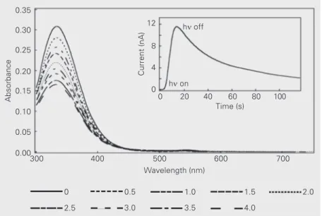
Changes in the absorption spectrum of 2 mM GSNO solutions previously saturated with oxygen were monitored as a function of light exposure. Figure 3 shows that after light exposure using 10 laser pulses of 50 mJ each a decrease in GSNO concentration oc-curred due to the homolytic fission of the S-N bond (Equation 1).
After homolysis of the S-N bond (Equa-tion 1), other processes can occur such as
reaction of the thiyl radical formed with GSNO to yield GSSG and further NO (Equa-tion 2), or with oxygen to yield the peroxy radical (Equation 3) with a rate constant close to the diffusion process (k = 3.0 x 10⁹
M-1 s-1). The peroxy radical formed reacts
with GSNO to yield GSSG and NO (Equa-tion 4) (4). Both processes leading to NO production present rate constants close to the diffusion limit.
GS· + GSNO = GSSG + NO (k = 1.7 x 10⁹
M-1 s-1) (Eq. 2)
GS· + O2 = GSOO
·
(Eq. 3) GSOO· + GSNO = GSSG + NO + O2 (k = 3.8
x 108 M-1 s-1) (Eq. 4)
The RSNO compounds studied showed a slow initial generation of NO due to ther-mal decomposition, which was nonsignifi-cant in our experiments. In contrast, NO release was accelerated by direct light exci-tation (hn on). The inset in Figure 3 shows
this behavior for GSNO which, after irradia-tion with a total laser energy of 0.5 J, showed an NO release of 221 ± 19 nM. When irra-diation was stopped (hn off) an
electrochemi-cal signal showing decay of NO started, following a second-order reaction which was most probably due to a relatively slow diffusion process (lifetime of NO 80 s) (14).
Another possible explanation for this slow decay of NO release can be due to NO auto-oxidation induced by molecular oxygen dis-solved in solution (15). The reaction with O2
consumes two NO equivalents and produces two nitrogen dioxide (·NO2) equivalents
(Equations 5-7). The radical-radical combi-nation reaction between NO2 and NO yields
an electrophilic nitrosating species known as dinitrogen trioxide (N2O3) (Equation 8).
The initial S-nitrosothiol, RSNO, can be re-generated by reaction of thiol nitrosation with N2O3 (Equation 9) (5,14,15).
NO + O2 = ONOO
·
(Eq. 5)
Figure 3. Ground-state absorption spectrum of S-nitroso-glutathione (GSNO; initial concen-tration = 2.0 mM) in an air-saturated PBS/HCl solution, pH 6.4, showing the decrease in absorption at 355 nm following irradiation with a total laser energy of 0.5, 1.0, 1.5, 2.0, 2.5, 3.0, 3.5, and 4.0 J. The inset displays the chronoamperogram of nitric oxide released by photolysis of 2 mM GSNO in an aerated 10 mM PBS/HCl buffer solution, pH 6.4, after irradiation at 355 nm with a total laser energy of 0.5 J.
Absorbance
0.35
0.30
0.25
0.20
0.15
0.10
0.05
0.00
300 400 500 600 700
Wavelength (nm)
Current (nA)
12
8
4
0
0 20 40 60 80 100
Time (s) hn off
hn on
0 0.5 1.0 1.5 2.0
ONOO· + NO = ONOONO (Eq. 6) ONOONO = 2 ·NO2 (Eq. 7)
·
NO2 + NO = N2O3 (k = 1 .1 x 1 09 M-1 s-1)
(Eq. 8) RSH + N2O3 = RSNO + NO2- + H+ (Eq. 9)
Analogous experiments were carried out for 2 mM NacySNO in buffer solutions at pH 6.4 and 7.4 (data not shown) using laser energy of 0.1 and 0.5 J and the results showed that NO release from NacySNO followed the same process as described previously for GSNO. The values of NO release obtained in these experiments are displayed in Figure 4, which shows that NO release is dependent on laser energy and pH. When laser energy was increased from 0.1 to 0.5 J, NO release by GSNO and NacySNO was increased about 1.8-fold. The measurements of NO release in more acidic medium (pH 6.4) showed higher values than in physiological medium (pH 7.4). We also observed that NO release from NacySNO was higher than from GSNO under the same experimental conditions.
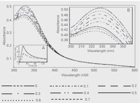
A similar procedure was followed in ho-mogeneous buffer solutions for GSNO and NacySNO for changes in NO release for flash photolysis to NacySNO in liposomes. The results obtained practically showed no difference from those for RSNO in homog-enous solution, as illustrated in Figure 4. The chronoamperogram (inset A, Figure 5) indi-cated that after photolysis was stopped NO concentration decreased in a second-order reaction in agreement with the mechanism described in Equations 5-9. It can also be observed that the release of NO in liposomal medium was dependent on the laser energy applied. When a higher intensity of irradia-tion was used in the process of NO genera-tion the result was a higher release of NO into the system (e.g., NO release from NacySNO liposomes was 176 ± 17 and 243 ± 0.03 nM for a laser energy of 0.1 and 0.5 J, respectively).
Ground-state absorption experiments for
NacySNO liposome at pH 7.4 (inset B, Fig-ure 5) showed a decrease in the absorption intensity band in the 300-400 nm region as a consequence of NacySNO decomposition. Another important aspect observed was a
Figure 5. Ground-state absorption spectrum of S-nitroso-N-acetylcysteine (NacySNO; initial concentration = 2.0 mM) in liposomes showing the decrease in absorption at 355 nm following irradiation with a total energy of 0.1, 0.2, 0.3, 0.4, 0.5, 0.6, and 0.7 J. Inset A
displays the chronoamperogram of nitric oxide released by photolysis of 2 mM NacySNO in liposomes using irradiation (355 nm) with 0.1 and 0.5 J. Inset B shows the enlargement of the ground-state absorption band in the spectrum of NacySNO in the 300-360 nm region. Figure 4. Nitric oxide concentration (nM) for S-nitroso-glutathione (GSNO) and S-nitroso-N -acetylcysteine (NacySNO) in PBS/HCl buffer, pH 6.4, and PBS, pH 7.4, after irradiation with a total laser energy of 0.01 and 0.05 J.
Nitric oxide concentration (nM)
0 50 100 150 200 250 300 350
GSNO pH 6.4 (10 mJ)
GSNO pH 6.4 (50 mJ)
NacySNO pH 6.4 (10 mJ)
NacySNO pH 6.4 (50 mJ)
NacySNO pH 7.4 (10 mJ)
NacySNO pH 7.4 (50 mJ)
Absorbance
0.5
0.4
0.3
0.2
0.1
Absorbance
0.50 0.48 0.46 0.44 0.42 0.40 0.38
300 310 320 330 340 350 Wavelength (nm)
Wavelength (nM)
300 350 400 450 500 550 600
120 80 40 0 0 4 8 12
Current (nA)
Time (s) A
B
0.1
· · · 0.2
· 0.3 · · 0.4 0.5
· 0.6 0.7
band shift centered between 300 and 305 nm (after application of a laser energy of 0.5 J), an indication of peroxynitrite generation (13). The peroxynitrite generation is due to the increased concentration of decomposition products leading to an increase in superox-ide concentration, which in turn reacts with NO leading to peroxynitrite formation (Equa-tion 10).
NO + O2·-®-OONO (k = 4-7 x 10⁹ M-1 s-1)
(Eq. 10) It was possible to measure NO release in liposomal medium and the NO produced diffused into a hydrophobic medium (phos-pholipids) and can be detected. This is due to the fact that NO, like O2, is somewhat
lipo-philic and has 6- to 8-fold higher solubility in nonpolar solvents and lipid membranes com-pared to water (5). Thus, the rates of NO reactions in hydrophobic environments can be dramatically increased (e.g., with O2)
com-pared to reactions in water due to its in-creased concentration. It has been clearly
demonstrated that the reaction of NO with O2 in a hydrophobic phase proceeds 300
times more rapidly than in the aqueous phase because NO and O2 concentrations are much
higher in the former than in the latter. This finding suggests that the auto-oxidation of NO and the subsequent reactions occur rap-idly within the membrane (16).
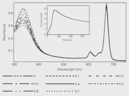
In our studies we also evaluated the re-lease of NO from 2.0 mM NacySNO in liposomal medium in the presence of 5.0 µM ZnPC, a well-known photosensitizer used in photodynamic therapy. Our results (Figure 6) showed that both probes in liposomal medium maintained their spectral profile af-ter photolysis, i.e., the typical absorption bands of phthalocyanines in the visible light region (600-700 nm), namely the Q band, and the Soret band in the violet or ultraviolet region (300-350 nm), and the typical absorp-tion band of NacySNO in the ultraviolet region (300-350 nm) and visible light region (500-600 nm). Changes in the absorption spectra were observed as a function of light exposure. After flash photolysis, a decrease in NacySNO concentration occurred accord-ing to the mechanism described earlier (Equa-tions 1-4).
The presence of ZnPC induced a crease of NO release by photochemical de-composition (Figure 6, inset). This behavior can be explained on the basis of a competi-tion process for the energy of flash photoly-sis (355 nm), since both sensitizers have absorption bands in this region. The oppo-site behavior was observed for photosensi-tizers such as aluminum phthalocyanine tetrasulfonate, hematoporphyrin and Rose Bengal, which show enhanced NO release from donor compounds (6). This behavior can be attributed to the energy transfer pro-cess between the triplet state of the photo-sensitizer (e.g., Rose Bengal labsorption = 550
nm) and the triplet of S-nitrosothiols (labsorption
= 550-600 nm) leading to an enhancement of the energy necessary for an efficient ho-molytic decomposition and to a consequent
Absorbance
0.8
0.4
0.2 0.6
300 400 500 600 700
Wavelength (nm)
0 50 100 150
Current (nA)
5 4 3 2
1
Time (s)
· · · ·
· · · · ·
· ·
0.1 0.2
0.3 0.4 0.5
0.6 0.7
Figure 6. Ground-state absorption spectrum of the S-nitroso-N-acetylcysteine-zinc phthalo-cyanine (NacySNO-ZnPC) complex in liposomes showing the decrease in absorption at 355 nm following irradiation with a total energy of 0.1, 0.2, 0.3, 0.4, 0.5, 0.6, and 0.7 J. The inset displays the chronoamperogram of nitric oxide released by photolysis of 2 mM NacySNO in liposomes using irradiation (355 nm) with a laser energy of 0.5 J.
0
increase of NO release (6).
During the photosensitization of ZnPC in liposomes, active oxygen species other than singlet oxygen could be generated (16). The use of ZnPC permits a synergistic action between both sensitizers (ZnPC and RSNO) resulting in the formation of reactive species such as peroxynitrite (ONOO-), superoxide
anion radical (O2·-), hydroxyl radical (·OH),
nitrogen dioxide (·NO2), as well as NO and
singlet oxygen that are extremely important for photodynamic therapy.
Singlet oxygen is produced by a type II mechanism, while a type I mechanism leads to the formation of superoxide anion, as shown in Equations 10-12. After irradiation, there is the formation of triplet ZnPC (Equa-tion 11), which collides with another ZnPC molecule in the ground state, leading to an electron transfer process (Equation 12). The anion radical produced, ZnPC·-, donates its
electron to dioxygen, generating O2·-
(Equa-tion 13). The process of ZnPC liposome photosensitization can generate the hydroxyl radical via reduction of hydrogen peroxide (Equations 14 and 15).
ZnPC + hn®1ZnPC ®3ZnPC (Eq. 11) 3ZnPC + ZnPC ® ZnPC·- + ZnPC·+ (Eq. 12)
ZnPC·- + O
2® ZnPC·+ + O2·- (Eq. 13)
O2·- + O2·- + 2H+® H2O2 + O2 (Eq. 14)
ZnPC·- + H
2O2 ® ZnPC +
·
OH + OH
(Eq. 15) Peroxynitrite, a potent oxidizing agent, is formed by the reaction of superoxide anion radical with NO, an extremely easy process since both have an unpaired electron (Equa-tion 16). The ONOO-, once protonated,
rap-idly decomposes to H+ and NO
3- through an
intermediate complex owing to the charac-teristics of both nitrogen dioxide and hy-droxyl radical (Equation 17). Peroxynitrite can react with a number of biomolecules, including thiols, amines, lipids and proteins (17). The concentration of CO2 in vivo is
high (ca. 1 mM), and the rate constant for reaction of CO2 with ONOO- is also high
(pH-independent k = 3 x 10⁴ M-1 s-1)
(Equa-tion 17). Consequently, the rate of reac(Equa-tion of peroxynitrite with CO2 is so fast that most
commonly used scavengers would need to be present at very high, near toxic levels in order to compete with peroxynitrite for CO2.
The mechanism of the reaction of CO2 with
ONOO- produces metastable nitrating,
nitrosating, and oxidizing species as inter-mediates. The role of CO2 is ambiguous. It is
postulated that it can serve as a catalyst for ONOO- degradation or can serve to activate
ONOO- as a nitrating agent (18).
HOONO ® [HO· + ·NO
2] ® NO3- + H+
(Eq. 16)
-OONO + CO
2® ONOOCOO- (k = 3 x 10⁴
M-1 s-1) (Eq. 17)
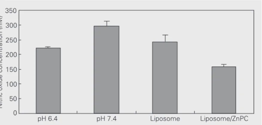
The data reported here demonstrate the behavior of NO-donating compounds in so-lution after flash photolysis under different experimental conditions. Changes in pH, la-ser energy, NO donor and environment me-dium were shown to release NO, as illus-trated in Figure 7, which shows that NO release from NacySNO after application of a laser energy of 0.5 J was higher at pH 7.4 than at pH 6.4 (19) and lower in the NacySNO/ZnPC complex.
An important observation reported here
Nitric oxide concentration (nM)
350
300
250
200
150
100
50
0
pH 6.4 pH 7.4 Liposome Liposome/ZnPC
is that incorporating the insoluble photosen-sitizer ZnPC into liposomal medium together with RSNO does not alter the photophysical properties of the two compounds. This be-havior permitted the study of the synergistic action of both active species (NO and ONOO-) produced under photolysis,
result-ing in the formation of reactive species which could react with a wide range of biological substrates, and which has been implicated in many pathophysiological conditions, increas-ing the photodynamic action of the RSNO and ZnPC complex.
References
1. Hoog N (2000). Forum: Therapeutic applications of reactive oxygen and nitrogen species in human disease. Free Radical Biology and Medicine, 28: 1478-1486.
2. Williams DLH (1999). The chemistry of S-nitrosothiols. Accounts of Chemistry Research, 32: 869-876.
3. Ceron PIB, Cremonez DC, Bendhack LM & Tedesco AC (2001). The relaxation induced by s-nitroso-glutathione and s-nitroso-n -acetyl-cysteine in rat aorta is not related to nitric oxide production. Journal of Pharmacology and Experimental Therapeutics, 298: 1-9. 4. Wood PD, Mutus B & Redmond RW (1996). The mechanism of
photochemical release of nitric oxide from s-nitrosoglutathione. Pho-tochemistry and Photobiology, 64: 518-524.
5. Ignarro LJ (2000). Nitric Oxide: Biology and Pathobiology. Academic Press, San Diego, CA, USA, 41-75 and 895-897.
6. Singh RJ, Hogg N, Joseph J & Kalyanaraman B (1995). Photosensi-tized decomposition of S-nitrosothiols and 2-methyl-2-nitrosopro-pane. Possible use for site-directed nitric oxide production. FEBS Letters, 360: 47-51.
7. Wink DA, Vodovotz Y, Cook JA, Krishna MC, Kim S, Coffin D, Degraff W, Delluca AM, Liebmann J & Mitchell JB (1998). Review: The role of the nitric oxide chemistry in cancer treatment. Biochem-istry, 63: 23-37.
8. Maree MD & Nyokong T (2001). Effect of oligomerization on the photochemical properties of silicon octaphenoxyphthalocyanine.
Journal of Photochemistry and Photobiology. A: Chemistry, 142: 39-46.
9. Hadjur C, Wagnières G, Ihringer F, Monnier PH & van den Bergh H (1997). Production of the free radicals O2·- and ·OH by irradiation of
the photosensitizer zinc (II) phthalocyanine. Photochemistry and Photobiology B, 38: 196-202.
10. Kassab K, Fabris C, Defilippis MP, Dei D, Fantetti L, Roncucci G, Reddi E & Jori G (2000). Skin-photosensitizing properties of Zn(II)-2(3),9(10),16(17),23(24)-tetrakis-(4-oxy-N-ethylpiperidinyl) phthalo-cyanine topically administered to mice. Journal of Photochemistry
and Photobiology. B: Biology, 55: 128-137.
11. Kremer JMH, von der Esker MWJ, Pathmamanoharan C & Wirsma PH (1977). Vesicles of variable diameter prepared by a modified injection method. Biochemistry, 16: 3932-3955.
12. ISO-NO & ISO-NO Mark II (1996). Isolated Nitric Oxide Meters and Sensors. Instruction Manual. World Precision Instrument, Inc., Sarasota, FL, USA.
13. Wang PG, Xian M, Tang X, Wu X, Wen Z, Cai T & Janczuk AJ (2002). Nitric oxide donors: chemical activities and biological applications.
Chemical Reviews, 102:1091-1134.
14. Zhelyaskov VR, Gee KR & Godwin DW (1998). Control of NO con-centration in solutions of nitrosothiol compounds by light. Journal of Photochemistry and Photobiology, 67: 282-288.
15. Oliveira MG, Shishido SM, Seabra AB & Morgon NH (2002). Thermal stability of primary S-nitrosothiols: roles of autocatalysis and struc-tural effects on the rate of nitric oxide release. Journal of Physical ChemistryA, 106: 8963-8970.
16. Ford PC, Bourassa J, Miranda K, Lee B, Lorkovic I, Boggs S, Kudo S & Laverman L (1998). Photochemistry of metal nitrosyl complexes. Delivery of nitric oxide to biological targets. Coordination Chemistry Reviews, 171: 185-202.
17. Nagano T & Yoshimura T (2002). Bioimaging of nitric oxide. Chemi-cal Reviews, 102: 1235-1269.
18. Augusto O, Bonini MG, Amanso AM, Linares E, Santos CCX & Menezes SL (2002). Nitrogen dioxide and carbonate radical anion: two emerging radicals in biology. Free Radical Biology and Medi-cine, 32: 841-859.