Photophysical studies of zinc
phthalocyanine and chloroaluminum
phthalocyanine incorporated into
liposomes in the presence of additives
Departamento de Química, Faculdade de Filosofia,
Ciências e Letras de Ribeirão Preto, Universidade de São Paulo, Ribeirão Preto, SP, Brasil
S.M.T. Nunes, F.S. Sguilla and A.C. Tedesco
Abstract
The photophysical properties of zinc phthalocyanine (ZnPC) and chloroaluminum phthalocyanine (AlPHCl) incorporated into lipo-somes of dimyristoyl phosphatidylcholine in the presence and absence of additives such as cholesterol or cardiolipin were studied by time-resolved fluorescence, laser flash photolysis and steady-state tech-niques. The absorbance of the drugs changed linearly with drug concentration, at least up to 5.0 µM in homogeneous and heteroge-neous media, indicating that aggregation did not occur in these media within this concentration range. The incorporation of the drugs into liposomes increases the dimerization constant by one order of magni-tude (for ZnPC, 3.6 x 10⁴ to 1.0 x 10⁵ M-1 and for AlPHCl, 3.7 x 10⁴
to 1.5 x 10⁵ M-1), but this feature dose does not rule out the use of this
carrier, since the incorporation of these hydrophobic drugs into lipo-somes permits their systemic administration. Probe location in bio-logical membranes and predominant positions of the phthalocyanines in liposomes were inferred on the basis of their fluorescence and triplet state properties. Both phthalocyanines are preferentially dis-tributed in the internal regions of the liposome bilayer. The additives affect the distribution of these drugs within the liposomes, a fact that controls their delivery when both are used in a biological medium, retarding their release. The addition of the additives to the liposomes increases the internalization of phthalocyanines. The interaction of the drugs with a plasma protein, bovine serum albumin, was examined quantitatively by the fluorescence technique. The results show that when the drugs were incorporated into small unilamellar liposomes, the association with albumin was enhanced when compared with organic media, a fact that should increase the selectivity of tumor targeting by these phthalocyanines (for ZnPC, 0.71 x 10⁶ to 1.30 x 10⁷
M-1 and for AlPHCl, 4.86 x 10⁷ to 3.10 x 10⁸ M-1). Correspondence
A.C. Tedesco
Departamento de Química FFCLRP, USP
Av. Bandeirantes, 3900 14040-901 Ribeirão Preto, SP Brasil
Fax: +55-16-633-8151 E-mail: tedesco@ffclrp.usp.br
Research supported by FAPESP.
Received January 9, 2003 Accepted September 9, 2003
Key words
•Zinc phthalocyanine •Chloroaluminum
phthalocyanine
•Bovine serum albumin •Liposomes
•Cholesterol •Cardiolipin •Dimyristoyl
Introduction
Photodynamic therapy (PDT) of cancer is a noninvasive treatment of small and su-perficial tumors that is currently being used in a number of countries (1). The therapy is based on the systemic administration of a tumor-localizing photosensitizer followed by illumination with light of appropriate wave-length. The resulting photodynamic reac-tions give rise to singlet oxygen (1O
2) and to
other active oxygen species that lead to tu-mor destruction (1). The efficiency of PDT depends on the development of new drugs and the ability of these drugs to accumulate selectively in tumor tissues in comparison with normal tissues. Several new classes of photosensitizers for PDT have reached the stage of clinical trials during the past few years (2). Among the more promising sec-ond-generation photosensitizers are phthalo-cyanines (Figure 1). There has been consid-erable interest in phthalocyanines for use in PDT, mainly because of their high absorb-ance coefficient (at 650-680 nm), with opti-mal tissue penetration by light (3). The photophysical properties of phthalocyanines are strongly dependent on the central metal ion. Among the metal phthalocyanines, Zn (II) and Al (III) complexes (zinc phthalocya-nine, ZnPC, and chloroaluminum phthalo-cyanine, AlPHCl) present the most favor-able photophysical properties for
applica-tion in PDT (4), i.e., relatively long-lived excited singlet states (ca. 3-8 ns) and long-lived triplet states that are produced in high quantum yields (5).
Unfortunately, these phthalocyanines (ZnPC and AlPHCl) are insoluble in water or biologically compatible solvents. Thus, they must be administered in vivo by means of
delivery systems (6). The transport of por-phyrins in the bloodstream via liposomes has been shown to provide a larger and more selective accumulation of the drugs in neo-plastic tissues (7), a fact that is a basic re-quirement for PDT. Their association with serum proteins can also enhance the prefer-ential uptake of hydrophobic photosensitiz-ers by tumor tissues, since serum albumin is one of the key components in blood that influences drug distribution. Since the in-trinsic fluorescence of proteins is usually quenched upon the binding of tetrapyrrolic compounds (8), this spectroscopic behavior provides a means to study the interaction between these drugs and bovine serum albu-min (BSA), permitting the deteralbu-mination of the binding constant, Kb, and of the binding
stoichiometry of the complex formed (9). Nevertheless, phthalocyanines are prone to self-aggregation. The deviation from lin-ear Beer-Lambert behavior for these drugs in solution caused by dimer formation is normally more pronounced in water than in organic solvents. Dimers are reported to be inactive or much more inefficient than mono-mers as photosensitizers (10). Unfortunately, phthalocyanines have been reported to dis-play a strong tendency to form dimers in water as a result of the large hydrophobic skeleton avoiding contact with the aqueous medium (11), leading to the dimerization process and affecting their photophysical and photosensitizing properties and photo-dynamic action (12). Aggregation was found to reduce the sensitizing ability of phthalo-cyanines in electron transfer reactions and in O2 (1∆g) photoproduction (13). Since
incor-poration of photosensitizers into liposomes
R
R R
R
N N
N N
N N N
N
M Figure 1. Typical phthalocyanine
(used as a drug delivery system) results in a high local concentration, information about the behavior of these drugs in these media is important for the understanding of their ac-tion in biological media.
In the present investigation we studied the aggregation parameters of ZnPC and AlPHCl in order to accurately determine the photophysical properties of these drugs. The predominant positions of these phthalocya-nines in dimyristoyl phosphatidylcholine (DMPC) liposomes are analyzed, as well as the triplet state lifetime and the binding of these exogenous drugs with BSA.
Material and Methods
Reagents
ZnPC, AlPHCl, methylviologen (MV2+),
9,10-anthraquinone-2-sulfonate (AQS-, so-dium salt), and 9,10-anthraquinone-2,6-disulfonate (AQDS2-, disodium salt) were
purchased from Aldrich Chemical Company Inc. (Milwaukee, WI, USA) and used with-out further purification. DMPC, cardiolipin, cholesterol, and phosphate-buffered saline (PBS) were obtained from Sigma (St. Louis, MO, USA). All other chemicals were com-mercially available reagents of at least ana-lytical grade.
All experiments were carried out with a phthalocyanine concentration of 5.0 µM. Stock solutions of ZnPC and AlPHCl (1.0 mM) were routinely prepared in ethanol and DMSO, respectively, and stored in the dark at 4ºC (ZnPC was dissolved in 0.1% pyridine in ethanol, v/v). The concentrations of the phthalocyanines were estimated spectropho-tometrically using ε678 = 2.93 x 10⁵ M-1 cm-1
for AlPHCl in DMSO and liposomes and
ε673 = 2.41 x 10⁵ M-1 cm-1 for ZnPC in
ethanol and liposomes. Stock solutions of MV2+ (0.3 M, ε = 20,500 M-1 cm-1 at 257 nm)
and AQDS2- (17 mM, ε = 6760 M-1 cm-1 at
328 nm) in water were stored in the dark at -15ºC. Stock solutions of AQS- (17 mM,
ε = 5450 M-1 cm-1 at 330 nm) in water were
stored in the dark at room temperature. The values of the extinction coefficients were obtained from the literature (14).
Liposome preparation
Small unilamellar liposomes of 0.7 mM DMPC were prepared on the basis of the injection method of Kremer et al. (15). Typi-cally, 0.380 ml of an ethanolic solution, which was 9.21 mM in DMPC and 66 µM in ZnPC or AlPHCl, was injected with a sy-ringe into 5 ml PBS, pH 7.4. The injection was performed at 46ºC under magnetic stir-ring and at a rate of 1 µl/s. Mixed liposomes were prepared by adding ethanolic solutions of cholesterol (50% DMPC, w/w) or cardio-lipin (30% DMPC, w/w) to the ethanolic solution.
Steady-state spectroscopic measurements
Absorbance spectra were recorded with a Hitachi U-3000 spectrophotometer and flu-orescence spectra were recorded with a Hi-tachi F-4500 spectrofluorometer. ZnPC and AlPHCl solutions were excited at 600 nm wavelength and their fluorescence emission was recorded in the 600-800 nm range. All measurements were made at 25 ± 2ºC. Band-widths were fixed at 5 nm for excitation and emission.
Determination of the dimerization equilibrium constants
In order to investigate the aggregation state of a phthalocyanine solution and to determine the equilibrium dimerization con-stant (KD), the absorbance spectra of a series
of phthalocyanine solutions were monitored with increasing phthalocyanine concentra-tions. KD for ZnPC and AlPHCl were studied
The KD for the equilibrium 2 M
↔
D isdefined as
KD = [D]/[M]2 (Eq. 1)
where [M] and [D] are the molar concentra-tions of the monomer and the dimer, respec-tively. The total concentration of the drug being equal to
C = [M] + 2[D] (Eq. 2) straightforward calculations lead to
(Eq. 3)
(Eq. 4)
The absorbance of a solution containing both the monomer and the dimer is given by
Abs = (εM[M] + εD[D])l (Eq. 5) where εM and εD are the extinction coeffi-cients of the monomer and the dimer, re-spectively, and l the optical path length.
Rearrangement of the above equations leads to
(Eq. 6) where C is the total concentration of the photosensitizer (monomer and dimer), εM
and εD are the extinction coefficients of the
monomer and dimer, respectively, and l is
the optical path length. KD was determined
by means of Equation 6 (16).
The plot of absorbance as a function of total drug concentration (C) according to Equation 6 was used to evaluate the pro-posed scheme. The values of KD, εM and εD
were computed by nonlinear regression based on the Lavenberg-Marquardt algorithm (16) using the Igor software. An estimated value of εM was obtained from measurements in organic solutions within the concentration range in which the Beer-Lambert law holds for each drug. The value of εM,determined independently, was fixed in Equation 6 and
the absorbance data were fitted (allowed to vary) to obtain εD and KD by a nonlinear least
square procedure (16).
Fluorescence quenching studies
The quenching of the fluorescence emis-sion by ZnPC and AlPHCl incorporated into unilamellar liposomes of DMPC in the pres-ence and abspres-ence of additives (cholesterol or cardiolipin) was studied using MV2+, AQDS
2-and AQS- as quenchers. Phthalocyanine so-lutions (5.0 µM) were excited at 600 nm and their fluorescence emission was recorded in the range of 640-750 nm. The fluorescence quenching data obtained were analyzed by Stern-Volmer formalism (17): F0/F = 1 + KSV
[Q], which relates the decrease in fluorescence intensity (F0/F) to quencher concentration [Q];
KSV is the Stern-Volmer quenching constant.
Interaction of ZnPC and AlPHCl with bovine serum albumin in organic and liposomal medium
The interaction of the phthalocyanines ZnPC and AlPHCl with BSA was studied spectrofluorometrically at 25ºC by the “double logarithmic plot” (18). For each drug tested, two sets of experiments were performed. In the first, solutions of phthalo-cyanines at low concentration in PBS were titrated with increasing BSA concentrations in order to determine maximum quenching, F∞. The relative fluorescence intensities of
BSA saturated with phthalocyanines, F∞,
were extrapolated from the experimental data by plotting 1/(F0 - F) against 1/[P], where F is
the measured fluorescence of a solution con-taining the protein and the phthalocyanine, F0 is the fluorescence of a solution of protein
ex-periments, the total dilution was kept below 2%. The fluorescence of BSA was excited at 280 nm and recorded between 300 and 450 nm, with excitation and emission bandpasses of 5 nm. Fluorescence data were treated according to the methods of Lehrer and Fashman (19) and Chipman et al. (20), with log [(F0 - F)/(F - F∞)] being plotted against
log [P]. The slope of the plot obtained gives N, the number of binding sites, and the value of log [P] at log [(F0 - F)/(F - F∞)] = 0 is equal
to the logarithm of the dissociation constant
Kdiss. The reciprocal of Kdiss is the binding
constant Kb.
Triplet state measurements
All measurements were made using ZnPC and AlPHCl (5.0 µM) in organic medium (pyridine and DMSO, respectively), and li-posomal medium in the presence and ab-sence of the additives cholesterol and car-diolipin. Triplet state was investigated using a laser flash photolysis apparatus which al-lows the simultaneous capture of the sient absorbance spectrum and of the tran-sient kinetics at a single wavelength. The system uses an Nd-YAG laser as the excita-tion source (SURELITE I-10 of Continuum, Santa Barbara, CA, USA), operating at 355 nm, ca. 50 mJ/pulse to give 10-ns pulses. The decay kinetics of the triplet state of ZnPC and AlPHCl was recorded at 480 and 490 nm, respectively - the triplet state absorbance maximum. Ten laser shots were averaged for each measurement. Decay profiles were fit-ted by an interactive nonlinear least-squares routine method using a data analysis soft-ware package from Edinburgh Instruments (Edinburgh, UK) on a personal computer.
Results and Discussion
Determination of the dimerization equilibrium constants
Like most phthalocyanines, ZnPC and
AlPHCl are prone to aggregation. In aque-ous solution, even the soluble phthalocya-nines (tetrasulfonated) display a typically aggregated spectrum even at low concentra-tions. Previous studies with another soluble photosensitizer (bacteriochlorin a) in PBS revealed that this hydrophilic photosensi-tizer is strongly aggregated in its dimeric form with a KD estimated to be 106 M-1 (21).
In the present study, it was not possible to determine the KD of the drugs ZnPC and
AlPHCl in aqueous solutions due to the lack of solubility in this medium. The solubiliza-tion of the drugs within DMPC liposomes induced dye monomerization and permitted us to evaluate the KD in DMPC medium.
The trend of the phthalocyanines ZnPC and AlPHCl to form aggregates and the equi-librium KD were analyzed and evaluated
us-ing absorbance spectroscopic analysis of the Q band at 673 nm. It was observed that, at least up to the concentration of 5.0 µM, the increase of absorbance with phthalocyanine concentration was linear in organic and lipo-somal medium (Figure 2). This behavior implies that, at least within this concentra-tion range (0 to ca. 5.0 µM), these phthalo-cyanines are in the monomeric state, in agree-ment with the Beer-Lambert law. In contrast, the behavior observed in liposomal medium
Figure 2. Variation in the absorbance (673 nm) as a function of zinc phthalocya-nine (ZnPC) concentration in DMPC liposomes and in organic solution (pyridine) in the inset.
Absorbance
1.2
1.0
0.8
0.6
0.4
0.2
2
1
Absorbance
5 10
5 10 15 x 10-6
[ZnPC] M
15 x 10-6
in the concentration range above 5.0 µM (5 to 15 µM) indicated that at higher concentra-tions a progressive change in the system occurred, indicating a typical aggregation process of the drugs. An example of the procedure for processing the data obtained is illustrated in Figure 2 for ZnPC. The points show the experimental results and were ad-justed by nonlinear regression in the full concentration range studied (0 to 15.0 µM). The KD of ZnPC and AlPHCl in organic
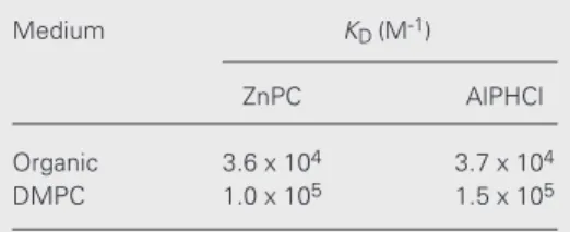
and liposomal medium obtained by nonlin-ear regression in the full concentration range studied (0 to 15.0 µM) are shown in Table 1.
KD for photosensitizers such as
porphy-rins and phthalocyanines range from 104 to
107 M-1 and depend on the solvent and
tem-perature (22). The results obtained confirmed that KD strongly depend on the medium,
since in liposome medium aggregation tends to be higher than in organic medium. In addition, the results indicate that the com-plexed metal ions zinc and aluminum do not interfere with the tendency to aggregate (same stability - same order of magnitude of KD
values for both drugs, i.e., ZnPC and AlPHCl, in liposomal medium). We observed that the incorporation of the drugs into liposomes increased the KD by one order of magnitude.
These results are in agreement with the be-havior expected for phthalocyanines incor-porated into liposomes. However, the in-crease of one order of magnitude in the KD of
these phthalocyanines after their incorpora-tion into liposomes is not a negative factor. A KD with an order of magnitude of 105 is not
a high value for these types of drugs (18). The advantage of the incorporation of these hydrophobic drugs into drug delivery sys-tems such as liposomes is the increase in their solubility, permitting their systemic administration.
Fluorescence quenching studies
The locations of ZnPC and AlPHCl in DMPC liposomes mixed with cholesterol (50% DMPC, w/w) or cardiolipin (30% DMPC, w/w) were evaluated and compared with those obtained for the drugs incorpo-rated into DMPC liposomes in the absence of these additives to assess how the distribu-tion of the phthalocyanines and the interac-tion mode of the quenchers with them are affected by the physicochemical properties of the liposomes in the presence of the addi-tives.
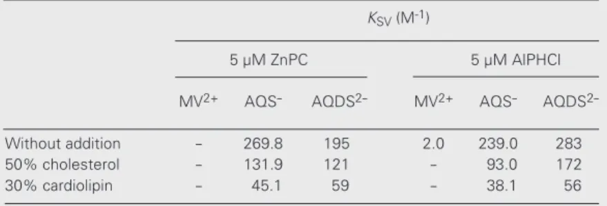
The quenching of ZnPC and AlPHCl by the quenchers was monitored by measuring the decrease in the emission intensity with excitation at 600 nm as a function of quencher concentration. Stern-Volmer plots were con-structed from the relative integrated fluores-cence intensity (640-750 nm). When ZnPC and AlPHCl were incorporated into DMPC liposomes in the presence and absence of additives at 25ºC, MV2+, AQS
-, and AQDS
2-yielded linear fluorescence quenching plots. Table 2 shows the quenching parameters obtained in these studies.
The quenchers used in the present inves-tigation did not penetrate the lipid bilayer and the quenching occurred by an electron transfer process (14). According to Ford and Tollin (14), in liposomal systems MV2+ is
oriented co-planar to the liposomal surface in order to maximize the contact between the polar and nonpolar parts of the quencher and the liposome. On the other hand, AQS- inter-acts perpendicularly to the bilayer surface,
Table 1. Dimerization constants of ZnPC and AlPHCl at 25ºC in organic solution and DMPC liposomes.
Medium KD (M-1)
ZnPC AlPHCl
Organic 3.6 x 10⁴ 3.7 x 10⁴
DMPC 1.0 x 10⁵ 1.5 x 10⁵
AlPHCl = chloroaluminum phthalocyanine; DMPC = dimyristoyl phosphatidylcholine; DMSO = di-methyl sulfoxide; KD = dimerization constant;
with the sulfonate group probably oriented in an opposite direction to the liposome. Thus, in liposomal systems MV2+ interacts
only with the drugs located in the outer part of the lipid bilayer, while AQS- and AQDS
2-interact with the drugs in both the inner and outer regions of the lipid bilayer. However, neither of these quenchers is able to diffuse into the lipid bilayer (quenching by an elec-tron transfer process). Thus, the KSV values
obtained for the quenching by MV2+ belong
to the drug population more exposed to the external medium, while KSV values obtained
for anthraquinone-type quenchers represent the weighted average of the KSV’s of both
classes of fluorophores (inner and outer). The lower KSV values for MV2+
com-pared with those for the anthraquinone quenchers (Table 2) in DMPC liposomes indicate that the drugs are incorporated into the bilayer structure, in the inner part of the liposomal structure, which lowers the acces-sibility of MV2+ to these drugs. The results
showed that AQS- and AQDS2- interacted
with ZnPC and AlPHCl in both the inner and outer lipid bilayers by an electron transfer process, although the efficiency of fluores-cence quenching of the phthalocyanines by AQDS2- was lower compared with AQS
-, as suggested by the drop of the KSV values
obtained for AQDS2- (Table 2). In this case,
the KSV values of AQDS2- are related to the
molecules of ZnPC and AlPHCl mainly lo-cated in the outermost regions of the lipid bilayer, and the efficiency of quenching was higher for AlPHCl than for ZnPC.
Different locations of the phthalocyanines in the lipid bilayer can be deduced from the changes in these KSV values under the same
experimental conditions. The AQS- quencher was more efficient for both probes. How-ever, AQDS2- showed a higher quenching
efficiency for AlPHCl than for ZnPC. These results indicate that AlPHCl was located closer to the bilayer/water interface than ZnPC. The KSV values for these compounds
are in agreement with the values found for
other photosensitizers in other drug delivery systems (23).
In the liposomes containing additives, the fluorescence quenching of the phthalo-cyanines by MV2+ was negligible. The
effi-ciency of quenching by AQDS2- in
liposo-mal medium with additives was also lower than the efficiency of quenching with AQS
-(Table 2). The presence of additives de-creased the KSV for both drugs studied.
Clusters of cholesterol appear in vesicles at mol fractions above 0.67. Mol fractions higher than 0.5 of cholesterol are required for cluster formation with phosphatidylcho-line (24-26). When the cholesterol content exceeds the saturation limit in phospholipid membranes, crystalline cholesterol mono-hydrate forms (27,28). In general, for cho-line phospholipids the saturation limit is
≈50% cholesterol, although a higher solubil-ity (65%) has been reported by Huang et al. (29) and a solubility higher than 60% was obtained by Guo and Hamilton (27). In this way, the cholesterol concentration used in the experiments (50% of total lipid, w/w) does not exceed the saturation limit in phos-phatidylcholine membranes and does not form crystalline cholesterol monohydrate.
In a membrane bilayer, cholesterol in-serts normal to the plane of the bilayer, with its hydroxyl group in close vicinity to the phospholipid polar heads and its alkyl side chain extending towards the bilayer center.
Table 2. Fluorescence quenching parameters of ZnPC and AlPHCl (5.0 µM) in DMPC liposomes in the presence and absence of additives at 25ºC.
KSV (M-1)
5 µM ZnPC 5 µM AlPHCl
MV2+ AQS- AQDS2- MV2+ AQS- AQDS
2-Without addition - 269.8 195 2.0 239.0 283 50% cholesterol - 131.9 121 - 93.0 172 30% cardiolipin - 45.1 59 - 38.1 56
AlPHCl = chloroaluminum phthalocyanine; AQDS2- =
9,10-anthraquinone-2,6-disulfonate; AQS- = 9,10-anthraquinone-2-sulfonate; K
SV =Stern-Volmer quenching
Cholesterol is intercalated in the membrane parallel to the phospholipid hydrocarbon chains, and the phospholipid carbons at po-sitions 2-10 have been estimated to lie in close proximity to the sterol tetracyclic ring structure (30,31). Although cholesterol can enhance lateral separation of lipids in bilay-ers consisting of a single lipid species, re-sulting in a higher water permeability in phosphatidylcholine bilayers and creating a cavity that was probably occupied by water molecules (32-34), the phthalocyanines did not become more accessible to the quenchers located in the external medium, as can be seen in Table 2 (lower KSV values in the
presence of cholesterol). Thus, the incorpo-ration of cholesterol into liposomes caused a change in the distribution of the molecules of ZnPC and AlPHCl from the outer to the inner region of the lipid bilayer. This choles-terol-induced redistribution of ZnPC and AlPHCl may be correlated with a change in the distribution of cholesterol itself (32-34). Above 30% DMPC (w/w), cholesterol pref-erentially dissolves in the inner lipid bilayer. When the liposomes contain equimolar amounts of phosphatidylcholine and choles-terol, the inner bilayer contains about 65% DMPC of cholesterol (w/w) and the external region of the bilayer contains about 40% DMPC (w/w) (32-34). If the drugs had been located near the bilayer/water interface, they would have become more accessible to the water-soluble quenchers (outer location) in liposomes containing cholesterol than in li-posomes without it (14). Consequently, these results prove the inner location of the drugs in the lipid bilayer with cholesterol, and not available to the quenchers, since a drop in quenching efficiency was noted after the addition of the additive.
Cardiolipin is found in the inner mem-brane of mitochondria (35). The results with cardiolipin demonstrate that the insertion of this lipid into the bilayer shows the same behavior as observed for cholesterol. This suggests a preferential distribution of ZnPC
and AlPHCl in cardiolipin-rich domains of the inner part of the lipid bilayer and its exclusion from DMPC-containing domains. The results obtained here show that the additives cholesterol and cardiolipin pro-mote a change of the drug molecules to-wards the inner part of the lipid bilayer, according to the lower KSV values obtained
after the incorporation of the additives into the liposomes (Table 2), but ZnPC continues to be more internalized than AlPHCl (lower
KSV values obtained for ZnPC when
com-pared with those obtained for AlPHCl for the quenching by AQDS2-) (Table 2). The
dis-placement of the drug molecules to the inner part of the lipid bilayer is probably related to their tendency to prefer hydrophobic envi-ronments.
Interaction of ZnPC and AlPHCl with bovine serum albumin in organic and liposomal medium
The interaction of serum albumin with phthalocyanines quenches the fluorescence of this protein, providing a means to assess binding quantitatively. The interaction be-tween ZnPC and AlPHCl with BSA can be followed by fluorescence spectroscopy, as illustrated in Figure 3. ZnPC and AlPHCl quench the fluorescence of BSA in propor-tion to the amount of drug added. The fluo-rescence intensity of BSA changes with the concentration of phthalocyanine in a way consistent with a reversible formation of a complex between BSA and the drugs.
The inset in Figure 4 provides a linear plot of F0/∆F versus 1/[P] for the binding of
ZnPC-BSA, used for the determination of the fluorescence intensity of BSA saturated with the phthalocyanines (F∞) ([P] =
phthalo-cyanine concentration). As described in the experimental section, Kdiss was calculated
from the slope and interception of the plot of log [(F0 - F) (F - F∞)] versus log [P]
addition, the slope of the plot gives the num-ber of the sites of the protein able to bind ZnPC and AlPHCl molecules.
The association constants obtained for the phthalocyanines studied are presented in Table 3, together with the binding stoichi-ometry of the complex formed.
Previous studies have indicated that phthalocyanines without SO3
groups adja-cent to iso-indole rings exhibit a high affinity binding site constant of 1-4 x 10⁴ M-1 (36,37).
Other studies (38) have reported mesoporphy-rin IX and magnesium mesoporphymesoporphy-rin parti-tioning between liposome and serum albumin with Kb of 2.5 x 10⁷ M-1 and 1.7 x 10⁷ M-1.
Our results agree with these data (Table 3). The present results show that when phthal-ocyanines are incorporated into DMPC lipo-somes, their association with BSA is in-creased in comparison with organic medi-um. Calorimetric titration studies indicated the binding of empty liposomes to the albu-min species. The albualbu-min molecules from different species adsorb strongly to phos-phatidylcholine liposomes due mainly to the action of hydrophobic dehydration forces and entropy gain (39). In this way, the lipo-somes themselves increase the Kb to BSA.
The encapsulation of the drug in the lipo-somes increases the already high association of the liposome/drug complex with BSA. The presence of a strong binding site and several weaker sites was observed for ZnPC and AlPHCl incorporated into liposomes. The present results indicate that the associa-tion of the drugs with lipid-based delivery systems affects their interaction with serum albumin. Liposomes of DMPC increase the binding of ZnPC and AlPHCl to BSA, which should increase the selectivity of tumor tar-geting by these phthalocyanines, since BSA can deliver the bound drugs to the vascular stroma of the tumors (7).
Triplet state measurements
The triplet state properties of ZnPC and
Figure 3. Effect of zinc phthalocyanine concentration on the fluorescence spec-trum of bovine serum albumin. From the top, in order of decreasing fluores-cence intensity with excitation at 290 nm, ZnPC concentration was 0.00, 0.13, 0.38, 0.64, 0.89, and 1.14 µM.
Figure 4. Double log plot of the quenching of protein fluorescence by zinc phthalocyanine in organic medium. Inset, Double reciprocal plot in organic medium (F = measured fluorescence of a solution containing the protein and the phthalocyanine, F0 is the fluorescence of a solution of protein alone, F∞ is the
fluorescence intensity of BSA saturated with ZnPC, and [PC] is the ZnPC concentration).
AlPHCl are similar. The transient spectra of ZnPC and AlPHCl in liposomal medium (in the presence and absence of additives) re-semble those obtained for a homogeneous solution (organic medium). Figure 5 illus-trates the transient spectrum of ZnPC (5.0 µM) incorporated into DMPC liposomes in the presence and absence of cholesterol and
Fluorescence intensity
2500
2000
1500
1000
500
0
300 350 400 450
Wavelength (nm)
Log [(F
0
- F)/(F - F
∞
)]
0.2
-1.0
0
1/(F
0
- F)
2 4 6
1/[ZnPC] µM-1
-5.4 Log [ZnPC]
2 4 6x10⁻³
cardiolipin, and also serves to illustrate the triplet-triplet absorbance centered at 480 nm, together with the Soret and Q band ground state bleaches.
Triplet lifetimes (τT) were calculated from
kinetic analysis of the transient decays, car-ried out using the fitting program of the apparatus itself and are shown in Table 4.
The triplet state reacts with forms of molecular oxygen (O2) by an energy transfer
process leading to singlet oxygen that is the key agent in cell damage in PDT. Transient absorbance spectra for the drugs obtained by laser flash photolysis showed the presence of transient species, which decayed mono-exponentially with the characteristic life-times shown in Table 4. As a consequence of the incorporation, triplet lifetimes are in-creased in liposomes in comparison with homogeneous solution. The increase in the triplet lifetimes is attributed to the ability of the bulk aqueous phase to interact with the sensitizer at the binding site. Therefore, we conclude that the increase in the triplet state lifetimes of ZnPC and AlPHCl as a function of the addition of cholesterol and cardiolipin to liposomes results from a progressive re-duction in the exposure of the sensitizer to the bulk aqueous phase, confirming the re-sults obtained in the quenching studies. Un-der identical conditions, the maximum trip-let lifetimes of ZnPC are shorter than those for AlPHCl, demonstrating that AlPHCl is more internalized than ZnPC.
The behavior observed in these studies implies that, at least up to ca. 5.0 µM, both phthalocyanines studied (ZnPC and AlPHCl) are in the monomeric state in both media (liposomal and organic), in agreement with the Beer-Lambert law. The increase of one order of magnitude in the dimerization con-stant of the phthalocyanines after their incor-poration into liposomes does not rule out the use of this carrier, considering the increased solubility of the drugs after incorporation into the drug delivery system, allowing their systemic administration, and the increased
Table 3. Equilibrium constants of ZnPC and AlPHCl binding to BSA.
Medium ZnPC AlPHCl
N Kb (M-1) N Kb (M-1)
Organic 1.05 0.71 x 10⁶ 1.37 1.30 x 10⁷
DMPC liposomes 1.07 4.86 x 10⁷ 1.11 3.10 x 10⁸
AlPHCl = chloroaluminum phthalocyanine; DMPC = dimyristoyl phospha-tidylcholine; Kb = binding constant; N = number of binding sites; organic
medium = pyridine for ZnPC and DMSO for AlPHCl; ZnPC = zinc phthalo-cyanine.
Table 4. Triplet lifetimes (τT) of ZnPC and AlPHCl in organic and liposomal medium in the presence of O2.
Medium ZnPC AlPHCl
Organic medium 0.30 0.80
DMPC 0.84 1.28
DMPC with cholesterol 1.35 4.62 DMPC with cardiolipin 1.28 2.50
AlPHCl = chloroaluminum phthalocyanine; DMPC = dimyristoyl phospha-tidylcholine; organic medium = pyridine for ZnPC and DMSO for AlPHCl; ZnPC = zinc phthalocyanine.
Absorbance
0.0
Absorbance
400 600 800 Wavelength (nm)
Wavelength (nm) 0.5
1.0
600 500
400 0.1
0.0
-0.1
-0.2
-0.3
Figure 5. Transient absorption spectra of zinc phthalocyanine (ZnPC) in organic (pyridine) and liposomal medium, in the presence and absence of the additives - liposome with cholesterol, and liposome with cardiolipin - after excitation at 670 nm (50 mJ per pulse), showing the absorption of the triplet state centered at λ = 480 nm and the bleach of the ground state centered at around 350 and 680 nm. Inset, Absorption spectra of ZnPC in organic medium.
Organic medium
Liposomal medium
Liposome with cholesterol
binding with BSA which should increase the selectivity of tumor targeting. As a conse-quence of the incorporation, triplet lifetimes are increased in liposomes compared with homogeneous solution. Thus, the results ob-tained clearly indicate that the incorporation of these drugs into DMPC liposomes maxi-mizes their photodynamic action, allowing an effective activity of the photosensitizer-liposome complex against cancer.
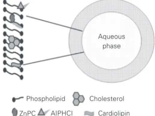
The results of the present study also indi-cate that the distribution of ZnPC and AlPHCl in unilamellar DMPC liposomes is heteroge-neous, with some predominant positions (Fig-ure 6). In the presence of additives there is a shift of the drug molecules towards the inner part of the lipid bilayer. The results obtained indicated that AlPHCl and ZnPC are prefer-entially distributed in the inner part of the phospholipid bilayer of DMPC liposomes in the presence and absence of cholesterol (50% DMPC, w/w) and cardiolipin (30% DMPC, w/w). The increased internalization of the drugs in liposomes containing additives is a positive aspect, since in this way the drugs can be protected while they circulate in the bloodstream after systemic injection.
Silicon (IV) phthalocyanine, the first phthalocyanine approved by the FDA for clinical trials, has the highest potential of this class of drugs for PDT (40). However,
silicon (IV) phthalocyanine needs to be used in a Cremophor emulsion, a fact that is an obstacle to the generalized use of this drug. Cremophor has been used clinically for the delivery of hydrophobic drugs. The possibil-ity of the use of hydrophobic phthalocya-nines such as ZnPC and AlPHCl encapsu-lated in liposomes that act as a drug delivery system, maintaining all the basic photophysi-cal and photochemiphotophysi-cal properties of the sen-sitizers, is an important result, since the lipo-somal vehicle is more economically viable than Cremophor. Thus, our results confirm the hypothesis that the liposomal medium represents an excellent vehicle for the ad-ministration of these drugs in biological sys-tems, making liposomal ZnPC and AlPHCl promising candidates for PDT from the pho-tophysical point of view.
Aqueous phase
Phospholipid Cholesterol
ZnPC AIPHCI Cardiolipin
Figure 6. Preferential locations of the photosensitizers ZnPC and AlPHCl in mixed liposomes of DMPC.
References
1. Pandey RK (2000). Recent advances in photodynamic therapy. Jour-nal of Porphyrins and Phthalocyanines, 4: 368-373.
2. Owens JW, Smith R, Robinson R & Robins M (1998). Photophysical properties of porphyrins, phthalocyanines and benzochlorins. Inorganica Chimica Acta, 279: 226-231.
3. Cahn W-S, Brasseur N, La Madeleine C, Quellet R & van Lier JE (2001). Current status of phthalocyanines in the photodynamic thera-py of cancer. European Journal of Cancer, 33: 1855-1860. 4. Urizzi P, Allen CM, Langlois R, Quellet R, La Madeleine C & van Lier
JE (2001). Low density lipoprotein-bound aluminium sulphophthalo-cyanine: targeting tumor cells for photodynamic therapy. Journal of Porphyrins and Phthalocyanines, 5: 154-160.
5. Ruch A, Beck G, Bachor R, Akgun N, Gschwence MH & Steiner R (1996). Dynamic fluorescence changes during photodynamic thera-py in vivo and in vitro of hydrophilic Al(III) phthalocyanine
tetrasul-phonated and lipophilic Zn(II) phthalocyanine administered in lipo-somes. Journal of Photochemistry and Photobiology. B, Biology, 36: 127-134.
6. Soncin M, Polo L, Reddi E, Jori G, Kenney ME, Cheng G & Rodgers MAJ (1995). Effect of the delivery system on the biodistribution of Ge(IV)octabutoxy-phthalocyanines in tumour-bearing mice. Cancer Letters, 89: 101-106.
7. Storm G & Crommelin DJA (1998). Liposomes: quo vadis. Pharma-ceutical Science Technology Today, 1: 19-31.
8. Aveline BM, Hasan T & Redmond RW (1995). The effects of aggre-gation, protein binding and cellular incorporation on the photophysi-cal properties of benzoporphyrin derivative mono acid ring A (BPDMA). Journal of Photochemistry and Photobiology. B, Biology, 30: 161-169.
hematoporphyrin with hemoglobin. Journal of Photochemistry and Photobiology. B, Biology, 41: 67-72.
10. Ball DJ, Wood SR, Vernon DI, Griffiths J, Dubbelman MAR & Brown SB (1998). The characterization of three substituted zinc phthalo-cyanines of different charge for use in photodynamic therapy. A comparative study of their aggregation and photosensitizing ability in relation to mTHPC and polyhaematoporphyrin. Journal of Photo-chemistry and Photobiology. B, Biology, 45: 28-35.
11. Dhami S & Phillips D (1996). Comparison of the photophysics of an aggregation and non-aggregating aluminium phthalocyanine sys-tem incorporated into unilamellar vesicles. Journal of Photochemis-try and Photobiology. A, ChemisPhotochemis-try, 100: 77-84.
12. Damoiseau X, Schuitmaker HJ, Lagerberg JW & Hoebeke M (2001). Increase of the photosensitizing efficiency of the Bacteriochlorin a by liposome-incorporation. Journal of Photochemistry and Photobi-ology. B, Biology, 60: 50-60.
13. Tanielian C & Heinrich G (1995). Effect of aggregation on the he-matoporphyrin-sensitized production of singlet molecular oxygen. Photochemistry and Photobiology, 61: 131-135.
14. Ford WE & Tollin G (1984). Chlorophyll photosensitized electron transfer in phospholipid bilayer vesicle systems: effects of choles-terol on radical yields and kinetic parameters. Photochemistry and Photobiology, 40: 249-259.
15. Kremer JMH, Esker MWJ, Pathmamanoharan C & Wiersema PH (1977). Vesicles of variable diameter prepared by a modified injec-tion method. Biochemistry, 16: 3932-3936.
16. Oulmi D, Maillard P, Vever-Bizet C, Momenteau M & Brault D (1998). Glycosylated porphyrins: characterization of association in aqueous solutions by absorption and fluorescence spectroscopies and determination of singlet oxygen yield in organic media. Photo-chemistry and Photobiology, 67: 511-518.
17. Lakiwicz JR (1999). Principles of Fluorescence Spectroscopy. 2nd edn. Kluwer Academic Publishing/Plenum, New York.
18. Bárdos-Nagy I, Galántai R, Kaposi AD & Fidy J (1998). Difference in the transport of metal and free-base porphyrins. Steady-state and time-resolved fluorescence studies. International Journal of Phar-macology, 175: 255-267.
19. Lehrer S & Fashman GD (1966). The fluorescence of lysozyme and lysozyme substrate complexes. Biochemical and Biophysical Re-search Communications, 23: 133-138.
20. Chipman DM, Grisaro V & Shanon N (1967). The binding of oligosac-charides containing N-acetylglucosamine and N-acetylmaramic acid to lysozyme. Journal of Biological Chemistry, 242: 4388-4394. 21. Damoiseau X, Tfibel F, Hoebeke M & Fontaine-Aupart MP (2002).
Effect of aggregation on bacteriochlorin a triplet-state formation: a laser flash photolysis study. Photochemistry and Photobiology, 76: 480-485.
22. Monahan AR, Brado JA & DeLuca AF (1972). The association of copper (II), vanadyl and zinc (II) 4,4', 4',-tetraalkylphthalocyanine dyes in benzene. Journal of Physical Chemistry, 76: 1994-1996. 23. Ricchelli F, Jori G, Gobbo S & Tronchin M (1991). Liposomes as
models to study the distribution of porphyrins in cell membranes. Biochimica et Biophysica Acta, 1065: 42-48.
24. Bittman R, Kasireddy CR, Mattjus P & Slotte JP (1994). Interaction of cholesterol with sphingomyelin in monolayers and vesicles. Bio-chemistry, 33: 11776-11781.
25. Slotte JP (1992). Enzyme-catalysed oxidation of cholesterol in mixed phospholipid monolayers reveals the stoichiometry at which free cholesterol clusters disappear. Biochemistry, 31: 5472-5477.
26. Epand RM (2003). Cholesterol in bilayers of sphingomyelin or dihydrosphingomyelin at concentrations found in ocular lens mem-branes. Biophysical Journal, 84: 3102-3110.
27. Guo W & Hamilton JA (1995). A multinuclear solid-state NMR study of phospholipid-cholesterol interaction. Dipalmitoylphosphatidylcho-line-cholesterol binary system. Biochemistry, 34: 14174-14184. 28. Guo W, Kurze V, Huber T, Afdhal NH, Beyer K & Hamilton JA (2002).
A solid state NMR study of phospholipid-cholesterol interactions: sphingomyelin-cholesterol binary systems. Biophysical Journal, 83: 1465-1478.
29. Huang J, Buboltz JT & Feigenson GW (1999). Maximum solubility of cholesterol in phosphatidylcholine and phosphatidylethanolamine bilayers. Biochimica et Biophysica Acta, 1417: 89-100.
30. Morrow MR, Singh D, Lu D & Grant CW (1995). Glycosphingolipid fatty acid arrangement in phospholipid bilayers: cholesterol effects. Biophysical Journal, 68: 179-186.
31. O’Leary TJ (1993). Vibrational spectroscopy of cholesterol-lipid in-teractions. In: Finegold L (Editor), Cholesterol in Membrane Models. CRC Press, Boca Raton, FL, USA, 175-196.
32. Bittman R (1997). Has nature designed the cholesterol side chain for optimal interaction with cholesterol? In: Bittman R (Editor), Sub-cellular Biochemistry Series. Vol. 28: Cholesterol: Its Functions and Metabolism in Biology and Medicine. Plenum Press, New York, 145-172.
33. Davis JH (1993). The molecular dynamics, orientational order and thermodynamic phase equilibria of cholesterol/phosphatidylcholine mixtures: 2H nuclear magnetic resonance. In: Finegold L (Editor), Cholesterol in Membrane Models. CRC Press, Boca Raton, FL, USA, 67-136.
34. Ohvo-Rekilã H, Ramstedt B, Leppimäki P & Slotte JP (2002). Cho-lesterol interactions with phospholipids in membranes. Progress in Lipid Research, 41: 66-97.
35. Jacobson J, Duchen MR & Heales SJR (2002). Intracellular distribu-tion of the fluorescent dye nonyl acridine orange responds to the mitochondrial membrane potential: implications for assays of car-diolipin and mitochondrial mass. Journal of Neurochemistry, 82: 224-233.
36. Gantchev TG, Oullet R & van Lier JE (1999). Binding interactions and conformational changes induced by sulfonated aluminum phthalocyanines in human serum albumin. Archives of Biochemistry and Biophysics, 366: 21-30.
37. Filyasova AI, Kudelina IA & Feafanov AV (2001). A spectroscopic study of the interaction of tetrasulfonated aluminum phthalocya-nine with human serum albumin. Journal of Molecular Structure, 565-566: 173-176.
38. Bárdos-Nagy I, Galántai R & Fidy J (2001). Effect of trehalose in low concentration on the binding and transport of porphyrins in lipo-some-human serum albumin system. Biochimica et Biophysica Acta, 1512: 125-134.
39. Dimitrova MN, Matsumura H, Dimitrova A & Neitchev V (2000). Interaction of albumins from different species with phospholipid liposomes. Multiple binding sites system. International Journal of Biological Macromolecules, 27: 187-194.
![Figure 2. Variation in the absorbance (673 nm) as a function of zinc phthalocya- phthalocya-nine (ZnPC) concentration in DMPC liposomes and in organic solution (pyridine) in the inset.Absorbance1.21.00.80.60.40.2 2 1Absorbance5 105 10 15 x 10 -6[ZnPC] M 15](https://thumb-eu.123doks.com/thumbv2/123dok_br/15813238.651758/5.918.381.786.761.1031/variation-absorbance-phthalocya-phthalocya-concentration-liposomes-absorbance-absorbance.webp)