Article
Zinc Binding to Lambda Phage DNA Studied by Voltammetric Techniques
Jurandir R. de Souza*, Clarissa S. P. de Castro and Carlos Bloch Jr.
Instituto de Química, Universidade de Brasília, CP 04394, 70.919-970, Brasília - DF, Brazil
A interação do zinco com o DNA do fago λ foi investigada utilizando a voltametria de pulso diferencial e a voltametria cíclica. Os métodos baseiam-se no monitoramento direto da corrente de redução e oxidação do zinco na ausência e na presença do DNA viral. Curvas de titulação do Zn2+
com o DNA do fago λ foram obtidas nos intervalos de concentração 3,57 x 10⁻¹² a 3,92 x 10⁻¹¹
mol L-1 e 6,97 x 10⁻¹² a 5,56 x 10⁻¹¹ mol L-1. Estes dados foram utilizados para o cálculo da
constante de dissociação do complexo e da estequiometria. O mecanismo da reação foi estudado utilizando a voltametria cíclica. Curvas do tipo I (corrente de oxidação do zinco) versus v1/2 (raiz
quadrada da velocidade de varredura) mostraram que o processo oxidação-redução do zinco deixa de ser difusional na presença do DNA do fago λ.
Binding of zinc to lambda phage DNA was investigated by differential pulse voltammetry and cyclic voltammetry. These methods rely on the direct monitoring of reduction and oxidation current of zinc in the absence and presence of this virion DNA. Titration graphs of Zn2+ with DNA were
obtained in the concentration ranges from 3.57 x 10⁻¹² to 3.92 x 10⁻¹¹ mol L-1 and 6.97 x 10⁻¹² to
5.56 x 10⁻¹¹ mol L-1. These data were used to calculate the dissociation constant of the complex and
the stoichiometry. The mechanism of this reaction was studied by cyclic voltammetry through curves I (oxidation current of Zn2+) versus v1/2 (square root of scan rate). These curves showed that the
oxidation-reduction process of Zn2+ was not controlled by diffusion in the presence of lambda
phage DNA.
Keywords: differential pulse voltammetry, cyclic voltammetry, zinc, lambda phage DNA
Introduction
Nucleic acids, which play critical roles in living or-ganisms in the storage, transmission, and expression of genetic information, are actually salts of metal ions. Inter-est in metal-nucleic acid interactions is based on the in-volvement of metal ions (Zn2+, Mg2+, Mn2+) in nucleic acid biosynthesis, processing, and degradation, as well as information transfer and expression, mutagenesis, chro-mosomal abnormalities, and carcinogenesis. Unfortu-nately, it is often difficult to determine the actual molecu-lar mechanism responsible for the observed effects, since many intracellular species, in particular some proteins, can act as mediators between the metal ion and nucleic acid in vivo. Before such complex systems can be under-stood, binding and conformation effects of the compo-nents have to be analyzed1,2.
Metal-ion binding to nucleic acid has been studied by several techniques including conductimetry3-5 (Mg2+ -DNA, Ca2+-DNA), UV-Vis spectroscopy5-16 (Mg2+-DNA, Ca2+-DNA, Cu2+-DNA, Mn2+-DNA, Ni2+-DNA, Cd2+ -DNA, Hg2+-DNA, Cr3+-DNA, Zn2+-DNA), equilibrium di-alysis17-19 (Mg2+-DNA, Ca2+-DNA, Co2+-DNA, Zn2+ -DNA) and X-ray crystallography20-23 (Ca2+-DNA, Mg2+ -DNA, Na+-DNA, Li+-DNA). However, all these techniques present considerable experimental difficulties or limita-tions. In conductivity experiments a problem arises because the contribution of the partially complexed macromol-ecules to the conductivity is neither negligible nor con-stant. The utilization of UV-Vis spectroscopy is difficult in general because many metal complexes show small changes in molar absorptivity on binding to DNA24. Dialysis ex-periments reach equilibrium only in solutions of high ionic strength and at this condition the small monovalent cat-ions which are commonly used compete with divalent cat-ions for binding sites, making it difficult to measure the diva-lent ion binding at low concentration. In X-ray crystallo-graphy studies it has been found that, in many cases, the Presented at the XI Simpósio Brasileiro de Eletroquímica e
conformation of nucleic acids in crystals and in solution are not identical because there are more intermolecular interactions in a solid phase than in dilute solution21-23. The electrochemical methods have been reported to have several advantages in these measurements, e.g., applica-bility in determining the strength of binding of the com-plex and the size of binding site (i.e., number of base pairs) by studying the voltammetry of the complex in the ab-sence and preab-sence of DNA and noting shifts in standard potential caused by the interaction15,25-27.
In this work, an attempt was made to determine and analyze the complex formation of lambda phage DNA with zinc by using differential pulse voltammetry and cyclic voltammetry. Lambda phage is a bacterial virus composed by a protein capsid divided into an icosahedrally symmet-ric head that contains a DNA genome and a flexible heli-cal tail that plays a role in attaching to the specific host bacterium (E. coli) and injecting DNA. The lambda ge-nome is a linear double-stranded DNA molecule having almost 50.000 base pairs (bp) with 12 bp single-stranded complementary 5’-ends (sticky ends). It is a common sub-strate for restriction endonucleases and for generating DNA size markers fragments.
This study was motivated by the thought that useful and interesting information concerning the structure and properties of the nucleic acids can be obtained by investi-gation of their metal ion complexes. Particularly, our in-terest is focused on the cleavage of DNA which is an im-portant biological process because, through the DNA frag-ments produced, it is possible to manipulate and map genes and also identify some specific viruses and parasite groups. Zinc is an important regulatory element in the cleavage of DNA, but details of its chemical interaction with DNA and nucleases and its potential role in the ligand binding pro-cess are not well characterized.
Experimental
Chemicals
Lambda phage DNA was purchased from Sigma Chemi-cal Co. It had been isolated from Escherichia coli Host Strain W3110 and had a high molecular weight MM = 3.2 x 10⁷ g mol-1. All other chemicals were reagent grade (Aldrich) and all solutions were prepared with triply- dis-tilled water from a quartz still (Quartex).
Apparatus
Differential pulse polarographic (DPP) measurements were carried out on a 646 Metrohm Voltammetric Analyzer Processor connected to a 647 Metrohm electrochemical
cell composed of a dropping mercury electrode (working electrode), an Ag/AgCl (KCl 3.0 mol L-1) electrode (refer-ence electrode) and a platinum electrode (auxiliary elec-trode). All DPP measurements were performed in the po-tential range 0.18 V (initial popo-tential, Ei) to –1.15 V (final potential, Ef) at the following settings: surface area of the mercury drop 0.4 mm2, pulse amplitude 50 mV and the scan rate v = 10 mV s-1.
Cyclic voltammetric (CV) measurements were per-formed using a PAR 173 Potentiostat/Galvanostat con-nected to a PAR 175 Universal Programmer and a Houston X-Y recorder at the following settings: initial potential Ei = -0.2 V, switching potential Ew = -1.6 V and scan rate range 2 mV s-1≤ v ≤ 50 mV s-1. A three electrode system with a hanging mercury electrode (Metrohm) as working electrode, an Ag/AgCl (KCl 4.0 mol L-1) electrode as ref-erence electrode and a platinum wire as the auxiliary elec-trode were used.
Procedure
Prior to voltammetric measurements, lambda phage DNA was exhaustively dialyzed against 0.1 mol L-1 EDTA to remove zinc and other metals and then further dialyzed against quartex water to remove all the EDTA and other salts. The concentration of lambda phage DNA was deter-mined by UV measurements at 260 nm through the ex-pression 1.0 A260nm DS DNA=50 µg/ml28. The purity (free-dom from bound protein) was assessed from the ratio of the absorbances at 260 and 280 nm28.
In order to avoid measurement interference due to DNA and zinc adsorption on the working surfaces of the elec-trode system, elecelec-trodes were submitted to periodic clean-ing with HNO3 20% (v/v) followed by a generous wash with triply-distilled water. All experiments were performed at room temperature and preceded by a gentle N2 bub-bling to prevent oxygen diffusion into the electrochemi-cal cell (10 minutes for the supporting electrolyte – KNO3 0.1 mol L-1 and 3 minutes after each DNA addition).
The amperometric titration of Zn2+ with lambda ph-age DNA by using differential pulse polarography and cyclic voltammetry was accomplished through additions of 5 or 10 µL of this virion DNA to the electrochemical cell containing 1.0 mL of 1.5 x 10⁻⁸ mol L-1 Zn2+ in 20 mL of 0.1 mol L-1 KNO
3 or 1.5 ml of 1.5 x 10⁻⁸ mol L-1 Zn2+ in 20 mL of 0.1 mol L-1 KNO3, respectively.
40 µL of 1.5 x 10⁻⁸ mol L-1 lambda phage DNA to the elec-trochemical cell containing 1.5 mL of 1.5 x 10⁻⁸ mol L-1 Zn2+ in 20 mL of 0.1 mol L-1 KNO
3.
Results
Differential pulse voltammetry: voltammograms, stoichiometry and dissociation constant
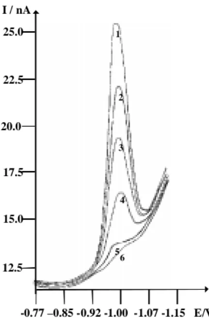
The addition of lambda phage DNA to a solution of Zn2+ (depolarizer) brings about a decrease in the diffusion current. The relative reduction in this current increases with DNA concentration and with its ratio to the concen-tration of the zinc. It decreases with increase in ionic strength which is maintained by the concentration of the supporting electrolyte26. The differential pulse voltam-mograms presented in Figure 1 show the dependence of the diffusion current at constant Zn2+ concentration on the concentration of added lambda phage DNA. Curve 1 (Figure 1) represents the reduction of 1.0 mL of 1.5 x 10⁻⁸ mol L-1 Zn2+ in 20 mL of 0.1 mol L-1 KNO3. Curves num-bered 2-6 (Figure 1) represent the effect of the addition of 7.1 x 10⁻¹², 1.4 x 10⁻¹¹, 2.1 x 10⁻¹¹, 2.9 x 10⁻¹¹ and 3.6 x 10⁻¹¹ mol L-1 DNA to the electrochemical cell. The re-sulting changes in reduction current attest to the interac-tion between Zn2+ and lambda phage DNA.
The amperometric titration curve of zinc with lambda phage DNA is shown in Figure 2. We found a complete decrease in the reduction current of Zn2+ after the addi-tion of 43 µL of DNA to the electrochemical cell. This volume of lambda phage DNA used in the complexation of zinc was determined through the inflection point (I.P.) of the titration curve and was employed to calculate the stoichiometry of the reaction. By this method, we found one lambda phage DNA per 23 Zn2+ ions.
I / nA
-0.77 –0.85 -0.92 -1.00 -1.07 -1.15 E/V 25.0
22.5
20.0
17.5
15.0
12.5
4 3
5 6 2 1
Figure 1. Differential pulse voltammograms of 1.0 mL of 1.5 x
10-8 mol L-1 Zn2+ in 20 mL of 0.1 mol L-1 KNO3 with different concentrations of lambda phage DNA. (1) Zn2+, no lambda phage DNA; (2) Zn2+ + 7.1 x 10⁻¹² mol L-1 lambda phage DNA; (3) Zn2+ + 1.4 x 10⁻¹¹ mol L-1 lambda phage DNA; (4) Zn2+ + 2.1 x 10⁻¹¹ mol L-1 lambda phage DNA; (5) Zn2+ + 2.9 x 10⁻¹¹ mol L-1 lambda phage DNA; (6) Zn2+ + 3.6 x 10⁻¹¹ mol L-1 lambda phage DNA. Ei = 0.180 V, Ef = -1.15 V, pulse amplitude = 50 mV, scan rate = 10 mV s-1, working electrode: DME.
0 10 20 30 40 50 60
-2 0 2 4 6 8 10 12
I.P.
I /
n
A
V l a m b d a P ha g e D N A / µl
Figure 2. Amperometric titration Graph of 1.0 mL of 1.5 x 10⁻⁸ mol L-1 Zn2+ in 20 mL of 0.1 mol L-1 KNO
3 with lambda phage DNA. Successive additions of 5.0 µL of 1.5 x 10⁻⁸ mol L-1 lambda phage DNA solution were made.
For a complex of the electro-active substance Zn2+ with lambda phage DNA, the electrochemical reduction reactions can be divided in two steps:
Zn2+ - DNA Zn2+ + DNA (1)
Zn2+ + 2e- Zn0 (2)
The dissociation constant (Kd) of the Zn2+-Lambda Phage DNA complex was obtained using the following equation29:
(
)
[
]
]
[
2 0 2 2
0 2
DNA
DNA
k
i
i
i
i
p p pd
p
=
−
+
−
(3)where: Kd = dissociation constant of the complex Zn2+ -Lambda Phage DNA; ip02 = reduction current of Zn2+ in the absence of lambda phage DNA; ip2 = reduction current of Zn2+ in the presence of lambda phage DNA; [DNA] = concentration of added lambda phage DNA in solution.
According to equation (3), ip0 and different values of ip are determined respectively by holding the concentra-tion of Zn2+ constant and varying the concentration of lambda phage DNA.
The curve ip2 versus
(
)
[
DNA]
i
i
2p2 0 p −
(Figure 3) for the
com-plex Zn2+- lambda phage DNA was plotted and one straight line was obtained. From the slope, the dissociation constant was determined as 3.44 x 10⁻¹¹ mol L-1.
5.5 6.0 6.5 7.0 7.5 8.0 8.5
0 20 40 60 80 100 120
i
2 x 1p
0
+18
/ A
2
[(i2p 0 - i2p) / [DNA]] x 10+ 6 / mol- 1 L A2
Linear Regression: Y = A + B * X
Param Value sd A -191.8243 10.86039 B 34.43641 1.51382 r = 0.99616
SD = 3.45712, N = 6 P = 0.00002
Figure 3. The plot ip2 vs.
(
)
[DNA]
i i 2p
2 0 p −
used to calculate the dissociation constant of Zn2+- lambda phage DNA complex.
Cyclic Voltammetry: voltammograms, stoichiometry, dissociation constant and reaction mechanism
The cyclic voltammograms of 1.5 mL of 1.5 x 10⁻⁸ mol L-1 Zn2+ in 20 mL of 0.1 mol L-1 KNO
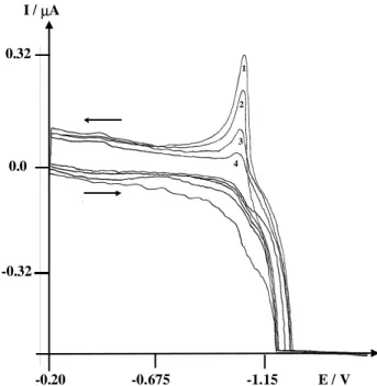
3 with differ-ent concdiffer-entrations of lambda phage DNA are shown in Figure 4. Curve 1 (Figure 4) represents the oxidation of zinc in 0.1 mol L-1 KNO3. Curves numbered 2, 3 and 4 (Figure 4) show the decrease of the oxidation current of zinc during incremental additions (20 µL) of 1.5 x 10⁻⁸ mol L-1 lambda phage DNA. The resulting changes in this current again demonstrate interaction between Zn2+ and lambda phage DNA.
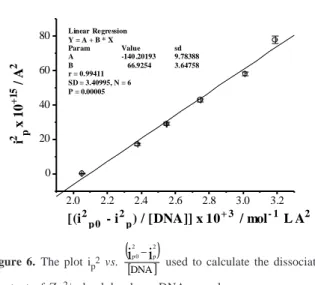
Figure 5 shows the amperometric titration curve of Zn2+ with lambda phage DNA. The addition of 4.4 x 10⁻¹¹ mol L-1 lambda phage DNA to the electrochemical cell decrease completely the oxidation current of zinc. This volume of lambda phage DNA used in the complexation of zinc was determined through the inflection point (I.P.) of the titra-tion curve and was employed to calculate the stoichiom-etry of the reaction. By this method, we found one lambda phage DNA per 22 Zn2+ ions.
The dissociation constant (Kd) of the Zn2+- lambda phage DNA complex was calculated to be 6.69 x 10⁻¹¹ mol L-1 again using the equation 3 (Figure 6).
The oxidation-reduction process of zinc was investi-gated through curves I (oxidation current) versus v1/2 (square
-0.20 -0.675 -1.15 E / V 1
4 3 2 I / µA
-0.32 0.32
0.0
Figure 4. Cyclic voltammograms of 1.5 mL of 1.5 x 10⁻⁸ mol L-1 Zn2+ in 20 mL of 0.1 mol L-1 KNO
3 with different concentrations of lambda phage DNA. (1) Zn2+, no lambda phage DNA; (2) Zn2+ + 2.8 x 10⁻¹¹ mol L-1 lambda phage DNA; (3) Zn2+ + 4.2 x 10⁻¹¹ mol L-1 lambda phage DNA; (4) Zn2+ + 5.6 x 10⁻¹¹ mol L-1 lambda phage DNA. Ei = -0.2 V, Ew = -1.6 V, scan rate = 10 mV s-1, working electrode: HMDE.
I /
µ
A
Figure 5. Amperometric titration Graph of 1.5 ml of 1.5 x 10⁻⁸ mol L-1 Zn2+ in 20 mL of 0.1mol L-1 KNO
3 with lambda phage DNA. Successive additions of fix volume of 10 µL of 1.5 x 10⁻⁸ mol L-1 lambda phage DNA solution.
root of scan rate). As described by the Randles-Sevcik equa-tion30, the oxidation current (I) of Zn2+ increases as v1/2 in the absence of lambda phage DNA. A plot of this equation yields a straight line, the slope of which can be used to determine the diffusion coefficient of the system (Figure 7, curve O ). However, in the presence of lambda phage DNA, the electrochemical behavior of Zn2+ changed and its oxi-dation-reduction process was no longer controlled by diffu-sion anymore (Figure 7, curve ).
0 20 40 60 80
0.0 0.1 0.2 0.3 0.4
I.P.
Discussion
Zinc regulates the gene expression machinery. It af-fects the structure of chromatin, the template function of its DNA, the activity of numerous transcription factors and of DNA and RNA polymerases1,2. In order to understand why Zn2+ ions in particular are required, many researchers have studied the interactions of zinc and DNA by several techniques16,19,31-32. In these studies an attempt was made to understand specific changes of DNA secondary struc-ture induced by zinc ion binding. Zn2+ ions, as other tran-sition metals bind preferentially to the phosphate sugar backbone, but they can also interact with bases (guanines and cytosines) after heat desnaturation. Zn2+ decreases the melting temperature of the DNA molecule at high Zn2+ : DNA-P ratio and aids the reformation of the DNA double helix upon cooling. These effects have been attributed to the formation of metal bridges between complementary bases (GC pair) of opposite strands in the unwound state
and were found to be directly and reversely related to GC content of the DNA. The Zn2+ : DNA-P ratio used in our experiments was one, so that we can infer that zinc ions are stabilizing the duplex (GC pairs) by their electrostatic interaction with the negative phosphate groups of lambda phage DNA.
The methodology developed in this work allowed de-termination of stoichiometry, dissociation constant and a possible mechanism of the interaction between Zn2+ and lambda phage DNA. The stoichiometry values obtained by differential pulse voltammetry (23:1) and cyclic voltammetry (22:1) are very close. However, considering the size of the DNA molecule under investigation and the potential number of phosphate groups available, one could expect a higher number of binding sites for Zn2+. Although on Zn2+ : DNA stoichiometries are surprisingly low, one has to take into account stereochemistry prob-lems due to overfolding of such a large molecule under the experimental conditions used. These results will be checked by further voltammetric experiments with a syn-thetic oligonucleotide. Using this smaller DNA, it will be possible to make an accurate measure of the stoichiometry and the binding sites of the reaction. The low Kd values found for the complex Zn2+- lambda phage DNA using differential pulse voltammetry (Kd = 3.44 x 10⁻¹¹ mol L -1) and cyclic voltammetry (K
d = 6.69 x 10⁻¹¹ mol L-1) are very close and imply that Zn2+ ions are indispensable for catalytic function and structural stability of zinc enzymes which participate in the replication, degradation and trans-lation of genetic material of all species. Moreover, they are probably interacting not only with the active site of the enzyme during these processes, as already well-known in the literature 1,2, but also with DNA.
The differential pulse voltammograms in figure 1 show that the addition of lambda phage DNA to the electro-chemical cell containing 1.0 mL of 1.5 x 10⁻⁸ mol L-1 Zn2+ did not shift the reduction peak of zinc (E
p = con-stant), in addition to that, this peak disappears after the complete complexation of Zn2+. We can also notice that the quantity of charge is constant (the width of the reduc-tion peak of zinc at h/2 is not changing with the addireduc-tion of DNA). These two evidences suggest that Zn2+ ion is the chemical species which is arriving on the Hg surface to undergo reduction. Curves I vs. v1/2 in Figure 7 show that the oxidation-reduction process of zinc is no longer con-trolled by diffusion in the presence of lambda phage DNA. It is known that DNA adsorbs onto mercury electrode in the potential range of about 0 to –1.3V at neutral pH and moderate ionic strength33, so that we can infer that lambda phage DNA is mediating the charge transfer of bulk Zn2+ ions (Zn2+ (b)) through its adsorption on mercury surface. 2.0 2.2 2.4 2.6 2.8 3.0 3.2
0 20 40 60 80
i
2 x 1p
0
+1
5 / A
2
[(i2p0 - i2p) / [DNA]] x 10+ 3 / mol- 1 L A2
Linear Regression Y = A + B * X
Param Value sd
A -140 .20193 9.78388
B 66.9254 3.64758
r = 0.99411 SD = 3.40995, N = 6 P = 0.00005
Figure 6. The plot ip2 vs.
(
)
[DNA]
i i 2p
2 0 p −
used to calculate the dissociation constant of Zn2+- lambda phage DNA complex.
0 1 2 3 4 5 6 7 8
0.0 0.1 0.2 0.3
I /
µ
A
v1 / 2 / (mV/s)1 / 2
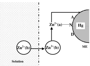
Unfortunately, the mechanism of this process is not known. Probably, the bulk Zn2+ ions (Zn2+ (b)) are replacing the zinc bounded to lambda phage DNA (Zn2+(a)), thereupon, this Zn2+ (a) is released to Hg surface to undergo reduc-tion (Figure 8).
Conclusion
Conductimetry, equilibrium dialysis, UV-Vis spectros-copy and X-ray crystallography have been used to study metal-nucleic acid interactions since 1960. However, they present some experimental difficulties or limitations which make them not suitable for measurement of metal-ion bind-ing to DNA. The electrochemical methods used in the present work overcame these difficulties. The results ob-tained in the study of Zn2+-DNA interaction by differen-tial pulse polarography and cyclic voltammetry pointed out the possibilities that voltammetric techniques now offer for determining quantitative data of metal-nucleic acid interactions. Dissociation constant and stoichiometry val-ues and also a possible mechanism of Zn2+-DNA interac-tion were determined successfully by using a simple and rapid voltammetric procedure. A quantitative knowledge of the degree and strength of Zn2+ binding to lambda phage DNA will be helpful in understanding the cleavage of deoxyribonucleic acids.
Acknowledgments
The authors wish to express their gratitude to Profes-sors A. C. Barbosa and L. Morhy for providing certain facilities, to Dr. Daniel Rigden for his help in revising the English text and to CNPq, CAPES, FAPDF for finan-cial support.
References
1. Blackburn, G. M.; Gait, M. J. Nucleic Acids in
Chemistry and Biology; Oxford University Press;
New York, 1996.
2. Hetch, S. M. Bioorganic Chemistry: Nucleic Acids; Oxford University Press; New York, 1996.
3. Shack, J.; Jenkins, R. J.; Trompsett, J. M. J. Biol.
Chem. 1953, 203, 373.
4. Felsenfeld, G.; Huang, S. L. Biochim. Biophys. Acta 1961, 51, 19.
5. Mathieson, A. R.; Olayemi, J. Y. Arch. Biochem.
Biophys. 1975, 169, 237.
6. Lyons, J. W.; Kotin, L. J. Am. Chem. Soc. 1964, 86, 3634.
7. Eichhorn, G. L.; Clark, P.; Becker, E. D. Biochemistry 1966, 5, 245.
8. Bryan, S. E.; Frieden, E. Biochemistry 1967, 6, 2728. 9. Burks, P. P. Diss. Abstr. Int. B 1971, 31, 5228. 10. Zimmer, C. H.; Luck, G.; Fritzsche, H.; Triebel, H.
Biopolymers 1971, 10, 441.
11. Sorokin, V. A. Biophysics 1994, 39, 1041.
12. Hua, E.; Wang, H.; Yang, P.; Yang, B. Polyhedron 1996, 15, 2067.
13. Sorokin, V. A.; Valeev, V. A.; Gladchenko, G. O.; Sysa, I. V.; Blagoi, Y. P.; Volchok, I. V. J. Inorg. Biochem. 1996, 63, 79.
14. Yamame, T.; Davidson, N. J. Am. Chem. Soc. 1961, 83, 2599.
15. Gulanowski, B.; Swiatek, J.; Kozlowski, H. J. Inorg.
Biochem. 1992, 48, 289.
16. Zimmer, C. H.; Luck, G.; Triebel, H. Bioploymers 1974, 13, 425.
17. Wiberg, J. S.; Neuman, W. F. Arch. Biochem. Biophys. 1957, 72, 66.
18. Shapiro, J. T.; Stannard, B. S .; Felsenfeld, G.
Biochemistry 1969, 8, 3233.
19. Banerjee, K. C.; Perkins, D. J. Biochim. Biophys. Acta 1962, 61, 1.
20. Carrabine, J. A. Diss. Abstr. Int. B 1970 , 31, 3230.
21. Berman, H. M.; Shieh, H.-S. In Topics in Nucleic
Acid Structure; Neidle, S., Ed.; Wiley; New York,
1981, p. 17.
22. Sarma, R. H.; Dhingra, M. M. In Topics in Nucleic
Acid Structure; Neidle, S., Ed.; Wiley; New York,
1981, p. 33.
23. Swaminathan, V.; Sundaralingam, M. Crit. Rev.
Biochem. 1979, 6, 245.
24. Kalsbeck, W.; Thorp, H. J. Am. Chem. Soc. 1993, 115, 7146.
D A
Zn2+(a)
N Hg
Zn2+(b) Zn2+(b)
Solution
DME
Figure 8. Mechanism proposed to explain the oxidation-reduction
25. Carter, M. T.; Rodriguez, M.; Bard, A. J. J. Am. Chem.
Soc. 1989, 111, 8901.
26. Bach, D.; Miller, I. R. Biopolymers 1967, 5, 161. 27. Elzanowska, H.; Van de Dande, J. H. Bioelectrochem.
Bioenerg. 1988, 19, 441.
28. Fasman, G. D. Handbook of Biochemistry and
Mo-lecular Biology; 3rd ed.; CRC Press; Cleveland, OH,
1975, p. 589.
29. Niu, J.; Cheng, G.; Dong, S. Electrochim. Acta 1994, 39, 2455.
30. Van Benschoten, J. J.; Lewis, J. Y.; Heineman, W. R.; Roston, D. A.; Kissinger, P. T. J. Chem. Educ. 1983, 60, 772. 31. Eichhorn, G. L.; Butzow, J. J.; Clark, P.; Tarien, E.
Biopolymers 1967, 5, 283.
32. Jia, X. Diss. Abstr. Int. B 1991, 52, 3058.
33. Krznaric, D.; Cosovic, B. Anal. Biochem. 1986, 156, 454.