ORIGINAL
ARTICLE
Authors
Clemax Couto Sant´Anna1
Christianne Mello Schmidt2
Maria de Fátima B Pombo March3
Susan Martins Pereira4
Maurício Lima Barreto5
1PhD; Associate Professor; Medical School of Universidade Federal do Rio de Janeiro - UFRJ 2Master’s Degree; Physician of the Hospital Universitário Antônio Pedro, Universidade Federal Fluminense - UFF 3PhD; Adjunct Professor of the Medical School, UFRJ 4PhD; Associate Professor of the Instituto de Saúde Coletiva of Universidade Federal da Bahia - UFBA 5PhD - Full Professor of the Instituto de Saúde Coletiva, UFBA
Submitted on: 05/14/2010 Approved on: 08/03/2010
Correspondence to:
Clemax Couto Sant´Anna Rua Cinco de Julho, 350/604
Copacabana, Rio de Janeiro, RJ clemax@vetor.com.br
Financial Support: CNPq
We declare no confl ict of interest.
ABSTRACT
Objective: To describe radiologic findings of pulmonary tuberculosis (TB) in adolescents. Methods: Retrospective, cross-sectional, observational study of 850 patients with TB, aged 10 to 19 years, and notifi ed to the Brazilian Ministry of Health. Data were collected from the TB notifi cation and medical records in the cities of Manaus, Amazonas State, and Salvador, Bahia State, in the 1996-2003 period. Data are shown in tables and analyzed using the chi-square and Mann-Whitney tests, with a 5% signifi cance level. Results: Mean age was 15.6 years; 443 (52.1%) patients were males. The most common radiologic lesion was the upper pulmonary lobe infi ltrate (53.3%), and isolated cavitation was found in 32.4% of the patients. Both lungs were affected in 29.2% of the patients. The fi nding of bilateral radiologic lesions was signifi cantly associated with longer disease duration (p = 0.0005). Conclusions: Pulmonary TB in adolescents has similar characteristics to TB in adults, evidencing the important role played by adolescents in community disease transmission.
Keywords: tuberculosis; adolescents; diagnosis; chest radiography.
[Braz J Infect Dis 2011;15(1):40-44]©Elsevier Editora Ltda.
INTRODUCTION
Although the real situation of tuberculosis (TB) in adolescents is not well-known, children and adolescents account for 3% to 25% of the TB cases registered in different countries, with high frequencies in areas of high disease burden.1
In developed countries, TB affects mainly the elderly, but, in developing countries, the produc-tive younger population is the most affected.2,3
Children play a limited role in TB transmis-sion in the community, but adolescents can de-velop bacilliferous, thus, transmissible, pulmo-nary TB.4 Adolescents account for around 20% of
the Brazilian population.5 At that age, the
indi-vidual is under development and undergoing be-havioral and emotional changes, which can make adherence to treatment of prolonged diseases, such as TB, diffi cult. This can lead to treatment discontinuation, resulting in perpetuation of TB transmission in the community and appearance of resistant strains. Adolescents have greater so-cial interaction and are more susceptible to ill-nesses and transmission of TB and other diseases.
This study aimed at assessing radiological as-pects of pulmonary TB in Brazilian adolescents based on data of two Brazilian capital cities.
METHODS
This is a retrospective, descriptive, observa-tional study. This study assessed TB notifi-cations (individual investigation sheet of the Ministry of Health) of all adolescents living in the cities of Manaus and Salvador, diag-nosed with TB and notified to the Brazilian Ministry of Health, from 1996 to 2005. Each notified case had the medical record located and scrutinized, along with the respective chest radiographic report present in the database of the BCG-Revac trial,6 which
al-lowed the analysis of the radiologic patterns and their distribution according to age and disease duration. Radiologic patterns were adapted from Marais et al.4
The defi nition of adolescence of the World Health Organization that includes individuals aged 10 through 19 years, was adopted. Data of the original database were stored in the sta-tistical software Epiinfo 6.0.
Statistical analysis was performed with the software SAS 6.04 (SAS Institute, Inc, Cary, North Carolina). The following tests were used: the Mann-Whitney non-parametric test for comparing continuous (numerical) variables
Radiologic findings of pulmonary
between two subgroups; and the chi-square test for com-paring categorical (categorical) variables. The signifi-cance level was 0.05.
The BCG-Revac trial was approved by the Committees on Ethics in Research of the Universidade Federal da Bahia
and by the London School of Hygiene and Tropical Medi-cine.6 As the present study used secondary data from the
original project, a new approval by a Committee on Ethics in Research was not required.
RESULTS
Initially, the records of 904 adolescents were evaluated and 850 (93.7%) patients who had chest radiography reports were included. Mean age was 15.6 years, and 443 (52.1%) patients were male.
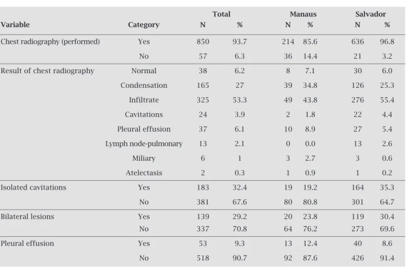
Table 1 shows the major characteristics of the chest ra-diographic fi ndings.
The most frequent types of radiologic lesion were as fol-lows: infi ltrates (53.3%); cavitation (32.4%); and condensa-tion (27%). Hilar lymph node enlargement was found in 18/548 (3.2%) of the cases, and atelectasis in 11/555 (1.9%)
of the cases. The TB radiologic lesions were in the right side in 220/476 (42%) patients, bilateral in 139/476 (29.2%), and in the left side in 137/476 (28.8%) patients.
Age and the duration of disease until diagnosis were as-sessed in relation to unilateral or bilateral pulmonary radio-logic involvement (Table 2).
A signifi cant difference in disease duration until diag-nosis was observed when the extension of the radiologic le-sion was considered. The subgroup with bilateral lele-sion had signifi cantly longer disease duration than the subgroup with unilateral lesion. On the other hand, no difference regarding age was observed in patients with unilateral or bilateral le-sion when the total study sample was considered.
In the city of Manaus, the subgroup with bilateral lesions were signifi cantly younger (13.7 years) than the subgroup with unilateral lesions (15.2 years). In the city of Salvador, pa-tients with bilateral TB lesions were slightly older (16.4 versus
15.9 years) and had shorter duration of disease (median of 60 versus 30 days) than the subgroup with unilateral lesions.
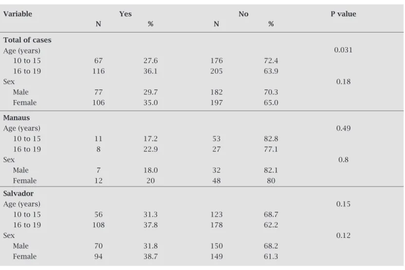
Radiologic cavitation was more frequently found in pa-tients aged from 16 to 19 years (36.1%). No difference was observed regarding sex (Table 3).
Table 1. Major findings in chest radiography of adolescents with pulmonary tuberculosis. Cities of Manaus and Salvador. 1996-2005
Total Manaus Salvador
Variable Category N % N % N %
Chest radiography (performed) Yes 850 93.7 214 85.6 636 96.8
No 57 6.3 36 14.4 21 3.2
Result of chest radiography Normal 38 6.2 8 7.1 30 6.0
Condensation 165 27 39 34.8 126 25.3
Infiltrate 325 53.3 49 43.8 276 55.4
Cavitations 24 3.9 2 1.8 22 4.4
Pleural effusion 37 6.1 10 8.9 27 5.4
Lymph node-pulmonary 13 2.1 0 0.0 13 2.6
Miliary 6 1 3 2.7 3 0.6
Atelectasis 2 0.3 1 0.9 1 0.2
Isolated cavitations Yes 183 32.4 19 19.2 164 35.3
No 381 67.6 80 80.8 301 64.7
Bilateral lesions Yes 139 29.2 20 23.8 119 30.4
No 337 70.8 64 76.2 273 69.6
Pleural effusion Yes 53 9.3 13 12.4 40 8.6
Table 2. Distribution of the adolescents with pulmonary tuberculosis according to age and duration of disease in the general sample and radiologic location (unilateral vs. bilateral), in the cities of Salvador and Manaus. 1996-2005
Variable Radiologic N Mean SD Median Minimum Maximum P value
location
Total
Age (years) Unilateral 337 15.8 2.2 16 10 19 0.14
Bilateral 139 16.0 2.3 16 10 19
Disease duration (days) Unilateral 266 58.5 67.3 30 2 730 0.0005
Bilateral 108 76.8 68.7 60 7 365
Manaus
Age (years) Unilateral 64 15.2 1.5 15 11 18 0.002
Bilateral 20 13.7 2.2 13.5 10 18
Disease duration (days) Unilateral 61 61.6 51.7 45 5 300 0.30
Bilateral 19 92.3 92.2 60 14 360
Salvador
Age (years) Unilateral 273 15.9 2.3 16 10 19 0.037
Bilateral 119 16.4 2.1 17 10 19
Disease duration (days) Unilateral 205 57.6 71.3 30 2 730 0.0004
Bilateral 89 73.5 62.8 60 7 365
SD, standard deviation
Table 3. Distribution of the radiologic finding cavitation according to age bracket and sex. 1996-2005
Variable Yes No P value
N % N %
Total of cases
Age (years) 0.031
10 to 15 67 27.6 176 72.4
16 to 19 116 36.1 205 63.9
Sex 0.18
Male 77 29.7 182 70.3
Female 106 35.0 197 65.0
Manaus
Age (years) 0.49
10 to 15 11 17.2 53 82.8
16 to 19 8 22.9 27 77.1
Sex 0.8
Male 7 18.0 32 82.1
Female 12 20 48 80
Salvador
Age (years) 0.15
10 to 15 56 31.3 123 68.7
16 to 19 108 37.8 178 62.2
Sex 0.12
Male 70 31.8 150 68.2
DISCUSSION
In Brazil, data on the incidence of TB in adolescents (individuals aged from 10 to 19 years) provided by the Ministry of Health became available in 1999. For that age bracket and from 1999 to 2005, the incidence of TB ranged from 91.53 to 36.44/100,000 inhabitants in the city of Salvador and from 70.12 to 45.65/100,000 inhabit-ants in the city of Manaus.7
The present study showed a predominance of character-istic lesions of re-infection or adult type TB in the adoles-cents assessed: 53% of chest x-rays had infi ltrates in the up-per third of the lungs, and 32% of the radiographs showed cavitations. In addition, most patients with cavitations were adolescents in the postpubertal stage (median of age, 16 years), a situation compatible with primo-infection occur-ring early in childhood.
Chest radiograph was largely used in the services of the National TB Control Program emphasizing the importance of imaging diagnosis in health care services in Brazil.
Radiologic patterns of pulmonary TB allow us to in-fer several aspects of the pathogenesis and clinical picture of the patients assessed. Classically, there are two pres-entations of TB: primo-infection or primary TB, and re-infection. The former is more commonly found during childhood, and is characterized by uni- or bilateral hilar lymph node enlargement either in association or not with pulmonary infi ltrates.4 Likewise, hematogenous
dissemi-nations, also found in TB primo-infection, radiologically expressed as disseminated micronodular infi ltrates, known as the miliary pattern.8 In the present study, the primary
TB presentation classifi ed as lymph node-pulmonary, pri-mary complex, and miliary added up to over 3% of the total. It is evident that the adolescents here studied had al-ready developed TB primo-infection prior to the disease that made them look for health care.
In Brazil and in other countries with a high TB burden, TB primo-infection and primary TB are more common in children than in adults, because of the high likelihood of contact with M. tuberculosis during childhood. In developed countries, the likelihood of developing TB primo-infection can be postponed to adolescence or adulthood.8,9
Individuals who had TB primo-infection or had been vaccinated with BCG develop a type of immunogenic de-fense that, when exposed to a bacillary burden originating from a contagious source, relies on the immune memory to trigger phagocytosis of the bacilli, which then entry a state of metabolic inactivity.10,11 If the immune system fails
re-infection or adult-type TB can occur. In such cases, the chest radiograph shows characteristic infi ltrates and cavitations in the upper pulmonary thirds, usually in the posterior segments. The most severe radiologic forms of re-infection TB appear as extensive bilateral lesions, cavita-tions, and bronchial dissemination of the disease.8
In this study, cavitations were more common in adoles-cents aged 16 years or older, while lymph nodes enlargement were more common in patients aged 15 years or less. This distribution confi rms the classical notion that more sugges-tive forms of primary TB occur in younger individuals and post-primary manifestations in older adolescents. The same analysis regarding sex, showed no difference.
Pleural effusion due to TB, more common in adolescents and adults than in children, was observed in 9% of the patients.
In this study, the most severe TB lesions were related to the longer duration of symptoms, possibly due to a diagno-sis delay in health services. The effectiveness of TB control programs can be assessed through the delay to establish TB diagnosis.11 In our study, the median of symptom duration
was 60 days, suggesting a high risk for patients to infect their families and close contacts. In addition, the maximum delay for the diagnosis was 365 days for patients with bilateral le-sions, and 730 days for patients with unilateral lesions. In the case of adolescents who can cough and expectorate, sputum bacilloscopy can provide earlier diagnosis.12
Patients from the city of Manaus had TB forms of faster evolution and at a younger age than patients from the city of Salvador. This may have been due to the high frequency of non-tuberculous mycobacteria in the Amazon region. In ad-dition, factors related to the host, low effectiveness of BCG vaccination, or, more likely, the characteristics of M. tuber-culosis could explain such fi ndings.6,13
On the other hand, in the city of Salvador, the disease lasted longer for older patients. This suggests that the health services are having problems to detect TB cases, which can result in a long interval for establishing the diagnosis.
This study had some limitations. One concern is the lack of information about some variables, such as radiographic reports and demographic data, that, however, had a small infl uence in the fi nal results, considering the large number of cases. Tuberculosis notifi cation in countries where TB is endemic offers innumerous diffi culties to TB control pro-grams.11 Similarly, comparison of our data with those
re-ported in the literature could not be done as most studies in countries where TB is endemic and affects adolescents do not allow for separate analysis of that age group. TB control programs around the world use the cut-off point of 15 years to categorize patients as children or adults, and, thus, data referring to adolescents (over 10 years of age) can not be retrieved. In addition, this study could not assess the social conditions of the adolescents analyzed based on the data found in their notifi cation sheet. However, it is known that, in Brazil and other countries where TB is endemic, most pa-tients belong to the most vulnerable extract of society.
In conclusion, most cases of TB in adolescents were simi-lar to those in adults: apical pulmonary infi ltrates, extensive lesions, and cavitations. Primary TB forms were rare.14 In
prolonged. This delay may have accounted for the fi nding of severe lesions in many patients. Adolescents belong to a group that deserves special attention from health care providers, either due to their diffi culty in adhering to prolonged treat-ments or to their reluctance to look for medical care. Thus, further efforts are recommended to improve the effi cacy of the health care network for diagnosing TB and to provide more information regarding the complaints suggestive of TB in adolescents, aiming at earlier diagnosis of the disease.
ACKNOWLEDGEMENTS
The authors thank Profs. Gesmar V Haddad and Adauto Dutra of the Post-graduation Program in Pediatrics of the
Universidade Federal Fluminense, city of Niterói, state of Rio de Janeiro.
REFERENCES
1. Hesseling AC, Schaaf HS, Gie RP et al. A critical review of diagnosis approaches in the diagnosis of childhood tuber-culosis. Int J Tuberc Lung Dis. 2002; 6:1038-1045.
2. Donald PR. Childhood tuberculosis: the hidden epidemic. Int J Tuberc Lung Dis. 2004; 8:627-629.
3. Marais BJ, Graham SM, Cotton MF, Beyers N. Diagnosis and management challenges for childhood tuberculosis in the era of HIV. J Infect Dis. 2007; 196,supl. 1:76-85. 4. Marais BJ, Gie RP, Schaaf HS et al. The natural history of
childhood intra-thoracic tuberculosis: a critical review of literature from the pre-chemotherapy era. Int J Tuberc Lung Dis. 2004; 8:392-402.
5. Brasil. IBGE/Censos demográficos. População total. Dis-tribuição (%) por faixa etária segundo unidade da feder-ação, 2006. Available at <http://tabnet.datasus.gov.br/cgi/ tabcgi.exe?idb2007/a01.def>. Accessed on: 1 Oct. 2009. 6. Rodrigues LC, Pereira SM, Cunha SS et al. Effect of BCG
revaccination on incidence of tuberculosis in school-aged children in Brazil: the BCG-REVAC cluster-randomised trial. Lancet 2005; 366:1290-1295.
7. Brasil. Ministério da Saúde, SVS, SINAM. Indicadores de morbidade e fatores de risco. Taxa de incidência de tu-berculose por faixa etária segundo Unidade da Federação. Brasília, 2007b. Available at <http://tabnet.datasus.gov.br/ cgi/tabcgi.exe?idb2007/d0202.def> Accessed on: 21 Oct. 2009.
8. Sant´Anna CC. Tuberculose na criança. J. Pediatr. (Rio J.) 1998; 74(Supl.1):S69-S75.
9. Styblo K. Estado del arte, I: epidemiologia de la tuberculo-sis. Bol UICT 1978; 53:145-147.
10. Barroso EW. Imunopatogenia. In: Sant´Anna CC (ed). Tu-berculose na infância e na adolescência. São Paulo: Athe-neu, 2002.
11. Procópio MJ (Coord.). Controle da Tuberculose - Uma Proposta de Integração Ensino – Serviço. Rio de Janeiro: EAD/ENSP, 2008.
12. Sociedade Brasileira de Pneumologia e Tisiologia. III Di-retrizes para tuberculose da Sociedade Brasileira de Pneu-mologia e Tisiologia. J Bras Pneumol. 2009; 35:1018-1048 13. Salem JI, Marója MF, Carvalho FF, Lima MO et al. Valor relativo do exame direto, após concentração e por cultivo de escarro no diagnóstico bacteriológico da tuberculose pulmonar no Amazonas. J. Pneumol. 1990; 16:133-136 14. Sant´Anna CC, March MF, Barreto M, Pereira S, Schmidt