The Brazilian Journal of
INFECTIOUS DISEASES
w w w . e l s e v i e r . c o m / l o c a t e / b j i d
Review article
Japanese encephalitis: a review of the Indian perspective
Sarika Tiwari, Rishi Kumar Singh, Ruchi Tiwari, Tapan N. Dhole
∗Department of Microbiology, Sanjay Gandhi Post Graduate Institute of Medical Sciences, Uttar Pradesh, India
a r t i c l e
i n f o
Article history:
Received 29 March 2012 Accepted 11 July 2012
Available online 8 November 2012
Keywords:
Epidemiology Pathogenesis Vector born diseases Epidemics
a b s t r a c t
Japanese encephalitis virus (JEV) causes Japanese encephalitis, which is a leading form of viral encephalitis in Asia, with around 50,000 cases and 10,000 deaths per year in children below 15 years of age. The JEV has shown a tendency to extend to other geographic regions. Case fatality averages 30% and a high percentage of the survivors are left with perma-nent neuropsychiatric sequelae. Currently, there is no cure for JEV, and treatment is mainly supportive. Patients are not infectious, but should avoid further mosquito bites. A num-ber of antiviral agents have been investigated; however, none of these have convincingly been shown to improve the outcome of JEV. In this review, the current knowledge of the epidemiology and the pathogenesis of this deadly disease have been summarized.
© 2012 Elsevier Editora Ltda. All rights reserved.
Introduction
Japanese encephalitis (JE) is a common mosquito borne fla-viviral encephalitis. It is one of the leading forms of viral encephalitis worldwide, mostly prevalent in eastern and southern Asia, covering a region with a population of over three billion.1 Most infections of JE are asymptomatic, but
if clinical illness develops, it causes significant morbidity and mortality. Though underreported, JE causes an estimated 50,000 cases and 15,000 deaths annually.2JE is a disease of
pub-lic health importance because of its epidemic potential and high fatality rate. In endemic areas, the highest age-specific attack rates occur in children of 3 to 6 years of age.3,4
Approxi-mately one third of patients die, and half of the survivors suffer severe neuropsychiatric sequelae from the disease.5
Japanese encephalitis virus (JEV) belongs to the family
flaviviridae and genus Flavivirus.6 It is a single stranded, positive-sense polarity RNA genome of approximately 11 kb in
∗ Corresponding author at:Department of Microbiology, Sanjay Gandhi Post Graduate Institute of Medical Sciences, Rai Barelly Road
Lucknow, 226014, Uttar Pradesh, India.
E-mail address:proftndhole@gmail.com(T.N. Dhole).
length. The virion of JEV contains three structural proteins – nucleocapsid or core protein (C), non-glycosylated membrane protein (M), and glycosylated envelope protein (E), as well as seven non-structural (NS) proteins – NS1, NS2A, NS2B, NS3, NS4A, NS4B, and NS.7JEV exists in a zoonotic cycle between
mosquitoes and pigs and/or water birds. This study reviewed JEV literature from 2000 to 2010, outlining the Indian sce-nario, clinical depictions, diagnosis, and the prevention of this deadly disease.
Historical perspective
The first outbreak of encephalitis attributed to JEV was reported in Japan in 1871. Major epidemics have been reported about every ten years; in 1924, over 6,000 cases were docu-mented in a severe epidemic in Japan.8In 1935, the prototype
Nakayama strain was isolated from the brain of a patient suffering from encephalitis. Thereafter, the virus had been
1413-8670/$ – see front matter © 2012 Elsevier Editora Ltda. All rights reserved.
classified with other flaviviruses as a group B arbovirus in the familyTogaviridae, Originally the term “type B” encephali-tis was used to distinguish this summer epidemic from von Economo’s lethargica/sleepy sickness, commonly known as type A encephalitis,5which occurs in winter with a different
clinical presentation. Later on, the designation “type B” was abandoned, and in 1985, JEV was designated under a sepa-rate familyFlaviviridae, as a member of genusFlavivirus.9The genusFlavivirushas been named after the prototype yellow fever virus (from the Latin wordflavi,), and is comprised of 70 small, enveloped viruses with single stranded positive-sense RNA.5
Epidemiological features
Global outlook
Japanese encephalitis is one of the most important forms of epidemic and sporadic encephalitis in the tropical regions of Asia, including Japan, China, Taiwan, Korea, Philippines, all of Southeastern Asia, and India; however, related neu-rotropic viruses are spread across the globe.10Countries with
proven epidemics of JE include India, Pakistan, Nepal, Sri Lanka, Burma, Laos, Vietnam, Malaysia, Singapore, Philip-pines, Indonesia, China, maritime Siberia, Korea, and Japan.11
In the past 50 years, the geographic areas affected by JEV have expanded (Fig. 1). Epidemic activity in Northern India, Central India, and Nepal has increased since the early 1970s. In the 1990s, the virus continued to spread in Pakistan,12in the
Kath-mandu valley of Nepal,13and also in continental Australia.14
JE is primarily found in Southeast Asian countries. Three epi-demiological regions can be distinguished. First, the endemic region composed of Southern India, Southern Vietnam, Southern Thailand, the Philippines, Malaysia, and Indone-sia. Secondly, the intermediary subtropical region, which includes Northern India, Nepal, North and Central Burma, Northern Thailand, Northern Vietnam, Southern China, and Bangladesh. Thirdly, the temperate epidemic region, span-ning Northern China, Korea, Japan, Taiwan, and the southern extremities of Russia. Transmission is variable, and is coupled with environmental temperature. During winter, mosquitoes are inactive, but huge epidemics can happen during summer and autumn. The geographical area of this disease is showing a trend towards expansion. Postulated explanations are bird migration, certain irrigation projects, animal smuggling, and global warming. Development of rice plantations is theoreti-cally foreseeable in other regions (Pakistan, Afghanistan, Nile Valley, Madagascar, and Oriental Africa), creating a favorable environment for further vector proliferation.15
Problem in India
In India, epidemics of JE are reported from many parts of the country, and it is considered a major pediatric problem. The first recognition of JE based on serological surveys was in 1955, in Tamil Nadu, India.16A total of approximately 65 cases were
reported between 1955 and 1966 in Southern India.17
Subse-quent surveys carried out by the National Institute of Virology
of Pune indicated that approximately half of the population in Southern India has neutralizing antibodies to the virus. Since 1955, many major outbreaks in different parts of the country have been reported. A major outbreak resulting in a 42.6% fatality rate was reported in the Bankura District of West Bengal in 1973. Subsequently, the disease spread to other states and caused a series of outbreaks in different parts of the country. In 1978, cases were reported from 21 states and union territories.15 In Uttar Pradesh, the first major JE
epi-demic occurred in Gorakhpur in 1978, with 1,002 cases and 297 deaths reported. Many outbreaks were reported in Gorakhpur after the 1978 JE outbreak, with varying intensity and mag-nitude. Since 1978 to 2005, this encephalitis has taken more than 10,000 lives in the state.18The 2005 epidemic surpassed
all previous reported outbreaks in the country. In that year, Uttar Pradesh faced a devastating outbreak of JE, mostly con-fined to Gorakhpur, with 6,061 cases and 1,500 deaths; another outbreak occurred in 2006, with 2,320 cases and 528 deaths. Similarly, JE cases in Uttar Pradesh were confined predomi-nantly to Gorakhpur during 2007, with 3,024 cases and 645 deaths,18and then onwards till 2007 there have been 103,389
reported cases in India, and 33,729 deaths.19 Approximately
597,542,000 people in India live in JE-endemic regions, and 1,500 to 4,000 cases are reported every year.20These figures are
based on total reported cases; it is possible that many cases are unreported and hence the actual magnitude of the threat of JE may be considerably higher, both in the Indian and in the global context. JE incidence during the past few years is given inTable 1.21The trend of JE suggests that the problem in
Northern India is escalating, and larger epidemics may occur in the future.22
Vector and transmission
The JEV is transmitted to vertebrates by mosquitoes. Mosquito transmission was suspected during the early 1930s; in 1938, Mitamura et al. reported isolation fromCulex tritaeniorynchus.23
The ecology of JEV has come from various studies carried out in Japan by Scherer et al.,24 and JEV ecology has been
the subject of several reviews.11,25,26 Many species of Culex
mosquitoes can transmit JE. For Southern Asia, Eastern Asia, and Southeastern Asia, the main vector of JE is C. tritae-niorhynchus. For Northern Australia, the main vector is C. annulirostris. However, various other secondary vectors may be important. Indian studies in particular have revealed a number of secondary vectors, includingMansonia indiana, C. pseudovishnui,C. whitmorei,C. gelidus,C. epidesmus,Anopheles subpictus,A. peditaeniatus, andM. uniform.27The natural cycle
of JE virus in Asia involves water birds andCulexmosquitoes. However, unlike many other mosquito-borne diseases, an amplifying host is important in the epidemiology of human JE. In Asia, pigs are considered to be the most important amplify-ing host, providamplify-ing a link to humans through their proximity to housing.28The life cycle of the virus is illustrated inFig. 2.
Russia
Mongolia
North Korea
South Korea
Taiwan China
Burma Laos
Thailand
Vietnam Bangladesh
India
Arabian Sea
Bay of Bengal
South China Sea
Philippine Sea
Indian Ocean
Japanese encephalitis risk areas
Areas with no known Japanese encephalitis risk
Sri Lanka
Singapore
W N
E S
Cambodia Brunei
Philippines Guam
Papua New Guinea
Indonesia
Timor-Leste
Australia
Guam
Malaysia Bhutan
Pakistan Afghanistan
Nepal
Japan
Saipan
Saipan
Fig. 1 – Global distribution of known Japanese encephalitis virus infection and risk areas.
Table 1 – Incidence of Japanese encephalitis in India.
Sl. No. Affected States/UTs 2004 2005 2006 2007 2008 2009(P) 2010(P)
C/D C/D C/D C/D C/D C/D C/D
1 Andhra Pradesh 7/3 34/0 11/0 22/0 6/0 14/0 132/1
2 Assam 235/64 145/52 392/119 424/133 319/99 462/92 274/59
3 Bihar 85/28 192/64 21/3 336/164 203/45 325/95 19/0
4 Delhi 17/0 6/0 1/0 0/0 0/0 0/0
5 Goa 0/0 4/0 0/0 27/0 39/0 66/3 23/0
6 Haryana 37/27 46/39 2/1 32/18 13/3 12/10 0/0
7 Karnataka 181/6 122/10 73/3 32/1 3/0 246/8
8 Kerala 9/1 1/0 3/3 2/0 2/0 3/0 19/5
9 Maharashtra 22/0 510 1/0 0/0 24/0 1/0 0/0
10 Manipur 0/0 1/0 0/0 65/0 4/0 6/0 111/5
11 Nagaland 0/0 0/0 0/0 7/0 0/0 9/2 11/6
12 Punjab 0/0 1/0 0/0 0/0 0/0 0/0
13 Uttrakhand 0/0 0/0 0/0 0/0 12/0 0/0
14 Tamil Nadu 88/9 51/11 18/1 37/0 144/0 265/8 242/3
15 Uttar Pradesh 1030/228 6061/1500 2320/528 3024/645 3012/537 3073/556 1065/172
16 West Bengal 3/1 12/6 0/0 16/2 58/0 0/0
Total 1714/367 6727/1682 2842/658 4024/963 3839/684 4482/774 1896/251
Reintroductions of infected mosquitoes or
vertebrates
Viral amplification
Vertical transmission
infected vertebrate
reservoir
Fig. 2 – Transmission cycle of the Japanese encephalitis virus.
Besides mosquitoes, birds also spread the virus to new geo-graphic areas.
Mortality and morbidity
JE’s mortality rate is approximately 25% to 30%.1,29 Although
intensive care support can reduce the mortality rate, patients often suffer significant long-term morbidity. Some effects, such as learning difficulties and behavioral problems, can be subtle and may remain undetected for several years.30,3150%
of those who recover suffer from neurological deficit.32Over
the past 60 years, it has been estimated that JEV has infected more than ten million people, of whom three million died and four million suffered long-term disabilities.29
Recurring pattern
Generally, two epidemiological patterns of JE are recognized.29,33 In northern temperate areas (Japan,
Tai-wan, China, Korea, Northern Vietnam, Northern Thailand, Nepal, and Northern India), large epidemics occur during the summer months, roughly from May to October.34In southern
tropical areas (Southern Vietnam, Southern Thailand, Indone-sia, MalayIndone-sia, Philippines, Sri Lanka, and Southern India), JE tends to be endemic; cases occur sporadically throughout the year, with a peak after the start of the rainy season (July to September). In India, the state of Karnataka experiences two epidemics each year, with a severe form from April to July and a milder one from September to December along with the rest of India.11
Target population
JE is mostly a disease of children and young adults. Rates of infection in the 3 to 15 year age group are five to ten times higher than in older individuals, because of high background immunity in older individuals. Epidemics in non-endemic regions have affected all age groups, but a bimodal age
distribution (young children and elderly) has appeared, indi-cating an increased risk in elderly people.23In endemic areas,
nearly all residents have sustained infection by young adult-hood. The ratio of unapparent to apparent infections is 200:1 to 300:1.35An excess of cases has been noted in males in many
outbreaks; presumably because of increased exposure in areas of rice cultivation.23
Prevalence of the disease
Almost half of the human population now lives in countries where the disease is endemic. The annual incidence of the disease is of 30,000 to 50,000 cases,1 and the annual
num-ber of deaths reported is 10,000 to 15,000.33,36 The disease
can cause irreversible neurological damage.36A fatality rate
of 30% to 50% has been attributed to JE in Southern and East-ern Asia. A large proportion of survivors, 30% to 60% of the cases, suffer from long-term neurological manifestations in the form of convulsions, tremors, paralysis, ataxia, and other such symptoms.28,34Annual incidence ranged between 1,765
and 3,428 cases and deaths ranged between 466 and 707 in India, according to the National Vector Borne Disease Control Programme of the Ministry of Health and Family Welfare.
Clinical depiction of JE
Pathogenesis
The incubation period of JEV ranges between six and 16 days. The factors determining who of all the infected develop the disease are unknown, but could include viral factors such as route of entry, titer, and neurovirulence of the inoculum, and host factors such as age, genetic make-up, general health, and pre-existing immunity. After the bite of an infected mosquito, the virus replicates in the skin and is then transported to regional lymph nodes. In mostFlavivirusinfections including dengue virus, and West Nile virus, Langerhans dendritic cells in the skin are reported to support viral replication.37,38Next,
it amplifies peripherally, causing a transient viremia before invading the central nervous system (CNS).39During primary
viremia, viral particles are seeded in the extraneural tis-sues. Major extraneural sites of replication include connective tissue, skeletal muscle, myocardium, smooth muscle, lym-phoreticular tissues, and endocrine and exocrine glands. From the blood, the virus penetrates into the CNS. The clinical man-ifestations of many infections are dependent on whether or not the virus gains access to susceptible cells within the CNS. If the infection is limited to extraneural tissues, the signs may be mild or inapparent; however, infection of neural tissues by the same agent leads to encephalitis. Therefore, the mechanism by which the virus penetrates the CNS is of prime impor-tance in understanding the pathogenesis of viral diseases.40,41
How JEV crosses the blood-brain barrier is unknown.5
How-ever, immunohistochemical staining of human postmortem material has shown diffuse infection throughout the brain, indicating a hematogenous route of entry.42,43Although
barrier in some flaviviruses, for JEV, passive transfer across the endothelial cells appears to be a more likely mechanism.5,40,41
Other factors that compromise the integrity of the blood-brain barrier have also been implicated as risk factors for neuroin-vasion. Several studies reported a disproportionate number of fatal cases had neurocysticercosis at necropsy.5,44
Clinical signs and symptoms
Infection due to JEV is most often asymptomatic.10On
aver-age, only one in 300 cases produce clinical symptoms. The first signs of infection appear after an incubation period between six and 14 days. It usually starts with a fever above 38◦ C,
chills, muscle pain, and meningitis-type headaches accompa-nied by vomiting. The initial presentation in children usually begins with gastrointestinal symptoms: nausea, vomiting, and abdominal pains similar to those found in an acute abdominal syndrome.23These may include confusion, paralysis,
Parkin-sonian movement disorders, abnormal posturing, seizures, and coma.45 A proportion of patients with JE have an acute
flaccid paralysis that is easily mistaken for poliomyelitis,46but
the majority present with a reduced level of consciousness, often heralded by generalized convulsions. Fatality is observed in 20 to 30% of the cases, with signs of acute cerebral edema or severe respiratory distress from pulmonary edema. Recovery usually leaves serious behavioral and neurological sequelae, most notably persistently altered sensorium, extrapyramidal syndrome, epileptic seizures, and severe mental retardation in children. The duration of the coma is associated with repetitive seizures, peduncular damage, or intracranial hyper-tension, which are considered poor prognostic factors, leading to fatality.47The course of disease may be divided into four
stages. The first is the prodromal stage, which is charac-terized by an abrupt onset of high fever accompanied by headache, with non-specific symptoms including malaise, anorexia, nausea, and vomiting. The second is acute stage, which includes changes in the level of consciousness ranging from mild clouding to stupors, semi-coma, or coma. Gener-alized or focal convulsions are common, with neck stiffness and weakness of extremities. In this stage, fatal cases progress rapidly and die. The third is a late stage characterized by defervescence with improved neurologic sequelae in uncom-plicated cases. The last stage is the sequelae phase, which includes complete recovery in mild cases, while severe cases also improve, but are left with neurological deficits.
Pathology
Several pathological findings in JE are documented. The main alteration is of the neurological system.8,48 In animal
mod-els, nonsuppurative encephalitis could be experimentally induced in piglets inoculated with JE.49 JE predominately
affects the thalamus, anterior horn cells of the spinal cord, cerebral cortex, and cerebellum.50 During the acute stage of
illness, congestion, edema, and herniation are found in the brain. Microscopic lesions include meningeal inflammation, perivascular lymphocytic cuffing, neuronal degeneration and neuronophagia, and microglial proliferation forming glial nodules. These changes usually occur in gray matter and predominantly affect diencephalic, mesencephalic, and
brainstem structures. Immunohistochemical studies of human fatal cases have shown a different topographic dis-tribution of JEV in the brain.43,51 JE virus antigen can be
immunohistochemically detected in the cytoplasm of the nerve cells in the cortex of the frontal and temporal lobes, and in the gray matter of the thalamus and midbrain.49At
necropsy, CNS findings in JE reflect the inflammatory response to widespread neuronal infection with a virus.5,8,13,43 The
leptomeninges are normal or hazy. The brain parenchyma is congested with focal petechiae or hemorrhage in the grey matter. Blotchy necrolytic zones are seen when survival is prolonged beyond seven days. The white matter usu-ally appears normal. In some patients, the grey matter of the spinal cord is confluent discolored, resembling that of poliomyelitis.5,52In humans, a characteristic involvement of
bilateral thalami can be seen by diffusion-weighted imaging. Magnetic resonance imaging lesions can also be detected in basal ganglia, midbrain, Pons, cerebellum, cerebral cortex, and subcortical white matter. After recovery from acute encephalitic illness, these cases usually manifest clinically with typical Parkinsonian features.53 In addition to brain
lesion, the involvement of anterior horn cells can be found as previously mentioned.54,55The distribution of cell types does
not vary between the first and the last day of hospitalization, is similar in fatal and nonfatal cases, and is unaffected by administration of steroids.56
Diagnosis
Patients with JE present vivid signs of acute encephalitic syn-drome. There are many possible causes of acute encephalitic syndrome; thus, laboratory confirmation is essential for the accurate diagnosis of JE, which is not a simple process due to the very low viremia.1Diagnosis of JE can be made by virus
isolation in cell/tissue culture, antigen detection, and antibody detection.
Culture
Japanese encephalitis virus can be isolated by intracerebral inoculation of clinical specimens in the suckling mouse brain. Various cell cultures that have been used more recently include primary chick, duck embryo cells, and lines of Vero, LLCMK2, C6/36, PK, and AP61 cells. The virus can be isolated from the blood of patients in the preneuroinvasive and neu-roinvasive phases of the illness, usually not later than six or seven days after the onset of symptoms.50,57
Antigen detection
Various studies have proved the efficacy of antigen detec-tion in CSF using reverse passive hemagglutinadetec-tion,58
immunofluorescence,59 and staphylococcal coagglutination
tests using polyclonal or monoclonal antibodies60 in rapid
diagnosis of JE. Modified techniques, such as M-IGSS, have been successfully used in the detection of antigen in mononuclear cells of peripheral blood and CSF of patients.61
antigens in the CNS. Histopathology examination is also very helpful for clinical correlation and diagnosis of JEV.
Antibody detection
IgM capture enzyme-linked immunosorbent assay (ELISA) has been the most widely used diagnostic methods for JEV anti-body detection.62 At present, much advancement has been
achieved with methods for the early detection of JEV, such as the dipstick method63and JEVCheX.64
PCR diagnosis
Real-time polymerase chain reaction (PCR) assays provide sen-sitivity and specificity equivalent to that of conventional PCR combined with Southern blot analysis, and since amplifica-tion and detecamplifica-tion steps are performed in the same closed vessel, the risk of releasing amplified nucleic acids into the environment is negligible. In general, both PCR and ampli-fied product detection are completed within an hour or less, which is considerably faster than conventional PCR detec-tion methods. By reverse transcriptase PCR, the viral genome can be amplified directly from tissue or blood.15,65 A novel
nested reverse transcription-polymerase chain reaction (RT-PCR)-based kit is described for detecting JEV, in which all reagents are lyophilized in reaction tubes and control RNA is included in each reaction to monitor false negative results.66
Another study described and evaluated a reverse transcrip-tion loop-mediated isothermal amplificatranscrip-tion (RT-LAMP) assay for detecting JEV. The sensitivity of the JEV RT-LAMP assay was in concordance with that real-time RT-PCR, and it was more sensitive than that of conventional PCR. The JEV RT-LAMP was highly specific; no cross-reactivity was found with dengue-2 virus, rabies virus, norovirus, astrovirus, and human enterovirus 71. The JEV RT-LAMP assay was simpler and less time-consuming compared to the conventional RT-PCR and real-time RT-PCR. The results suggest that the RT-LAMP assay can be applied as a practical molecular diagnostic tool for JEV infection and surveillance.67
Treatment
There is no cure for JE and treatment is mainly supportive. Patients are not infectious, but should avoid further mosquito bites.31A number of antiviral agents have been investigated,
including INF alfa-2a68 and diethyldithiocarbamate (a low
molecular weight dithiol).69 However, none of these have
convincingly been shown to improve the outcome of JE. Effec-tive supporEffec-tive management has been shown to improve the outcome.50 The standard management of viral encephalitis
should be used.70Mannitol might be used to reduce
intracra-nial pressure. A significant research on minocycline as an anti-JEV drug is anin vivostudy that showed that minocy-cline reduces neuronal apoptosis, microglial activation, active caspase activity, proinflammatory mediators, and viral titer markedly on the ninth day after infection.71 Another
com-pound that has shown inhibition of JEV replication completely
in vitrois an N-methyl isatin-b thiosemicarbazone derivative.72
Supportive nursing care and prevention of infection during hospitalization are important. Close monitoring is necessary for the physiological disturbances during hospitalization and for sequelae after discharge.
Prevention and control
The prevention of JE is based largely on two interventions; mosquito control, and by an immunization system.
Vector control
Vector control is important in primary prevention. To control the vector population, classical methods such as insecti-cide and bed nets are widely applied in endemic areas.50
Thermal fogging with ultra low volume insecticides such as pyrethrum or malathion has been recommended for the pre-vention of local transmission during epidemics, particularly in peri-urban areas with marshes. However, the vastness of breeding areas makes larvicidal measures currently imprac-ticable. Effective measures undertaken in some countries to prevent or inhibit larval development include novel water management and irrigation practices such as periodic lower-ing of the water level, intermittent irrigation, and constant flow systems. Vector control alone cannot be relied upon to prevent JE since it is almost impossible to control mosquito density in the rural areas, which are the worst affected due to poor socioeconomic conditions. Thus, JE control through vec-tor control methods is limited by the sustainability and cost effectiveness of the program.73
Immunization
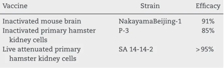
To prevent JE, it is necessary to implement a large-scale immunization of the susceptible human population. Vac-cination provides active immunity against JEV. There are several groups of vaccines which are currently in use: puri-fied, formalin-inactivated mouse-brain derived, cell-culture derived inactivated, and cell-culture derived live attenuated.15
Formalin-inactivated vaccines have been safe and effec-tive against JEV for at least 30 years.74 Of these, the
most widely produced and internationally distributed is the mouse-brain derived inactivated vaccine. The efficacy and the strain from which these are produced are given in
Table 2.
Table 2 – Vaccines against Japanese encephalitis.
Vaccine Strain Efficacy
Inactivated mouse brain NakayamaBeijing-1 91% Inactivated primary hamster
kidney cells
P-3 85%
Live attenuated primary hamster kidney cells
Purified, formalin-inactivated
mouse-brain-derived JE vaccine
Mouse-brain derived inactivated vaccines are based on the Nakayama and Beijing-1 strains (seroconversion rate 80% to 90%). This is the only vaccine against JE approved by the World Health Organization. The vaccine produced from the original Nakayama strain is manufactured in Japan, and was licensed in 1954. It is available internationally under the Biken label.11,23 This vaccine is also independently
pro-duced in China, India, Thailand, and Taiwan. The Central Research Institute in Kasauli is the manufacturer in India. It is available in lyophilized form, in which gelatin and sodium glutamate are used as stabilizers, and thimerosal is used as a preservative.75
The primary vaccination is done between the ages of 1 and 3 at doses of 0.5 mL to 1 mL (0.25 to 0.5 with children under age 3) subcutaneously. The dose regimen consists of one injection on days zero, seven, and 30 with a booster after one year and thereafter every three years until age 10. The protective effi-cacy is above 90%.15Due to its high production cost, lack of
long-term immunity, and adverse allergic reactions, this vac-cine is not practical to be administered in poor rural areas, where it is urgently needed. These difficulties have led to the development of improved vaccines.
Inactivated hamster kidney cell-culture-derived
JE vaccine
This vaccine is based on the Beijing-3 strain of JEV.15In China,
an inactivated vaccine produced in primary hamster kidney (PHK) cell culture was developed and has been in use since 1967. It has relatively fewer side effects and is easy to man-ufacture. In an extensive randomized field trial in China, its efficacy was found to range between 76% and 90%.34In the
last decade, a Vero cell-culture based inactivated vaccine using various local JEV isolates has also been developed and is undergoing clinical trials. A Vero-cell culture derived formalin-inactivated vaccine is being developed using an attenuated SA14-14-2 strain, and it has induced high titers of neutralizing antibodies in mice after two injections.76Recently, Vero-cell
culture derived formaldehyde inactivated JE vaccine using P20778 (Indian isolate) has been developed, and has gen-erated high titers of anti-JEV antibodies in mice; sera from immunized mice neutralized different JEV strains with varying efficacies.77
Cell-culture derived live attenuated JE vaccine
The ChimeriVax-JE has been available since 2001.78Live
atten-uated vaccine appears to offer great prospects for future vaccine development, since less virus is needed to trigger a satisfactory immune response, which makes the vaccine cheaper, and fewer doses are required, which makes it easy to administer.5
Vaccine based on the SA14-14-2 strain
This is an attenuated and genetically stable strain that in large-scale case–control studies in China has shown 95% pro-tection after two doses with an interval of one year.15In the
1980s, China developed this live attenuated vaccine named SA 14-14-2 by passaging the SA14 strain of JEV in PHK cells. Six amino acid changes in E protein and three in NS genes were associated with the attenuation.79 Recently, another
case–control study in Nepal showed that a single dose of this vaccine induced an efficacy of 98%.80A study on the long-term
efficacy (over five or ten years) is needed to know if the sin-gle dose is sufficient or if boosters are necessary for long-term immunization of the targeted population.
Adverse reactions
There are several side effects of JE vaccination. Local side effects include tenderness, redness, and swelling. Sometimes systematic adverse reactions are also noted after vaccination, such as headache, myalgia, abdominal pain, or skin rash.15
Occasionally local hypersensitivity reactions (erythema or edema at the injection site) can be observed in some chil-dren. Other reactions, such as generalized urticaria, facial angioedema, and respiratory distress have been reported in a few people from non-endemic zones after vaccination.15Some
recipients of the vaccine had, very rarely, major neurologi-cal side effects (1 to 2.3 per million recipients: encephalitis, seizures, and peripheral neuropathy).10
Other JE vaccines under development
Several vaccines are still in various stages of development. These include: recombinant protein based vaccines, recombi-nant virus based/chimeric vaccine, and DNA vaccines. Second generation recombinant vaccines are in development with the aim of improving immunogenecity and decreasing adverse reactions.50
JE vaccination in India
The JE vaccination campaign was launched during 2006 wherein 11 of the most sensitive districts in Assam, Kar-nataka and Uttar Pradesh were covered. Altogether, 86 JE endemic districts in the states of Assam, Andhra Pradesh, Bihar, Haryana, Goa, Karnataka, Kerala, Maharashtra, Tamil Nadu, Uttar Pradesh, and West Bengal have been covered. Re-orientation training course on AES/JE case management is a continuing process. Such orientating training courses were carried out in Andhra Pradesh, Assam, Haryana, Karnataka, Tamil Nadu, Uttar Pradesh, and West Bengal during 2008 and 2009.18
Conclusion
Asia, because epidemics are typically noticed only after out-breaks, and because the disease may go largely unobserved in endemic regions. Environmental and ecological factors are responsible for the spread of JEV. There is no specific treatment for JE; only prevention can control the disease. Control may be possible only after developing a strong surveillance system together with a high-quality immunization program. Imple-mentation of a vaccination program for young children, as well as modified agricultural practices, pig vaccination, rigor-ous monitoring, vector control, and improved living standards can reduce the number of JE cases.
Conflict of interest
All authors declare to have no conflict of interest.
Acknowledgements
The author Sarika Tiwari is thankful to the Indian Council of Medical Research, New Delhi for providing financial assis-tance.
r e f e r e n c e s
1. Ghosh D, Basu A. Japanese encephalitis-a pathological and clinical perspective. PLoS Negl Trop Dis. 2009;3:e437. 2. Tsai TF. Factors in the changing epidemiology of Japanese
encephalitis and West Nile fever. In: Saluzzo JF, editor. Factors in the Emergence of Arboviral Diseases. Amsterdam: Elsevier; 1997. p. 179–89.
3. Grossman RA, Edelman R, Chiewanich P, Voodhikul P, Siriwan C. Study of Japanese encephalitis virus in Chiangmai valley, Thailand. II. Human clinical infections. Am J Epidemiol. 1973;98:121–32.
4. Hoke Jr CH, Vaughn DW, Nisalak A, et al. Effect of high-dose dexamethasone on the outcome of acute encephalitis due to Japanese encephalitis virus. J Infect Dis. 1992;165:631–7. 5. Solomon T, Dung NM, Kneen R, et al. Japanese encephalitis. J
Neurol Neurosurg Psychiatry. 2000;68:405–15.
6. Karabatsos N. International catalogue of arboviruses. San Antonio, Texas: The American Society of Tropical Medicine and Hygiene; 1985.
7. Chambers TJ, Hahn CS, Galler R, Rice CM. Flavivirus genome organization, expression, and replication. Annu Rev Microbiol. 1990;44:649–88.
8. Miyake M. The pathology of japanese encephalitis. a review. Bull World Health Organ. 1964;30:153–60.
9. Westaway EG, Brinton MA, Gaidamovich S, et al. Flaviviridae. Intervirology. 1985;24:183–92.
10. Solomon T. Viral encephalitis in southeast Asia. Neuro Infect Epidemiol. 1997;2:191–9.
11. Vaughn DW, Hoke Jr CH. The epidemiology of Japanese encephalitis: prospects for prevention. Epidemiol Rev. 1992;14:197–221.
12. Igarashi A. Japanese encephalitis virus. In: Webster RG, Granoff A, editors. Encyclopedia of virology. San Diego: Academic Press; 1994. p. 746–51.
13. Zimmerman MD, Scott RM, Vaughn DW, et al. Short report: an outbreak of Japanese encephalitis in Kathmandu, Nepal. Am J Trop Med Hyg. 1997;57:283–4.
14. Hanna JN, Ritchie SA, Phillips DA, et al. Japanese encephalitis in north Queensland, Australia, 1998. Med J Aust.
1999;170:533–6.
15. Diagana M, Preux PM, Dumas M. Japanese encephalitis revisited. J Neurol Sci. 2007;262:165–70.
16. Namachivayam V, Umayal K, editors. Proceedings of the National Conference on Japanese Encephalitis. New Delhi: Indian Council of Medical Research; 1982. p. 30–3.
17. Carey DE, Myers RM, Pavri KM. Japanese encephalitis studies in Vellore, South India. II. Antibody response of patients. Indian J Med Res. 1968;56:1319–29.
18. NVBDC P. Directorate General of Health services Ministry of Health and Family Welfare. New Delhi. [cited April 4 2009]. Available from:http://nvbdcp.gov.in/je-cd.html
19. Dhillon GP, Raina VK. Epidemiology of Japanese encephalitis in context with Indian scenario. J Indian Med Assoc. 2008;106:660–3.
20. Kabilan L. Control of Japanese encephalitis in India: a reality. Indian J Pediatr. 2004;71:707–12.
21. NVBDC P. Directorate General of Health Services. Ministry of Health and Family Welfare. New Delhi. 2010. Available from:
http://nvbdcp.gov.in/je-cd.html. 22 Saxena SK, Mishra N, Saxena R, Singh M, Mathur A. Trend of Japanese encephalitis in North India: evidence from thirty-eight acute encephalitis cases and appraisal of niceties. J Infect Dev Ctries.
2009;3(7):517–30.
23. Burke DS, Monath TP. Flaviviruses. In: Knipe DM, Howkey PM, editors. Fields Virolgy. 4th edition. Philadelphia, PA:
Lippincott- Ravin Publishers; 2001. p. 1043–125.
24. Buescher EL, Scherer WF, Mc CH, et al. Ecologic studies of Japanese encephalitis virus in Japan. IV. Avian infection. Am J Trop Med Hyg. 1959;8:678–88.
25. Burke DS, Leake CJ. Japanese encephalitis. In: Monath TP, editor. The arboviruses: epidemiology and ecology. Florida: CRC, Boca Raton; 1988. p. 63–92.
26. Endy TP, Nisalak A. Japanese encephalitis virus: ecology and epidemiology. Curr Top Microbiol Immunol. 2002;267:11–48.
27. Kanojia PC, Shetty PS, Geevarghese G. A long-term study on vector abundance & seasonal prevalence in relation to the occurrence of Japanese encephalitis in Gorakhpur district, Uttar Pradesh. Indian J Med Res. 2003;117:104–10. 28. Kabilan L, Rajendran R, Arunachalam N, et al. Japanese
encephalitis in India: an overview. Indian J Pediatr. 2004;71:609–15.
29. Mackenzie JS, Gubler DJ, Petersen LR. Emerging flaviviruses: the spread and resurgence of Japanese encephalitis, West Nile and dengue viruses. Nat Med. 2004;10 12 Suppl: S98–109.
30. Schneider RJ, Firestone MH, Edelman R, Chieowanich P, Pornpibul R. Clinical sequelae after japanese encephalitis: a one year follow-up study in Thailand. Southeast Asian J Trop Med Public Health. 1974;5:560–8.
31. Lam K, Tsang OT, Yung RW, Lau KK. Japanese encephalitis in Hong Kong. Hong Kong Med J. 2005;11:182–8.
32. Kaur R, Vrati S. Development of a recombinant vaccine against Japanese encephalitis. J Neurovirol. 2003;9: 421–31.
33. Erlanger TE, Weiss S, Keiser J, Utzinger J, Wiedenmayer K. Past, present, and future of Japanese encephalitis. Emerg Infect Dis. 2009;15:1–7.
34. Tsai TF, Chang GJ, Yu XY. Japanese encephalitis vaccine. In: Plotkins SA, Orenstein WA, editors. Vaccines. Philadelphia: W.B. Saunders; 1999. p. 684–710.
35. Benenson MW, Top Jr FH, Gresso W, Ames CW, Altstatt LB. The virulence to man of Japanese encephalitis virus in Thailand. Am J Trop Med Hyg. 1975;24 6 Pt 1:974–80.
36. Solomon T. Control of Japanese encephalitis - within our grasp? N Engl J Med. 2006;355:869–71.
38. Byrne SN, Halliday GM, Johnston LJ, King NJ. Interleukin-1beta but not tumor necrosis factor is involved in West Nile virus-induced Langerhans cell migration from the skin in C57BL/6 mice. J Invest Dermatol. 2001;117:702–9.
39. Monath TP, Cropp CB, Harrison AK. Mode of entry of a neurotropic arbovirus into the central nervous system. Reinvestigation of an old controversy. Lab Invest. 1983;48:399–410.
40. Dropulic B, Masters CL. Entry of neurotropic arboviruses into the central nervous system: anin vitrostudy using mouse brain endothelium. J Infect Dis. 1990;161:685–91.
41. Liou ML, Hsu CY. Japanese encephalitis virus is transported across the cerebral blood vessels by endocytosis in mouse brain. Cell Tissue Res. 1998;293:389–94.
42. Desai A, Shankar SK, Ravi V, Chandramuki A, Gourie-Devi M. Japanese encephalitis virus antigen in the human brain and its topographic distribution. Acta Neuropathol.
1995;89:368–73.
43. Johnson RT, Burke DS, Elwell M, et al. Japanese encephalitis: immunocytochemical studies of viral antigen and
inflammatory cells in fatal cases. Ann Neurol. 1985;18: 567–73.
44. Liu YF, Teng CL, Liu K. Cerebral cysticercosis as a factor aggravating Japanese B encephalitis. Chin Med J. 1957;75:1010–7.
45. Solomon T, Vaughn D. Clinical features and pathophysiology of Japanese encephalitis and West Nile virus infections. In: McKenzie JSB, AD Deubel V, editors. Japanese encephalitis and West Nile viruses. New York: Springer-Verlag; 2002. p. 171–94.
46. Solomon T, Kneen R, Dung NM, et al. Poliomyelitis-like illness due to Japanese encephalitis virus. Lancet. 1998;351:1094–7. 47. Solomon T, Dung NM, Kneen R, et al. Seizures and raised
intracranial pressure in Vietnamese patients with Japanese encephalitis. Brain. 2002;125 Pt 5:1084–93.
48. Hirafuku I. Vitral encephalitis in Japan, pathology of Japanese encephalitis. Showa Igakkai Zasshi. 1963;23:23–5.
49. Yamada M, Nakamura K, Yoshii M, Kaku Y. Nonsuppurative encephalitis in piglets after experimental inoculation of Japanese encephalitis flavivirus isolated from pigs. Vet Pathol. 2004;41:62–7.
50. Tiroumourougane SV, Raghava P, Srinivasan S. Japanese viral encephalitis. Postgrad Med J. 2002;78:205–15.
51. Desai A, Murali- Krishna K, Ramireddy B, Ravi V, Manjumnath R.In vivoclearance of Japanese encephalitis virus by
adoptively transferred virus specific cytotoxic T lymphocytes. J Biosci. 1997;22:33–45.
52. Haymaker W, Sabin AB. Topographic distribution of lesions in central nervous system in Japanese B encephalitis; nature of the lesions, with report of a case on Okinawa. Arch Neurol Psychiatry. 1947;57:673–92.
53. Kalita J, Misra UK. Comparison of CT scan and MRI findings in the diagnosis of Japanese encephalitis. J Neurol Sci.
2000;174:3–8.
54. Pradhan S, Pandey N, Shashank S, Gupta RK, Mathur A. Parkinsonism due to predominant involvement of substantia nigra in Japanese encephalitis. Neurology. 1999;53:1781–6. 55. Solomon T. Recent advances in Japanese encephalitis. J
Neurovirol. 2003;9:274–83.
56. Sato Y, Hachiya N, Kuno H, Asoh T, Oizumi K. Cerebrospinal fluid atypical lymphocytes in Japanese encephalitis. J Neurol Sci. 1998;160:92–5.
57. Nguyen-Thi K-T, Lam-Thi C-T, Ngo Thi V, Nguyen Xuan Q. Isolation of a strain of Japanese encephalitis virus B from the blood of a young patient suffering from cardiovascular collapse. Bull Soc Pathol Exot Filiales. 1974;67:341–6. 58. Ravi V, Premkumar S, Chandramuki A, Kimura-Kuroda J. A
reverse passive haemagglutination test for detection of
Japanese encephalitis virus antigens in cerebrospinal fluid. J Virol Methods. 1989;23:291–8.
59. Zhang YH, Yu WF, Cai J, et al. A rapid method for detection of flavivirus antigens: staphylococcal co-agglutination test using monoclonal antibodies to Japanese encephalitis virus. Acta Virol. 1989;33:24–31.
60. Raghava PV, Badrinath S. Detection of Japanese encephalitis cell associated antigen in CSF by indirect
immunofluorescence. Ann Natl Acad Med Sci (India). 1998;34:207–11.
61. Deng YC, Su XC, Feng YQ. Immunocytochemical study of mononuclear cells in peripheral blood and cerebrospinal fluid of patients with Japanese B encephalitis. Zhonghua Bing Li Xue Za Zhi. 1994;23:20–2.
62. Burke DS, Nisalak A, Ussery MA. Antibody capture immunoassay detection of japanese encephalitis virus immunoglobulin m and g antibodies in cerebrospinal fluid. J Clin Microbiol. 1982;16:1034–42.
63. Shrivastva A, Tripathi NK, Parida M, et al. Comparison of a dipstick enzyme-linked immunosorbent assay with commercial assays for detection of Japanese encephalitis virus-specific IgM antibodies. J Postgrad Med. 2008;54:181–5. 64. Ravi V, Desai A, Balaji M, et al. Development and evaluation of
a rapid IgM capture ELISA (JEV-Chex) for the diagnosis of Japanese encephalitis. J Clin Virol. 2006;35:429–34.
65. Swami R, Ratho RK, Mishra B, Singh MP. Usefulness of RT-PCR for the diagnosis of Japanese encephalitis in clinical samples. Scand J Infect Dis. 2008;40:815–20.
66. Jeong YE, Jeon MJ, Cho JE, et al. Development and field evaluation of a nested RT-PCR kit for detecting Japanese encephalitis virus in mosquitoes. J Virol Methods. 2011;171:248–52.
67. Chen Z, Liao Y, Ke X, et al. Comparison of reverse transcription loop-mediated isothermal amplification, conventional PCR and real-time PCR assays for Japanese encephalitis virus. Mol Biol Rep. 2011;38:4063–70. 68. Solomon T, Dung NM, Wills B, et al. Interferon alfa-2a in
Japanese encephalitis: a randomised double-blind placebo-controlled trial. Lancet. 2003;361:821–6.
69. Saxena SK, Mathur A, Srivastava RC. Inhibition of Japanese encephalitis virus infection by diethyldithiocarbamate is independent of its antioxidant potential. Antivir Chem Chemother. 2003;14:91–8.
70. Bossi P, Tegnell A, Baka A, et al. Bichat guidelines for the clinical management of viral encephalitis and
bioterrorism-related viral encephalitis. Euro Surveill. 2004;9:E21–2.
71. Mishra MK, Basu A. Minocycline neuroprotects, reduces microglial activation, inhibits caspase 3 induction, and viral replication following Japanese encephalitis. J Neurochem. 2008;105:1582–95.
72. Sebastian L, Desai A, Shampur MN, et al.
N-methylisatin-beta-thiosemicarbazone derivative (SCH 16) is an inhibitor of Japanese encephalitis virus infectionin vitro
andin vivo. Virol J. 2008;5:64.
73. Wiwanitkit V. Cerebrospinal fluid examination and interpretation. Buddhachinaraj Med J. 2000;17:42–52. 74. Tsai TF. New initiatives for the control of Japanese
encephalitis by vaccination: minutes of a WHO/CVI meeting, Bangkok, Thailand, 13–15 October 1998. Vaccine. 2000;18:1–25. 75. Bharati K, Vrati S. Japanese encephalitis: development of new
candidate vaccines. Expert Rev Anti Infect Ther. 2006;4:313–24.
76. Srivastava AK, Putnak JR, Lee SH, et al. A purified inactivated Japanese encephalitis virus vaccine made in Vero cells. Vaccine. 2001;19:4557–65.
of Japanese encephalitis virus grown in Vero cells. Vaccine. 2004;22:3669–75.
78. Jones T, ChimeriVax-J.E. Acambis. Curr Opin Investig Drugs. 2003;4:1019–22.
79. Ni H, Burns NJ, Chang GJ, et al. Comparison of nucleotide and deduced amino acid sequence of the 5′non-coding region
and structural protein genes of the wild-type Japanese
encephalitis virus strain SA14 and its attenuated vaccine derivatives. J Gen Virol. 1994;75 Pt 6:
1505–10.