Effect of hydroxyurea on the
intracellular multiplication of
Toxoplasma gondii
,
Leishmania
amazonensis
and
Trypanosoma cruzi
Laboratório de Biologia Celular, Centro de Biociências e Biotecnologia, Universidade Estadual do Norte Fluminense, Campos dos Goytacazes, RJ, Brasil E.J.T. Melo and
H.J. Beiral
Abstract
Toxoplasma gondii, Leishmania amazonensis and Trypanosoma cruzi are obligate intracellular parasites that multiply until lysis of host cells. The present study was undertaken to evaluate the effect of hydroxyurea (an inhibitor of cell division at the G1/S phase) on the multiplication of L. amazonensis, T. gondii, and T. cruzi in infected host cells. Infected cells were treated with hydroxyurea (4 mM) for 48 h. Hydroxyurea arrested intracellular multiplication of all infective forms of the parasites tested. In treated cultures, the percent of infected host cells decreased (50-97%) and most intracellular parasites were eliminated. Ultrastructural observations showed no morphologic change in host cells while intracellular parasites presented drastic morpho-logic alterations or disruption. The results strongly suggest that hy-droxyurea was able to interfere with the multiplication of intracellular parasites, leading to an irreversible morphological effect on L. amazo-nensis, T. gondii, and T. cruzi without affecting the host cells.
Correspondence E.J.T. Melo
Laboratório de Biologia Celular CBB, Universidade Estadual do Norte Fluminense
Av. Alberto Lamego, 2000
28013-600 Campos dos Goytacazes, RJ Brasil
Fax: +55-022-2726-1514 E-mail: [email protected]
Received May 29, 2001 Accepted October 14, 2002
Key words
·Toxoplasma gondii ·Trypanosoma cruzi ·Leishmania amazonensis ·Hydroxyurea
The protozoan parasites Toxoplasma
gondii, Leishmania amazonensis and Trypa-nosoma cruzi are etiologic agents that cause toxoplasmosis, leishmaniasis and Chagas disease, respectively (1-3). These parasites invade the host while being localized within a vacuole known as the parasitophorous
vacu-ole (PV). L. amazonensis and T. gondii
de-velop within the PV whereas T. cruzi escapes
to the cytoplasm of host cells. It is important to point out that the latter multiplies inside the cell cytoplasm and is released into the intercellular space after rupture of the host cell (4). In experimental animals, control of infection with therapeutic drugs generally
causes drastic effects on the host cells (5-7). Hydroxyurea is a drug that specifically in-hibits the cell cycle in the G1/S phase inacti-vating the enzyme ribonucleotide reductase and the synthesis of DNA (8). Different con-centrations of hydroxyurea have been used for the treatment of several types of cancer (8,9) and various concentrations have also been tested against trypomastigotes and
epimastigotes of T. cruzi, promastigotes of
Crithidia luciliae (5-20 mM) (10),
tachy-zoites of T. gondii (4 mM) (11), CD4 cell
infected with HIV (100-200 mg/kg) and
has been used to synchronize the cell and parasite cultures without causing toxic ef-fects (10,12). We have recently shown that hydroxyurea, an inhibitor of DNA synthesis, inhibited the intracellular multiplication of T. gondii followed by tachyzoite elimination (11). We have extended this study by also using hydroxyurea against the intracellular
parasites L. amazonensis and T. cruzi.
Macrophages were collected from Swiss mice and cultivated for 24 h in medium 199 supplemented with fetal calf serum (FCS) at
37ºC under a continuous flow of 5% CO2
(13). L. amazonensis (MHUM/Br/175/Josefa
strain) was cultivated for 7 days at 25ºC. The culture was infected for 1 h, washed with sodium phosphate-buffered saline (PBS) to remove extracellular parasites, and incubated for 72 h in medium 199 at 37ºC under a
continuous flow of 5% CO2. After this
pro-cedure, most cells were infected. For
infec-tion with T. cruzi and T. gondii, Vero cells
(kidney fibroblasts from African green mon-keys) were obtained through periodic pas-sages and used when semi-confluent on Fal-con plates or in flask cultures (11). The
cultures were infected with T. cruzi
trypo-mastigotes (Y strain obtained from a culture of Vero cells infected for 6-7 days at 37ºC) or T. gondii tachyzoites for 1 h. The culture
was washed and incubated for 72 h (for T.
cruzi) or 48 h (for T. gondii)at 37ºC in the
presence of medium 199 supplemented with 5% FCS (11,13). All parasites were assumed to have interacted with the cells because the parasite:host cell relationship was 10:1.
Hydroxyurea (Merck Chemical Co., Darmstadt, Germany) was dissolved in PBS (4 M stock solution) and the solution was mixed with medium 199 in cultured cells at a final concentration of 4 mM. This drug con-centration has been established for synchro-nization of the cell cycle, and has no toxic effects on the cells (10-13). The drug was added to the infected cells during intense parasite proliferation. After incubation with hydroxyurea, the cultures were washed with PBS, fixed with Bouins fixative, stained with Giemsa and observed under a light microscope (63X objective Axioplan, Zeiss, Jena, Germany). The percentage of infected cells and the mean number of intracellular parasites were determined by examination of at least 400 cells.
Statistical analysis was carried out using
the Student t-test, with the level of
signifi-cance set at P<0.5. The data shown are
rep-resentative of five experiments in triplicate. The cultures in flasks were infected with parasites and incubated with the drug as described above for transmission electron microscopy. The cultures were fixed for 60 min at room temperature in a solution con-taining 1% glutaraldehyde, 4%
paraformal-dehyde, 5 mM CaCl2 and 5% sucrose in 0.1
M cacodylate buffer, pH 7.2. Cells were post-fixed for 1 h in a solution containing 1% osmium tetroxide, 0.8% potassium
fer-rocyanide and 5 mM CaCl2 in 0.1 M
cacody-late buffer, pH 7.2. The cells were rinsed in 0.1 M cacodylate buffer, dehydrated in ac-etone and embedded in Epon. Thin sections
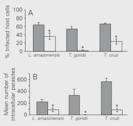
% Infected host cells
100 80 60 40 20 0
*
*
*
L. amazonensis T. gondii T. cruzi
A
when compared with untreated control cells (Figure 1A, filled columns). A similar sig-nificant decrease was observed in the mean number of intracellular parasites in the three cultures (Figure 1B, open columns) when compared with control untreated cells (Fig-ure 1B, filled columns). A drastic reduction in the number of infected cells was also
observed in host cells infected with T. gondii.
These results clearly show that the presence of hydroxyurea reduced both the percentage
of infected cells and the number of intracel-lular parasites.
Light and electron microscopy was used to observe the effects of the drug on parasite morphology and ultrastructure. Tachyzoites are elongated and curved, while amastigotes of T. cruzi and L. amazonensis have a spheri-cal shape. Light microscopy demonstrated
that treated Vero cells infected with T. gondii
showed normal morphology, although intra-cellular parasites, when present, were
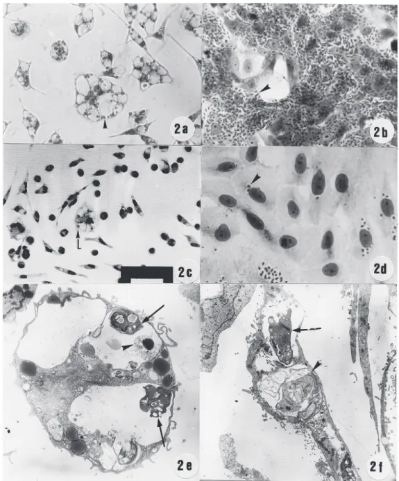
dis-Figure 2. Effect of hydroxyurea on the intracellular multiplication of T. gondii, L. amazonensis and
T. cruzi. a-d, Giemsa-stained host cells infected with para-sites. Macrophages infected with L. amazonensis (a) or Vero cells infected with T. cruzi (b) were treated (c,d) with 4 mM hydroxyurea for 48 h. A large number of parasites are ob-served in the cytoplasm of un-treated cultures (a,b, arrow-head). After incubation for 48 h in the presence of 4 mM hy-droxyurea, very few cells were infected with L. amazonensis (c, arrowhead) or T. cruzi (d, arrow-head). e-f, Ultrastructural obser-vation of macrophages or Vero cells infected with L. amazonen-sis (e) and T. cruzi (f), and treated with 4 mM hydroxyurea for 48 h. After incubation with 4 mM hydroxyurea, amastigotes of L. amazonensis with morpho-logical changes (arrow) or dis-rupted (arrowhead) were ob-served. Vero cells infected with
rupted (data not shown), in agreement with earlier observations (11). Culture of
macro-phages infected with L. amazonensis showed
intense parasite proliferation (Figure 2a, ar-rowhead). When infected macrophages were treated with hydroxyurea, a larger number of uninfected cells with normal morphology was observed (Figure 2c), while intravacuolar parasites, present in some infected cells, did not proliferate (Figure 2c, arrowhead). Ul-trastructural aspects of parasites showed the typical eukaryotic organelles and nucleus. When infected macrophages were observed by transmission electron microscopy, prolif-erating amastigotes were observed in the PV (data not shown). Treated infected cells dem-onstrated intravacuolar amastigotes with drastic ultrastructural alterations (Figure 2e, arrow) or disrupted (Figure 2e, arrowhead). The cytoplasm of the organelles in the host cells did not present any alterations (data not
shown). Intense T. cruzi multiplication was
observed inside the cytoplasm of Vero cells (Figure 2b). When infected cells were treated with hydroxyurea, most uninfected cells pre-sented normal morphology (Figure 2d), and the intracellular parasites were not prolifer-ating (Figure 2d, arrowhead). When observed by transmission electron microscopy, the in-tracellular parasites presented ultrastructural alterations (Figure 2f, arrow) or were dis-rupted (2f, arrowhead). These results sug-gest that host cells treated with hydroxyurea were preserved and intracellular parasites showed an arrested cell cycle or were dis-rupted.
T. gondii, L. amazonensis and T. cruzi
multiply within host cells, with parasite divi-sion occurring at different times. This fact can result in a variation of the mean number of intracellular parasites destroyed during incubation with the drug (Figure 1B). The effect of hydroxyurea is specific for the cell cycle and some studies have demonstrated that hydroxyurea used in synchronization experiments does not produce morphologi-cal or ultrastructural alterations in treated cells (10,12,14,15). Hydroxyurea arrests DNA replications without interfering with the S phase. In fact, DNA synthesis is a process that occurs independently of other aspects of cell growth. For this reason, hy-droxyurea has been used in cell synchroniza-tion studies and for cancer treatment (16). Recently, several hydroxyurea concentrations
(5-20 mM) were tested on T. cruzi
demon-strating that, although the drug inhibited DNA synthesis in the nucleus and kinetoplast, other metabolic processes in the cell were not affected (10).
The present results demonstrated that treatment with hydroxyurea for 48 h arrested parasite multiplication, causing drastic mor-phologic or ultrastructural alterations or com-plete elimination of the parasites. During incubation with the drug, the host cell de-fense may be activated or become more ef-fective against these parasites. This possibil-ity has already been investigated in our
labo-ratory. Studies of the in vivo action of
hy-droxyurea against infection with different parasites are also currently being conducted in our laboratory.
ence, 39: 1171-1175.
6. Brener Z, Tafuri WL & Almeida Maria T (1969). An electron micro-scope study of Trypanosoma cruzi intracellular forms in mice treated with an active nitrofuran compound. Revista do Instituto de Medi-cina Tropical de São Paulo, 11: 245-249.
7. Teixeira ARL, Silva R, Cunha Neto E, Santana JM & Rizzo LV (1990). Malignant non-Hodgkin’s lymphomas in Trypanosoma cruzi-infected rabbits treated with nitroagents. Journal of Comparative Pathology, 103: 37-48.
8. Yarbro JW (1992). Mechanism of action of hydroxyurea. Seminars in Oncology, 19: 1-10.
9. Melo EJT, Mayerhoffer RO & De Souza W (2000). Hydroxyurea inhibits intracellular Toxoplasma gondii multiplication. FEMS Micro-biology Letters, 185: 79-82.
10. Galanti N, Dvorak JA, Grenet J & McDaniel JP (1994). Hydroxyurea-induced synchrony of DNA replication in the Kinetoplastida. Experi-mental Cell Research, 214: 225-230.
11. Simonelli C, Nasti G, Vaccher E, Tirelli U, Zanussi S, Depoali P,
Comar M & Giacca M (1996). Hydroxyurea treatment in HIV-infected patients. Journal of Acquired Immune Deficiency Syndromes and Human Retrovirology, 13: 462-464.
12. Benchimol M (1999). Hydrogenosome autophagy: an ultrastructural and cytochemical study. Biology of the Cell, 91: 165-174.
13. Carvalho TMU & De Souza W (1989). Early events related with the behaviour of Trypanosoma cruzi within an endocytic vacuole in mouse peritoneal macrophages. Cell Structure and Function, 14: 383-392.
14. Hofer A, Ekaren JT & Thelander L (1998). Allosteric regulation of
Trypanosoma brucei ribonucleotide reductase studied in vitro and in vivo. Journal of Biological Chemistry, 273: 34098-34104.
15. Taylor WR, Agarwal ML, Agarwal A, Stacey DW & Stark GR (1999). P53 inhibits entry into mitosis when DNA synthesis is blocked.
Oncogene, 18: 283-295.