Changes in cell shape and desmin
intermediate filament distribution are
associated with down-regulation of
desmin expression in C2C12 myoblasts
grown in the absence of extracellular Ca
2+
Departamento de Histologia e Embriologia, Instituto de Ciências Biomédicas, Universidade Federal do Rio de Janeiro, Rio de Janeiro, RJ, Brasil
C.S. Mermelstein, L.M. Amaral, M.I.L. Rebello, J.S.N. Reis, R. Borojevic and M.L. Costa
Abstract
Desmin is the main intermediate filament (IF) protein of muscle cells. In skeletal muscle, desmin IFs form a scaffold that intercon-nects the entire contractile apparatus with the subsarcolemmal cytoskeleton and cytoplasmic organelles. The interaction between desmin and the sarcolemma is mediated by a number of membrane proteins, many of which are Ca2+-sensitive. In the present study,
we analyzed the effects of the Ca2+ chelator EGTA (1.75 mM) on the
expression and distribution of desmin in C2C12 myoblasts grown in culture. We used indirect immunofluorescence microscopy and reverse transcription polymerase chain reaction (RT-PCR) to ana-lyze desmin distribution and expression in C2C12 cells grown in the presence or absence of EGTA. Control C2C12 myoblasts showed a well-spread morphology after a few hours in culture and became bipolar when grown for 24 h in the presence of EGTA. Control C2C12 cells showed a dense network of desmin from the peri-nuclear region to the cell periphery, whereas EGTA-treated cells showed desmin aggregates in the cytoplasm. RT-PCR analysis revealed a down-regulation of desmin expression in EGTA-treated C2C12 cells compared to untreated cells. The present results sug-gest that extracellular Ca2+ availability plays a role in the regulation
of desmin expression and in the spatial distribution of desmin IFs in myoblasts, and is involved in the generation and maintenance of myoblast cell shape.
Correspondence
C.S. Mermelstein
Departamento de Histologia e Embriologia, ICB, UFRJ 21941-590 Rio de Janeiro, RJ Brasil
Fax: +55-21-2237-0844 E-mail: [email protected] Research supported by CNPq, FAPERJ, FUJB-UFRJ, and PRONEX-Brasil.
Presented at the XI Congresso Brasileiro de Biologia Celular, Campinas, SP, Brazil, July 15-18, 2004.
Received July 7, 2004 Accepted March 2, 2005
Key words
•Extracellular Ca2+ •EGTA
•Myoblasts
•Desmin
•Intermediate filaments
•RT-PCR
Introduction
Desmin (53 kDa) is the main intermediate filament protein of striated muscle cells. This abundant protein polymerizes to form an extensive network of 10-nm intermediate
direct or indirectly link this protein to the nuclear membrane, Z-disk components and the sarcolemma. It has been shown that desmin is present in subsarcolemmal costa-meres in association with membrane and membrane-associated proteins such as the dystrophin protein complex (2), spectrin, ankyrin, integrins, vinculin, and α-actinin (3). Mutational analysis indicates that it is the head domain of desmin that is specialized for its association with the sarcolemma (4). The association of desmin with the sarcolemma is supposed to be dependent on the versatile cytoskeletal linker protein plectin (5), which is a very large size polypeptide (>500 kDa) abundant in the subsarcolemmal region. Many transmembrane components of these com-plexes are Ca2+-sensitive in their intra- and
extracellular domains. Variations in extracel-lular Ca2+ concentration are one of the
regu-latory mechanisms involved in the activation of these membrane complexes through con-formational changes (6,7). One of the evolu-tionary advantages of Ca2+ ions as regulators
of protein structure and function is the pos-sibility of rapid changes in their concentra-tion (8).
To investigate the function of desmin in all muscle types in vivo, desmin null mice were generated by homologous recombina-tion (9). Although a considerable number of these mice are viable and fertile, they present a multisystem disorder involving cardiac, skeletal and smooth muscle that begins early during postnatal life. Histological and elec-tron microscopic analysis of both heart and skeletal muscle tissues reveals severe disrup-tion of muscle architecture and degenera-tion, including perturbation of myofibril an-chorage to the sarcolemma. The study of desmin is especially relevant because a num-ber of desmin-related pathologies have been described (10). Desmin-myopathies consist of alterations in the distribution of desmin, particularly the presence of desmin aggre-gates and inclusions in the cytoplasm of myogenic cells (11).
Previous work from our group suggested a role for extracellular Ca2+ in the regulation
of the spatial distribution of microfilaments, microtubules, intermediate filaments, and adhesion sites in myoblasts (12). The present investigation was undertaken to study the effects of extracellular Ca2+ withdrawal on
cell shape and on the expression and distribu-tion of desmin in myoblasts grown in culture.
Material and Methods
Antibodies and fluorescent probes
Rabbit polyclonal anti-desmin antibody was purchased from Sigma (St. Louis, MO, USA) and fluorescein isothiocyanate-goat anti-rabbit IgG antibody was purchased from Jackson Immunoresearch Laboratories (West Grove, PA, USA). The nuclear dye DAPI (4’6-diamino-2-phenylindole dihydrochlo-ride) was purchased from Molecular Probes Inc. (Eugene, OR, USA).
Cell cultures
The mouse skeletal muscle cell line C2C12 was obtained from the Rio de Janeiro Cell Bank (PABCAM, Federal University of Rio de Janeiro, Rio de Janeiro, RJ, Brazil). Cells were routinely grown in Dulbecco’s modi-fied Eagle’s medium containing 20% fetal calf serum, 1% L-glutamine and 1% penicil-lin-streptomycin (all from Sigma), in a hu-midified 5% CO2 atmosphere at 37ºC. Cells
were grown at a low density of 5 x 10⁵ cells/
fluo-rescence microscopy or to semi-quantitative polymerase chain reaction (RT-PCR).
Immunofluorescence and phase contrast microscopy
For immunofluorescence microscopy, cultures were rinsed with phosphate-buff-ered saline (PBS) and fixed with 2% formal-dehyde in PBS for 3 min at room temperature (14). They were then permeabilized with 0.5% Triton-X 100 (Sigma) in PBS three times for 10 min. The same solution was used for all subsequent washing steps. Cells were incubated with anti-desmin polyclonal antibody at 1:100 dilution for 1 h at 37ºC in a humid chamber. After incubation, cells were washed for 30 min and incubated with sec-ondary antibody for 1 h at 37ºC in a humid chamber, and then washed for 30 min. After a 30-min wash with PBS, the nuclear dye DAPI was added at 0.1 µg/ml in 0.9% NaCl for 5 min. Cells were washed for 5 min with 0.9% NaCl and specimens were mounted in glycerol containing, by weight, 5% n-propyl gallate, 0.25% 1,4-diazabicyclo[2.2.2]octane and 0.0025% para-phenylenediamine (all from Sigma). Cells were examined with an Axiovert 100 epifluorescence inverted light micro-scope (Carl Zeiss, Oberkochen, Germany) using filter sets that were selective for fluo-rescein or blue wavelength channel. Images were acquired with a C2400i integrated CCD camera (Hamamatsu Photonics, Shizuoka, Japan) using an Argus 20 image processor (Hamamatsu Photonics). Digitalized images were transferred to a Dell OptiPlex GL 575 computer (Dell Corporate, Round Rock, TX, USA) and plates were mounted using Adobe Photoshop software(Adobe Systems Incor-porated, San Jose, CA, USA).
Control experiments with no primary antibody showed only a faint background staining (data not shown).
Live C2C12 cells grown on collagen-coated aclar coverslips were examined and images were acquired by phase contrast
microscopy using the same microscope and digital system described above.
Digital image processing and analysis
Phase contrast images were analyzed using the public domain NIH Image program (developed at the US National Institutes of Health and available on the Internet at http:// rsb.info.nih.gov/nih-image). Pixels with a gray-level over 125 were set to black, and cells were initially isolated with 4 series of erosions and dilations. Cells were then manu-ally separated from their neighbors for proper identification as objects. The parameters area, perimeter, major axis, minor axis, angle, and axis ratio (ratio between the major and the minor axis of cells) were measured. Only cells over 200 pixels in area were considered in order to avoid computing parts of cells. Images were calibrated at 1.02 pixels/µm using a slide with a grating. After automati-cally measuring all the cells/objects, data were exported to Excel (Microsoft Corpora-tion, Redmond, WA, USA) and to Statistic for Windows (StatSoft Inc., Tulsa, OK, USA) for statistical analysis.
RNA extraction, reverse transcription, amplification and RT-PCR
Total cellular RNA from control and 24-h EGTA-treated C2C12 cells was isolated using TRIzol® reagent. The RNA and the
cDNA were quantified by spectrophotom-etry (Spectronic, Genesys 2PC, Rochester, NY, USA). cDNA was synthesized from 3 µg total RNA. RNA was primed with 0.5 µg oligo-(dT)12-18 primer (reaction volume: 20
Amplification reactions consisted of a 0.2 µM primer for desmin (5' primer sequence: TCTCCCGTGTTCCCT/3' primer sequence: ATACGAGCTAGAGTGGCA; size of PCR product: 571 base pairs), 10 mM of each dNTP, PCR buffer, 5 µl of the cDNA reac-tion, and 0.5 U Taq DNA polymerase. The primer sequence for glyceraldehyde-3-phos-phate dehydrogenase (GAPDH) was ATCA CCATCTTCCAGGAGCG (sense) and CCTG CTTCACCACCTTCTTG (antisense). cDNAs from control and EGTA-treated cells were subjected to PCR amplifications with serial dilutions at 0.1, 0.01, and 0.001 µg. GAPDH expression was used to standardize cDNA under non-saturating conditions at 28 cycles. Amplification was carried out during 33 PCR cycles for desmin, each cycle consisting of a denaturation step at 94ºC for 1 min, another denaturing step at 94ºC for 30 s, an annealing step at 60ºC for 30 s, and an extension step
at 72ºC for 30 s. After the last cycle, incuba-tion for another 5 min at 72ºC was per-formed. The PCR products (10 µl) were analyzed by electrophoresis in 1% agarose gels with Tris-borate-EDTA buffer, stained with 1 µg/ml ethidium bromide for 10 min and analyzed with the thermal imaging sys-tem FTI-500 (Pharmacia, Uppsala, Swe-den). Gels were scanned with a Silver Scan-ner II (LaCie Limited, Hillsboro, OR, USA), at 400 dpi, and densitometric profiles were obtained using the public domain NIH Image program (developed at the US National Insti-tutes of Health and available on the Internet at http://rsb.info.nih.gov/nih-image). Areas cor-responding to the GAPDH and desmin gel bands from each profile were selected and the number of pixels in each peak was measured. For normalization, desmin ex-pression (number of pixels in the gel band) was divided by GAPDH expression (for 0.1 and 0.01 µg cDNA dilution) for both control and EGTA-treated samples, and then plotted on a column chart. Data related to 0.001 µg cDNA were not submitted to normalization and analysis since they were considered to be below the minimum level of expression and visualization in the gels.
Results
To better understand the role of extracel-lular Ca2+ in the expression and distribution
of the muscle-specific protein desmin, skel-etal muscle C2C12 cells were grown in the presence or in the absence of the Ca2+
-chelator EGTA (1.75 mM) and then ob-served by phase contrast microscopy. C2C12 cells grown in normal medium without EGTA (control cells) were well spread (Figure 1A), whereas the majority of C2C12 cells grown in medium with EGTA for 24 h drastically changed to a bipolar morphology (Figure 1B).
Since there were major observable changes in the overall shape of cells treated with EGTA for 24 h, we decided to quantify
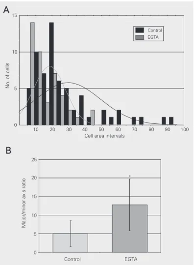
these changes using digital processing of phase contrast microscopy images. Figures 1C and 1D show the digitally processed images from control C2C12 cells and EGTA-treated C2C12 cells. The parameters area, perimeter, major axis, minor axis, angle, and axis ratio (ratio between the major and the minor axis of cells) were computed for the isolated objects from both processed im-ages. The major significant (P < 0.001) differences between control and EGTA-treated cells concerned the area, minor axis and axis ratio. Area measurements were 29.79 ± 19.78 µm2 for control cells and
18.30 ± 9.70 µm2 for EGTA-treated cells.
Minor axis measurements were 3.04 ± 1.41 µm for control cells and 1.39 ± 0.36 µm for EGTA-treated cells. Axis ratio measurements were 5.03 ± 3.47for control cells and 12.78 ± 6.98 for EGTA-treated cells. The other parameters (perimeter, major axis and angle) were not significantly different between con-trol and EGTA-treated cells (data not shown). It is possible to see that the areas of control C2C12 cells were larger than those of EGTA-treated cells (Figure 2A). Not only were the EGTA-treated cells smaller, but they also varied less in shape, as shown in the histogram in Figure 2A and by comparing the standard deviation (SD) of the cell areas described above.
The elongation of the cells can be quan-tified by the axis ratio (the ratio between the major and the minor axis of cells). The axis ratio of EGTA-treated C2C12 cells was more than two-fold greater than that of control cells (Figure 2B), showing that treated cells were more elongated than control cells.
To investigate whether the observed cell shape changes in EGTA-treated C2C12 were related to the distribution of the cytoskeleton, we analyzed the organization of the interme-diate filament desmin in these cells, since it has been postulated that desmin has a major structural role in maintaining the shape of myogenic cells (1,15). Indirect immunofluo-rescence microscopy using a polyclonal
an-Figure 2. Histogram and best-fit plot of control and EGTA-treated cell area (A), and major/minor axis ratio of control and EGTA-treated cells (B). Axis ratio data from control and EGTA-treated groups were compared statistically (*P < 0.0001, t-test for independent samples). Both histograms were obtained from data described in the Results section.
tibody against desmin revealed a characteris-tic desmin filament organization spanning the distance from a highly concentrated area around the nuclear envelope to the plasma membrane (Figure 3A), while EGTA-treated cells showed a less intense desmin staining, which was visible mainly as dots and short filaments (Figure 3C). The nuclear staining with DAPI showed a normal nuclear pattern in both control cells (Figure 3B) and in cells treated with EGTA for 24 h (Figure 3D), confirming that EGTA-treated cells were viable.
Figure 3. Fluorescence microscopy of C2C12 cells grown in the absence of EGTA (control cells, A and B) or in the presence of 1.75 mM EGTA for 24 h (C and D). Cells were stained with anti-desmin antibody (A and C) and with the nuclear probe DAPI (B and D). Desmin is present as a dense network of filaments in control cells (A), as opposed to fragmented filaments and dots observed in EGTA-treated cells (C). Bar in B corresponds to 10 µm and bar in D corresponds to 5 µm.
Figure 4. RT-PCR analysis of the expression of desmin in control (A) and in 24-h EGTA-treated C2C12 cells (B).
Lanes 1-3 of both agarose gels (A and B) correspond to GAPDH expression (0.1 µg, 0.01 µg, and 0.001 µg cDNA, respectively). Lanes 4-6 of both agarose gels correspond to desmin expression (0.1 µg, 0.01 µg and 0.001 µg cDNA, respectively). C, Comparative expression of desmin in control and EGTA-treated cells after normalization of PCR data (for details, see Material and Methods). GAPDH = glyceraldehyde-3-phosphate dehydrogenase.
of EGTA-treated cells desmin appeared to have a less intense staining, we decided to further analyze desmin expression by RT-PCR. In agreement with the immunofluores-cence analysis, we confirmed that desmin expression was down-regulated in C2C12 cells treated with EGTA for 24 h (Figure 4B) when compared to untreated C2C12 cells (control) that showed a comparatively higher expression of desmin (Figure 4A). These results can be better visualized in the normal-ized data obtained by densitometric analysis of the agarose gels shown in Figure 4C.
Discussion
The present study analyzed the involve-ment of extracellular Ca2+ in the generation
and maintenance of cell shape and its relation with the expression and distribution of the intermediate filament desmin in the mouse skeletal muscle cell line C2C12.
between the major and the minor axis of cells. The cell area of control cells was larger than that of EGTA-treated cells, and the axis ratio of EGTA-treated cells was more than two-fold larger than that of control myo-blasts. The lack of differences in perimeter between control and treated cells was due to the elongation of EGTA-treated cells, which is clearly demonstrated by the axis ratio showing that treated cells were thinner and longer than control cells.
Ca2+ depletion from the medium by EGTA
influences a number of Ca2+-sensitive
mem-brane proteins. One major Ca2+-dependent
transmembrane protein found in the sarco-lemma of myoblasts is integrin. Previous work from our group showed a change in the distribution of α5-integrin adhesion sites in C2C12 cells after EGTA treatment (12). Using indirect immunofluorescence, α 5-in-tegrin was detected in fibrillar adhesions at the terminations of stress fibers in both control and 24-h EGTA-treated C2C12 cells. Besides, a dense α5-integrin labeling was seen inside most treated cells, indicating a possible internalization of integrin after EGTA exposure (12). The effects of calcium deple-tion on desmin distribudeple-tion observed in the present study may have been initiated by various cell surface components that require extracellular Ca2+, such as α5-integrin, which
in turn can influence intracellular signaling pathways. It has been shown that desmin is present in the subsarcolemmal region in as-sociation with membrane and membrane-associated proteins, such as integrins (3).
In the present work, we found desmin aggregates in the cytoplasm of EGTA-treated C2C12 cells. Similar desmin dots and aggre-gates have been described as the major struc-tural alteration in several desminopathies (10).
Desmin-related myopathies are character-ized by the accumulation of desmin in certain inclusions, sarcoplasmic bodies, cytoplas-mic bodies, and granulofilamentous material (11). The mechanisms that control the for-mation and aggregation of desmin filaments in desminopathies are still unclear. A chaper-one protein, αB-crystalline, is involved in the correct assembly of desmin filaments and an impaired chaperone function could promote the increase in desmin aggregation (11).
The relation between changes in extracel-lular Ca2+ concentration and the expression
and distribution of cytoskeletal proteins (such as desmin) could be directly mediated by changes in Ca2+-binding membrane molecules
(such as integrins). Another possibility is that changes in extracellular Ca2+ concentration
could indirectly change the intracellular Ca2+
concentration, which in turn could have an effect on the cytoskeletal components. One consequence of changes in intracellular Ca2+
concentration is the activation of Ca2+
-acti-vated enzymes, such as calpains, which are major proteolytic systems in skeletal muscle (16). Calpains are proteases that can selec-tively cleave cytoskeletal proteins, and desmin has been shown to be one of their major and first substrates both in vivo and in vitro; and calpains are found in association with the sarcolemma (17). We speculate that in the present study Ca2+ depletion might have
induced a calpain-dependent cytoskeletal re-modeling including desmin cleavage, result-ing in the presence of desmin dots and aggregates in the cytoplasm.
The results presented in this paper sug-gest the requirement of extracellular Ca2+ for
References
1. Herrmann H & Aebi U (2000). Intermediate filaments and their associates: multi-talented structural elements specifying cytoarchi-tecture and cytodynamics. Current Opinion in Cell Biology, 12: 79-90.
2. Hijikata T, Murakami T, Ishikawa H & Yorifuji H (2003). Plectin tethers desmin intermediate filaments onto subsarcolemmal dense plaques containing dystrophin and vinculin. Histochemistry and Cell Biology, 119: 109-123.
3. Ervasti JM (2003). Costameres: the Achilles’ heel of Herculean muscle. Journal of Biological Chemistry, 278: 13591-13594. 4. Cary RB & Klymkowsky MW (1994). Differential organization of
desmin and vimentin in muscle is due to differences in their head domains. Journal of Cell Biology, 126: 445-456.
5. Wiche GJ (1998). Role of plectin in cytoskeleton organization and dynamics. Journal of Cell Science, 111: 2477-2486.
6. Mould AP, Garratt AN, Puzon-McLaughlin W, Takada Y & Humphries M (1998). Regulation of integrin function: evidence that bivalent-cation-induced conformational changes lead to the unmasking of ligand-binding sites within integrin α5ß1. Biochemical Journal, 331: 821-828.
7. Stuiver I, Ruggeri Z & Smith JW (1996). Divalent cations regulate the organization of integrins alpha V beta 3 and alpha V beta 5 on the cell surface. Journal of Cell Physiology, 168: 521-531. 8. Maurer P, Hohenester E & Engel J (1996). Extracellular
calcium-binding proteins. Current Opinion in Cell Biology, 8: 609-617. 9. Capetanaki Y, Milner DJ & Weitzer G (1997). Desmin in muscle
formation and maintenance: knockouts and consequences. Cell
Structure and Function, 22: 103-116.
10. Goebel HH & Warlo I (2000). Gene-related protein surplus myopa-thies. Molecular Genetics and Metabolism, 71: 267-275. 11. Carlsson L & Thornell LE (2001). Desmin-related myopathies in
mice and man. Acta Physiologica Scandinavica, 171: 341-348. 12. Mermelstein CS, Rebello MIL, Amaral LM & Costa ML (2003).
Changes in cell shape, cytoskeletal proteins and adhesion sites of cultured cells after extracellular Ca2+ chelation. Brazilian Journal of
Medical and Biological Research, 36: 1111-1116.
13. Cho WJ, Kim EJ, Lee SJ, Kim HD, Shin HJ & Lim WK (2000). Involvement of SPARC in in vitro differentiation of skeletal myo-blasts. Biochemical and Biophysical Research Communications, 271: 630-634.
14. Mermelstein CS, Guma FCR, Mello TG, Fortuna VA, Guaragna RM, Costa M & Borojevic R (2001). Induction of the lipocyte phenotype in murine hepatic stellate cells: reorganisation of the actin cytoskel-eton. Cell and Tissue Research, 306: 75-83.
15. Mermelstein CS, Costa ML, Chagas C & Moura Neto V (1996). Intermediate filaments in TPA-treated skeletal muscle cells in culture. Journal of Muscle Research and Cell Motility, 17: 199-206. 16. Goll DE, Thompson VF, Li H, Wei W & Cong J (2003). The calpain
system. Physiological Reviews, 83: 731-801.