Prospective Study of the Surgical Techniques
Used in Primary Rhinoplasty on the Caucasian
Nose and Comparison of the Preoperative and
Postoperative Anthropometric Nose
Measurements
Cezar Augusto Sarraf Berger
1,2,3Renato da Silva Freitas
4Osvaldo Malafaia
5,6José Simão de Paula Pinto
7Evaldo Dacheux Macedo Filho
3,8Marcos Mocellin
3Marina Serrato Coelho Fagundes
91Fellowship Program on Facial Plastic Surgery, IPO Hospital, Curitiba,
Paraná, Brazil
2MSc in Surgical Practice, Universidade Federal do Paraná (UFPR),
Curitiba, Paraná, Brazil
3Department of Otolaryngology, UFPR Clinical Hospital (HC/UFPR),
Curitiba, Paraná, Brazil
4Chief of Plastic Surgery Procedure, UFPR, Curitiba, Paraná, Brazil 5Titular Professor of Surgery, UFPR, Curitiba, Paraná, Brazil 6Institute of Medical Research and of the Postgraduate Program; MSc
and PhD on Surgery Principles, Evangélico University Hospital, Curitiba, Paraná, Brazil
7Department of Science and Information Management, UFPR,
Curitiba, Paraná, Brazil
8Department of NEP, IPO Hospital, Curitiba, Paraná, Brazil
9Department of Otolaryngology, IPO Hospital, Curitiba, Paraná, Brazil
Int Arch Otorhinolaryngol 2015;19:34–41.
Address for correspondence Cezar Augusto Sarraf Berger, MD, MSc, Department of Otolaryngology, Hospital Paranaense de
Otorrinolaringologia IPO, Av. República Argentina, 2069 Bairro Agua Verde Curitiba, Paraná 80620010, Brazil (e-mail: [email protected]).
Keywords
►
rhinoplasty
►
prospective studies
►
measurements
Abstract
Introduction
The knowledge and study of surgical techniques and anthropometric
measurements of the nose make possible a qualitative and quantitative analysis of
surgical results.
Objective
Study the main technique used in rhinoplasty on Caucasian noses and
compare preoperative and postoperative anthropometric measurements of the nose.
Methods
A prospective study with 170 patients was performed at a private hospital.
Data were collected using the Electronic System Integrated of Protocols software
(Sistema Integrado de Protocolos Eletrônicos, SINPE©). The surgical techniques used in
the nasal dorsum and tip were evaluated. Preoperative and 12-month follow-up photos
as well as the measurements compared with the ideal aesthetic standard of a Caucasian
nose were analyzed objectively. Student
t
test and standard deviation test were applied.
Results
There was a predominance of endonasal access (94.4%). The most common
dorsum technique was hump removal (33.33%), and the predominance of sutures
received May 26, 2014
accepted after revision August 13, 2014 published online December 2, 2014
DOI http://dx.doi.org/ 10.1055/s-0034-1393721. ISSN 1809-9777.
Copyright © 2015 by Thieme Publicações Ltda, Rio de Janeiro, Brazil
Introduction
Jack Gunter in his classic book Dallas Rhinoplasty: Nasal Surgery by the Mastersmentions that the access incisions in rhinoplasty should not be seen only as external or internal accesses.1Every nasal access necessarily acts in specific areas of the nose with higher or lower trauma, exposition, possi-bility of complex surgical maneuvers, and use of grafts and/or sutures.
Several surgical maneuvers currently known in open rhinoplasty can be performed in closed rhinoplasty, which results in less morbidity, reduced surgery time, and predict-able results. The knowledge and analysis of surgical techni-ques as well as the anthropometric measurements of the nose make possible the quantitative analysis of the results, which is of great importance for the surgeon and for the services regarding facial plastic surgery. The record of surgical tech-niques performed is important for the study of successes and the side effects of rhinoplasty.
High-quality clinical trials are essential for continuous scientific development. They allow safe access to new infor-mation resulting in improvement of knowledge, target plan-ning, spread of evaluation of procedures, and professional conduct.
The Electronic System Integrated of Protocols (SINPE) is a computer program created for manage database, ideal-ized in the beginning of the 1990s by Dr. Osvaldo Malafaia, whose purpose is the development of electronic protocols for fact gathering for its subsequent utilization in clinical study.
In addition to enabling the construction of protocols, SINPE also provides a data analysis module, which performs the descriptive statistical evaluation. This information visu-alization interface, called SINPE Analyzer, is able to generate graphs and statistics, save results, and export data.2
The present study aims to evaluate the main techniques used in rhinoplasty on Caucasian noses, comparing preoper-ative and postoperpreoper-ative anthropometric measurements of the nose.
Methods
This research was conducted in a private hospital. The work was approved by the ethics committee of the institution (2528.135/20116), and an explained and free consent form was provided to the patients.
Data collection was performed prospectively from Febru-ary 2010 to March 2011 by the researchers, using the SINPE software. A specific protocol called Rhinoplasty was created
based on 954 items, being part of the SINPE Analyzer module, which quickly gathers the information in the protocols given by the data collection.3,4The generation and storage of graphs make possible a quick analysis of extensive protocols, and it is also possible to copy each graph to the analysis sheet and include comments and references, creating a sequence of self-analysis. This protocol was developed by Dr. Cezar Berger in 2011 for his master’s thesis.5
The SINPE Analyzer and statistical analysis (Studentttest) were used for the validation of the protocol through the analysis of the existing data in the database; p<0.05 was
considered significant.
The research started with 170 patients who underwent surgery; 58 patients, however, were not present throughout the times evaluated, which makes 112 the total number of patients who participated in all the phases of the study.
The information was collected in four phases: (D1) imme-diately postoperative and at (D2) 3-month, (D3) 6-month, and (D4) 12-month follow-up. Patients’clinical conditions were followed according to the specific need of each case. For the purpose of this research, however, only the cases with 3, 6, and 12 months’ postoperative follow-up were taken into account.
The data evaluated corresponded to the primary rhino-plasties of the researcher. The surgical techniques performed on the nasal tip, on the nasal dorsum, and on the nasal base as well as the D1, D2, D3, and D4 records were evaluated. Preoperative and follow-up photos at 12 months (D4) were analyzed objectively, and the measurements were compared with the ideal aesthetic standard of a Caucasian nose. All the photos were taken by the same researcher with the same camera and standardization: model Sony Cyber shot DSCW125 7.2 Megapixels, 6.0 (Sony, Japan)fixed zoom at a distance of 1.5 m between the camera and the volunteer to provide uniformity for the purpose of scale and measure-ments. The positions were anteroposterior and direct profile. Inclusion criteria were primary rhinoplasties, Caucasian noses, and age from 15 to 55 years. Exclusion criteria were previous surgical interventions on the nose and on the face, non-Caucasian noses (Mestizo, Asian, Negroid), and patients who did not return for follow-up.
The quantitative evaluation of the anthropometric measure-ments of the nose was performed by comparing preoperative and 12-month follow-up (D4) photos. The following measure-ments were analyzed and compared with the ideal aesthetic standard6,7: front view (anteroposterior;►Fig. 1): (1) intercan-thal distance, (2) interalar distance; side view (direct profile;►Fig. 2): (3) nasolabial angle, (4) nasal tip projection (through Goode’s method;►Fig. 3), and (5) nasofrontal angle.
According to the nature of the analyzed data, Studentttest and the standard deviation test were applied. The level of significance adopted wasp<0.05.
Results
Main Techniques Used in Rhinoplasty
Access (Approach)
The predominant access to the nose was endonasal (94.4%). The closed access without inferior lateral cartilage exposure was predominant in 92.63% of cases against 7.37% with delivery.
Surgical Techniques on the Nasal Dorsum
Removal of osteocartilaginous hump was the most frequent surgical technique performed on the nasal dorsum (33.33%). Removal of the nasal hump did not happen in all the cases. Release of the superior lateral cartilage was not systematically performed. It was necessary to use grafts in some noses, which featured a change in the type of nose to be operated—in other words, not all the noses needed a reduction operation (►Fig. 4). Of the grafts, 41.94% of cases had camouflage type added (onlay graft), followed by the spreader graft, which was
Fig. 2 Side view (direct profile). Abbreviations: ANF, nasofrontal angle; ANL, nasolabial angle.
Fig. 3 Side view (direct profile). Note: Measurement of the nasal tip projection through Goode’s method, which entails measurement of the perpendicular line from the tip point (1) to the line of the facial plane (2) divided by the measurement of the line from the nasion to the tip point.
used in 35.48% of cases (►Fig. 5). In 60.67% of cases, a lateral osteotomy was performed, followed by paramedian osteot-omy in 15.33% of cases (►Fig. 6). Cartilage was the most frequent graft donor site, with 90.32% of cases (►Fig. 7); among them, the septal graft showed the highest frequency at 82.14% (►Fig. 8).
Surgical Techniques on the Nasal Tip
The use of sutures on the tip (24.76%) for definition and refinement was the most frequent, followed by La Garde maneuver (19.44%) for detachment and adjustment of the
skin; then the resection of the membranous septum (15.67%) for cephalic rotation, the placement of grafts (10.66%) for structure and refinement, and then the inter-posing maneuver between the septum and the medial crura (tongue-in-groove; 9.72%) for support of the tip. Cephalic resection of the inferior lateral cartilage occurred in only 5.02% of cases.
Resection of the membranous septum (15.67%) was twice as frequent as caudal septum shortening (6.27%), which confirmed the lesser need of cartilaginous removal from the tip of the noses (►Fig. 9).
Carlage
17.86%
82.14%
Ear
Septum 17.86%
82.14%
Fig. 8 Surgical maneuvers on the nasal dorsum—graft donor site cartilage.
3.23% 6.45% 3.23% 35.48%
9.68% 41.94%
0.00% 10.00% 20.00% 30.00% 40.00% 50.00%
Other Buerfly wing
Turkish graSpreader gra
Pea-pod gra
Onlay gra
Shape
Series1
Fig. 5 Surgical maneuvers on the nasal dorsum—grafts.
0%
5.33% 0% 0%
15.33% 4%
11.33% 3.33%
Osteotomy
Transverse - 0 Complete mobilizaon - 8Percutaneous - 0
Intermediate (oblique) - 0
61.67%
Osteotomy
-
- 0
Paramedian (medial) - 23
Lateral - 91
Double - 6
Release of the periosteum - 17
Incomplete mobilizaon (green branch) - 5
Fig. 6 Surgical maneuvers on the nasal dorsum—osteotomies.
3.23%
6.45%
Donor Site
Alloplasc material (describe) Other (describle)
Allodern
90.32%
Temporal fascia
Rib
Carllage
Fig. 7 Surgical maneuvers on the nasal dorsum—graft donor site. 33.33%
27.40% 27.85%
15.00% 20.00% 25.00% 30.00% 35.00%
Dorsum
33.33%
27.40% 27.85%
0.00% 5.00% 10.00% 15.00% 20.00% 25.00% 30.00% 35.00%
Removal of the Release of the superior Detachment carlaginous humplateral
carllage
Dorsum
Fig. 4 Surgical maneuvers on the nasal dorsum.
58.51%
41.49%
10.00% 20.00% 30.00% 40.00% 50.00% 60.00% 70.00%
Septo-columellar suture
total 94
58.51%
41.49%
0.00%
Basal point (low) Cephalic rotaon (high)
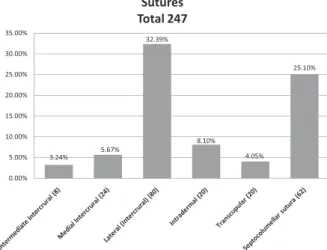
The lateral intercrural was the most frequent suture on the nasal tip, used in 32.39% of cases. Next was the septocolu-mellar suture at 25.1% (►Figs. 10 and 11). The highest frequency of the basal septocolumellar suture (low) was 58.51%, followed by the rotation septocolumellar suture (high), which was observed in 41.49% of cases.
The type of suture thread used in the surgical technique on the nasal tip is shown in►Fig. 12, with the polydioxanone suture 4–0 being used in the majority of cases (70%).
The columellar strut was the most frequent graft added to the nasal tip (56.86%), with the cartilage of the nasal septum the most often used (81.08%).
Cephalic resection of the inferior lateral cartilage was predominantly performed through McIndoe eversion tech-nique or retrograde (70%).
Surgical Techniques on the Nasal Base
The three areas to be observed in the operation of the nasal base are: (1) the nostrils (nasal vestibule), (2) the distance from the nasal base in relation to the intercanthal distance, and (3) the alar-flare, in other words, the external outline of the lateral wall of the inferior lateral cartilage right above the facial groove.
More than one technique can be performed on the same patient depending on the alterations found. The most com-mon techniques used were the correction of asymmetric
nostrils (28.38%), followed by cerclage (25.68%) to reduce the alarflare, and resection of the alar base (22.97%) to reduce the width of the nasal base. For the internal correction of the
flaring of the lateral inferior wall, the technique of Cinelli was used in 10.81% of cases. Release of the periosteum from the premaxilla (1.35%) and alar wall debulking (1.35%) were performed in only one patient. It was not necessary to perform larger operations such as the V-Y advancement (0%) in any of the patients. There was no incidence of dermabrasion, a procedure to reduce scars (►Fig. 13).
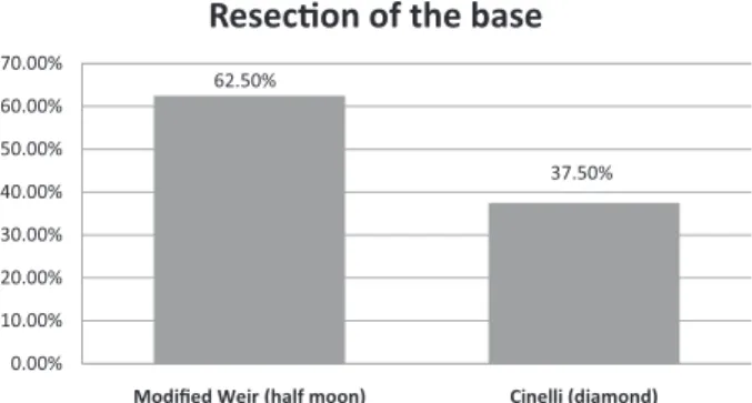
The modified Weir’s technique for the resection of the alar base was the most frequent (62.5%;►Fig. 14).
In the cerclage of the nasal base, the most commonly used thread was Mononylon 4–0 (83.33%) (Johnson & Johnson Medical, São Paulo, Brazil).
32.39% 8.10% 25.10% 10.00% 15.00% 20.00% 25.00% 30.00% 35.00% Sutures Total 247 3.24% 5.67% 32.39% 8.10% 4.05% 25.10% 0.00% 5.00%
Fig. 10 Surgical maneuvers on the tip of the nose—sutures.
Septocollumelar suture
Total 94
58.51% 41.49% 40.00% 50.00% 60.00% 70.00% 58.51% 41.49% 0.00% 10.00% 20.00% 30.00%Basal point (low) Cephalic rotaon (high)
Fig. 11 Surgical maneuvers on the tip of the nose—medial intercrural suture. 2% 0% 14% 2% 0% 12%
Thread
PDS 5–0 PDS 4–0 Vicryl 8–0 Vicryl 4–0 70% 0% Prolene 5–0 Nylon 4–0 Nylon 5–0 Nylon 4–0 Total: 50Fig. 12 Surgical maneuvers on the tip of the nose—threads.
22.97% 10.81% 28.38% 4.05% 4.05% 25.68% 5 00% 10.00% 15.00% 20.00% 25.00% 30.00%
Nasal Base
22.97% 10.81% 4.05% 4.05%1.35% 0.00% 1.35% 1.35%
25.68%
0.00%
Resecons of the ala base
Internal correcon of the lateral wall Asymetric nostrils Resecon of the skin of the vesbule Resecon of the nasal base skin Alar wall debulking Removal of the medial intercrural ssue
Release of the periosteum
Cerclage 22.97%
10.81%
4.05% 4.05%
1.35% 0.00% 1.35% 1.35%
25.68% of
One hundred twelve patients returned in 12 months for follow-up and participated in all the phases of the study. Fifty-eight patients did not complete the study.
Comparative Study of the Preoperative and
Postoperative Anthropometric Measurements of the Nose at 12-Month Follow-Up
Nasal Tip Projection
Goode’s method was used through the measurement of the perpendicular line from the tip point to the line of the facial plane divided by the measurement of the line from the nasion
to the tip point (►Fig. 3). It is recognized that the value should be from 0.55 to 0.66. We found an increase in the ratio in both sexes (►Table 1). There are significant differences in the averages of the projection coefficient between preoperative and postoperative phases in the total group and in both male and female subjects.
Nasolabial Angle
The nasolabial angle is the angular inclination of the colu-mella at the point where it meets the superior lip (►Fig. 2). A nasolabial angle that varies between 90 and 120 degrees is considered ideal. The average found for the nasolabial angle was higher for both sexes (►Table 2).
There were significant differences in the averages of the nasolabial angle between preoperative and postoperative phases in the total groups and in male and female patients.
Nasofrontal Angle
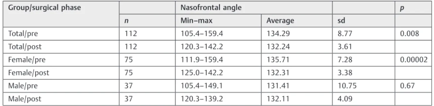
The nasofrontal angle is found by tracing a tangent line to the glabella through the nasion, which crosses a line traced tangent to the nasal dorsum (►Fig. 2). The ideal measurement varies between 115 and 130 degrees. We found an increase in the angle in the male sex and a decrease in the angle of the female sex (►Table 3). The differences of the nasofrontal angle averages between preoperative and postoperative phases in the total group and in male and female patients were significant.
62.50%
37.50%
0.00% 10.00% 20.00% 30.00% 40.00% 50.00% 60.00% 70.00%
Modified Weir (half moon) Cinelli (diamond)
Resecon of the base
62.50%
37.50%
Fig. 14 Surgical maneuvers on the nasal base—resection of the alar base.
Table 1 Preoperative and 12-month postoperative projection coefficient
Group/surgical phase n Projection coefficient p
Min–max Average sd
Total/pre 112 0.60–0.68 0.63 0.02 0.00001
Total/post 112 0.61–0.68 0.65 0.01
Female/pre 75 0.61–0.68 0.63 0.02 0.00001
Female/post 75 0.61–0.68 0.65 0.02
Male/pre 37 0.60–0.68 0.63 0.02 0.000001
Male/post 37 0.62–0.68 0.65 0.01
Abbreviations: min–max, minimum and maximum value; post, postsurgical; pre, presurgical; sd, standard deviation.
Table 2 Preoperative and 12-month postoperative nasolabial angle
Group/surgical phase n Nasolabial angle p
Min–max Average sd
Total/pre 112 65.3–138.5 97.50 12.86 <0.0001
Total/post 112 80.3–120.9 102.77 9.41
Female/pre 75 78.5–126.2 98.73 11.38 <0.0001
Female/post 75 80.3–120.9 104.08 9.16
Male/pre 37 65.3–138.5 95.00 15.30
Male/post 37 80.4–120.3 100.10 9.48
Discussion
In a Brazilian journal, Patrocínio et al showed the importance of the surgical record and the knowledge of the surgical techniques performed in rhinoplasty.8 In our study, we recorded all the surgical techniques performed and we eval-uated the most frequently used ones on the nasal dorsum, on the nasal tip, and on the nasal base. Because noses in our region are mostly Caucasian, which were the aim of our study, our results are different regarding the accesses, the types of grafts on the nasal tip, and the frequency of surgery on the nasal base compared with thefindings by Patrocínio et al.8
Thefindings regarding the evolution of patients made it possible for us to identify the best techniques to achieve excellence in our results. The postoperative follow-up of the patient is essential for the analysis of the results; the doctor-patient relationship is important even in the presence of unsatisfactory results.
Standardized photographic documentation is essential; preoperative and postoperative photos serve to document the results.
The scarring process of the nose is directly related to the surgical trauma, to the type of skin, the type of cartilage, and postoperative care.
International magazines on facial plastic surgery advise the need to restrict qualitative evaluations and to increase quantitative evaluations more objectively with the use of tools such as the Rhinoplasty Outcomes Evaluation.9
Nasal measurements, despite providing objective data of the evaluation, are not more important than the patient’s
satisfaction. Both pieces of information are important for the patient’s postoperative follow-up.
According to a study performed by McKiernan et al in 2001,10the impact of rhinoplasty in the quality of life can be evaluated through tools such as the Glasgow Inventory. In the last decade, the quality of life has been a common matter of discussion, and rhinoplasty has a highly positive impact on the functional purpose as well as on the aesthetic one.
Leong and White stated that the interalar and intercanthal ratio is calculated through the division of the interalar distance by the intercanthal distance (►Fig. 1).6 The ideal interalar-to-intercanthal ratio of the Caucasian nose is con-sidered as 1, that is 100% equivalent (►Table 4). In our study, the average postoperative proportion in the male group, the female group, and total group was lower when compared with the same group preoperatively (p>0.05), approaching
the ideal values.
According to Goode’s method, the ideal nasal projection is a 0.67 ratio. In our study, we observed increased nasal projection at 12 months postoperatively (p<0.05), being
closer to the aesthetic ideal of the nose.
Several authors stated that the ideal nasolabial angle should be found between 90 and 120 degrees, but some authors suggest that it should be kept between 90 and 105 degrees. It is a consensus, however, that the measurement of the male nose should have a more angle and the female nose should have a more obtuse angle, promoting higher rotation on the nasal tip in women.11–14
In a population study in the city of Dundee, Scotland, Leong and White found a value between 67 and 116 degrees.6In our Table 3 Preoperative and 12-month postoperative nasofrontal angle
Group/surgical phase Nasofrontal angle p
n Min–max Average sd
Total/pre 112 105.4–159.4 134.29 8.77 0.008 Total/post 112 120.3–142.2 132.24 3.61
Female/pre 75 111.9–159.4 135.71 7.28 0.00002 Female/post 75 125.0–142.2 132.31 3.38
Male/pre 37 105.4–149.1 131.41 10.75 0.67 Male/post 37 120.3–139.2 132.11 4.09
Abbreviations: min–max, minimum and maximum value; post, postsurgical; pre, presurgical; sd, standard deviation.
Table 4 Preoperative and 12-month postoperative interalar and intercanthal distances
Proportion of interalar and intercanthal distances
Group/surgical phase n Min–max Average sd p
Total/pre 112 0.75–1.58 1.12 0.15 0.00004
Total/post 112 0.75–1.42 1.08 0.13
Female/pre 75 0.81–1.55 1.10 0.15 0.002
Female/post 75 0.81–1.41 1.07 0.12
Male/pre 37 0.75–1.58 1.15 0.16 0.009
Male/post 37 0.75–1.42 1.09 0.14
study, the average of value was 97.5012.86 preoperatively and 102.779.41 postoperatively (p<0.05), showing an
increase in the nasolabial angle and in conformity with the ideal aesthetic values for a Caucasian nose.
In our study, the average nasofrontal angle postoperatively in the female group was lower than the average preoperative value (p>0.05), which probably occurred due to the
reduc-tion of the nasal dorsum through surgical maneuvers such as the removal of the osteocartilaginous hump, approaching the ideal values for the Caucasian nose mentioned by Leong and White.6
It is important to remember that the facial static morphol-ogy, despite being the dominant factor in the aesthetic criteria, is not the only factor to be considered. Therefore, dynamic proportions, skin texture, and color as well as the appearance of teeth are also important in facial aesthetic.7For that reason, a multidisciplinary team becomes necessary with the aid of dermatologists, orthodontists, and bucomaxillofa-cial surgeons, among others. In addition, in thefinal evalua-tion, more than the standard aesthetic measurements, the most important thing is that the patient’s desire in sync with the surgeon’s aesthetic sense.
The learning curve is based on the development of tactile skills and on the surgeon’s judgment. Surgical success is based on the systematization of surgical steps, the level of search for excellence of new technical details, and surgical maneuvers that are being improved, through sutures and grafts, always striving for the best refinement for the Caucasian nose.
This study begins the research of the records of surgical maneuvers in rhinoplasty and of the standardized objective evaluations of the surgical results at the hospital.
Conclusion
The main surgical techniques in rhinoplasty on Caucasian noses were studied and are currently vast. The anthropomet-ric evaluation at 12-month follow-up of the nose showed quantitatively the efficacy of the procedures performed when
compared with the ideal aesthetic measurements for a Cau-casian nose.
References
1 Gunter J. Dallas Rhinoplasty: Nasal Surgery by the Masters. St. Louis: Quality Medical Publishing; 2006:2279
2 Pinto JSP. Interface de visibilização de informações para o sistema integrado de protocolos eletrônicos [Tese]. Paraná, Brazil: Uni-versidade Federal do Paraná, Setor de Ciências da Saúde; 2005:111 3 Maniglia AJ, Maniglia JJ, Maniglia JV. Rinoplastia Estética Funcional
e Reconstrutora. São Paulo, Brazil: Revinter; 2002:129–50 4 Tardy M, Brown RJ. Surgical Anatomy of the Nose. New York, NY:
Raven Press; 1990:12–66
5 Berger C. Electronic data collection for the analysis of surgical maneuvers on patients submitted to rhinoplasty. Int Arch Oto-rhinolaryngol 2012;16(4):497–501
6 Leong SCL, White PS. A comparison of aesthetic proportions between the healthy Caucasian nose and the aesthetic ideal. J Plast Reconstr Aesthet Surg 2006;59(3):248–252
7 Farkas LG, Katic MJ, Forrest CR, et al. International anthropometric study of facial morphology in various ethnic groups/races. J Craniofac Surg 2005;16(4):615–646
8 Patrocínio LG, Carvalho PM, de Souza HM, Couto HG, Patrocínio JA. Surgical maneuvers performed on rhinoplasty procedures carried out at an otorhinolaryngology residency program. Braz J Otorhi-nolaryngol 2006;72(4):439–442
9 Luce EA. Outcome studies and practice guidelines in plastic surgery [editorial]. Plast Reconstr Surg 1999;104(4):1187–1190 10 McKiernan DC, Banfield G, Kumar R, Hinton AE. Patient benefit
from functional and cosmetic rhinoplasty. Clin Otolaryngol Allied Sci 2001;26(1):50–52
11 Doddi NM, Eccles R. The role of anthropometric measurements in nasal surgery and research: a systematic review. Clin Otolaryngol 2010;35(4):277–283
12 Piccardi GH. Assessing facial beauty through proportion analysis by image processing and supervised learning. Int J Hum Comput Stud 2006;64(12):1184–1199
13 Gruber RP, Weintraub J, Pomerantz J. Suture techniques for the nasal tip. Aesthet Surg J 2008;28(1):92–100
14 Guyuron B, Behmand RA. Nasal tip sutures part II: the interplays. Plast Reconstr Surg 2003;112(4):1130–1145, discussion 1146–