ORIGIN
AL RESEAR
CH
Validation of a topography system for evaluation
spine in sagittal plane for children in diferent
nutrient proiles
Validação de um sistema de topograia para avaliação da coluna vertebral no plano sagital de
crianças em diferentes peris nutricionais
Validez de un sistema de topografía para evaluación de la columna vertebral en el plano
sagital de niños con distintos periles nutricionales
Juliana Adami Sedrez1, Claudia Tarragô Candotti2, Maria Izabel Zaniratti da Rosa3,
Fernanda da Silva Medeiros3, Mariana Tonietto Marques1, Jeferson Fagundes Loss2
Mailing address: Juliana Adami Sedrez – Universidade Federal do Rio Grande do Sul – Av. Domingos de Almeida, 2187, Areal – Pelotas (RS), Brazil – CEP: 96085-470 – E-mail: [email protected] – Financing source: Nothing to declare – Conlict of interest: Nothing to declare – Presentation: Apr. 2015 – Accepted for publication: Jun. 2016. Approved
Study developed on the Laboratory of Exercise Research (LAPEX) from School of Physical Education of the Federal University of Rio Grande do Sul (UFRGS) – Porto Alegre (RS), Brazil.
1Master’s degree in Human Movement Science from Federal University of Rio Grande do Sul (UFRGS) – Porto Alegre (RS), Brazil. 2PhD in Human Movement Science Federal University of Rio Grande do Sul (UFRGS) and professor in the Program of Physiotherapy
and Physical Education, Master and PhD by UFRGS – Porto Alegre (RS), Brazil.
3Academic of the Program of Physiotherapy of Federal University of Rio Grande do Sul (UFRGS) – Porto Alegre (RS), Brazil. ABSTRACT | The objective of this study was to
determine the validity, repeatability and inter-evaluator reproducibility of the Vert 3D system in evaluating thoracic and lumbar curvatures of children with diferent nutritional proiles. A total of 115 children participated, which were divided into 3 groups: low weight and eutrophic, overweight, and obese. Each child underwent a panoramic radiography exam of the spine in right proile, from which we obtained Cobb angles for thoracic kyphosis and lumbar lordosis, in addition to being evaluated with the Vert 3D topography system, ive times in the same day, immediately after radiological evaluation. Evaluations were conducted by three independent evaluators and provided the Vert angles for thoracic kyphosis and lumbar lordosis. Using the SPSS software, data were submitted to Pearson Product-moment Correlation Coeicient, Intra-class Correlation Coeicient, paired t-test and one-way ANOVA. Vert 3D system presented excellent levels of repeatability and inter-evaluator reproducibility, regardless of the nutritional proile evaluated, for angles of thoracic kyphosis and lumbar lordosis, but showed low correlation with Cobb angle for thoracic kyphosis and moderate correlation for lumbar lordosis. These results indicate that this system can be used in the clinical follow-up of postural alterations of spine in sagittal plane, of children with all nutritional proiles, but cannot be used
163
as a means of diagnosis or for the purpose of estimating the Cobb angle.
Keywords | Reproduicibility of Results; Spine; Child.
e moderada para lordose lombar. Esses resultados sinalizam que esse sistema pode ser utilizado no acompanhamento clínico de alterações posturais da coluna vertebral no plano sagital de crianças de todos os peris nutricionais, mas não pode ser utilizado como forma de diagnóstico ou com o objetivo de estimar o ângulo de Cobb.
Descritores | Reprodutibilidade dos Testes; Coluna Vertebral; Criança.
RESUMEN | Este estudio se propone a veriicar la validez concurrente, la repetibilidad y la reproductibilidad interevaluadora del sistema Vert 3D en la evaluación de las curvaturas torácica y lumbar de niños con distintos periles nutricionales. Han participado del estudio 115 niños, y se los dividieron en tres grupos: bajo peso y eutróicos, sobrepeso y obesos. Cada participante realizó una radiografía panorámica de la columna vertebral en el lateral derecho, de la cual se obtuvieron los ángulos de Cobb de cifosis torácica y lordosis torácica. Además, a cada participante se les evaluaron con
el sistema de topografía Vert 3D cinco veces al día, tras evaluarles radiológicamente. Las evaluaciones las realizaron tres evaluadores independientes, las cuales fornecieron los ángulos Vert de cifosis torácica y lordosis lumbar. En el software
SPSS, se sometieron a los datos al coeiciente de correlación de Pearson, coeiciente de correlación interclase, prueba t pareada y ANOVA One-Way. El sistema Vert 3D presentó excelentes niveles de repetibilidad y reproductibilidad interevaluadora, independiente del peril nutricional evaluado, para los ángulos de cifosis torácica y lordosis lumbar, en cambio, presentó una débil correlación con el ángulo de Cobb para cifosis torácica y moderada correlación para lordosis lumbar. Esos resultados apuntan que este sistema puede utilizarse en el seguimiento clínico de alteraciones posturales de la columna vertebral en el plano sagital de los niños de todos periles nutricionales, en cambio, no se lo puede como forma de diagnosticar o de proponer el ángulo de Cobb.
Palabras clave | Reproducibilidad de Resultados; Columna Vertebral; Niño.
INTRODUCTION
he vertebral column, in physiological conditions, consists of a succession of harmonious sagittal curves of opposite directions: lumbar lordosis, thoracic kyphosis, and cervical lordosis1. Alterations of curvatures in sagittal plane are characterized by increase or reduction of magnitudes of these curves and often there are reports of functional impairments associated with, for example, increased thoracic kyphosis and with reduction of lumbar lordosis. In several studies, increased thoracic curvature has been associated with the reduction of spine mobility2, to the presence of back pain3, and increased risk of fractures4 and falls5, in addition to causing reduced quality of life2,6; and increased mortality7. Reduction of lumbar lordosis has also been associated to the presence of lumbar pain8, to higher risk of falls9 and to reduction of quality of life6.
herefore, spinal evaluations have a relevant role, in clinical and school environment, as well as in research. Clinically, it assists in choosing treatment techniques, because the therapies are proposed based on the degree of curvature or progression of the same. In the school context, it is a screening tool for early detection of
alterations, and, in research, evaluation of curvatures is fundamental so the efects of treatments in studies can be reported adequately10.
However, tools of easy handling that enable quantifying sagittal curvatures of spine are scarce and not studied adequately11. To ill this gap, a three-dimensional scanning system was developed for topographic evaluation of dorsum, named Vert 3D. his system enables radiation-free examination and provides a three-dimensional view of the surface of the back, allowing to estimate the position of spine12. However, no reference was found indicating that this system has been submitted to validation procedures.
version 1 in evaluating thoracic and lumbar curvatures of children with diferent nutritional proiles.
METHODOLOGY
Sample
Sample size was determined based on the study of hometz et al.16, assuming a 5% margin of error and conidence interval of 95%. Participants attended schools registered in a Family Health Strategy (FHS) of Porto Alegre, and those who had medical referral for panoramic Radiology exam of the spine were invited to participate in the study. hey should meet the following inclusion criteria: chronological age from 6 to 13 years, conditions to remain in orthostasis, no history of spinal surgery, medical request for spinal radiography exam, and participate in ive exams with Vert 3D. Initially, the study had the participation of 119 children, but there was loss of four: two due to radiological examination with poor positioning and two due to no completion of ive exams with Vert 3D. hus, the sample comprised 115 children, mean age 10.93±2.50 years, 53.9% (n=62) male, mean weight 42.5±14.5kg, and mean height 1.43±0.15 m.
he children were divided into three groups: 1) low weight and eutrophic; 2) overweight; and 3) obese. hey were stratiied according to the state percentage of children in these nutritional proiles17.
his study was approved by the Research Ethics Committee of the Federal University of Rio Grande do Sul, under number 19685 and was conducted according to Resolution No. 466/12 of the National Health Council. he children were included after agreeing to participate in the study and after their parents or guardians signed the informed consent.
Anthropometric evaluation
Measurement of mass and stature of the children, to calculate body mass index (BMI). BMI classiication followed the international standard, stratiied according to age18.
Radiographic evaluation
Digital panoramic radiography exams of spine in right proile were conducted with children in orthostasis,
with lexion of shoulder and elbows to avoid overlapping the humerus on the spine.
Based on the radiographs, Cobb angles were calculated in MATLAB® 7.9. To calculate thoracic kyphosis, we marked the upper vertebral plateau of T1 and the lower vertebral plateau of T1219 and, to calculate lumbar lordosis, the upper vertebral plateau of L1 and the lower vertebral plateau of L520. However, if the ends of the selected vertebrae were not clearly visible, the adjacent vertebrae above or below were used as alternatives to deine the curvature angle.
All these calculations were carried out by two independent evaluators, and when the measures between the evaluators difered by more than ive degrees, a third evaluator conducted a new evaluation.
Topographic evaluation
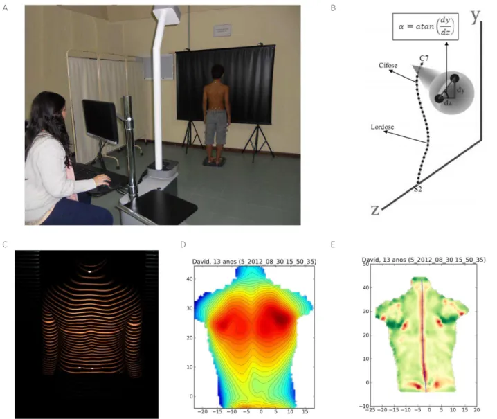
he children were positioned facing away from the Vert 3D equipment in orthostatic posture, with bare back, pending arms along the body, barefoot and positioned with the aid of a positioner (Figure 1A). We palpated and marked the spinous processes of the seventh cervical vertebra (C7) and of the second sacral vertebra (S2), as well as the posterior-superior iliac spines (PSIS) right and left.
hese evaluations were performed by three independent evaluators, properly trained, and each child was evaluated ive times on the same day, immediately after radiological evaluation.
Vert 3D system (Miotec Biomedical Equipment Ltda, Porto Alegre)
Statistical treatment
Performed in software SPSS 17, through descriptive data analysis, Intra-class Correlation Coeicient (ICC), Pearson Product-moment Correlation Coeicient (r), paired t-test, and one-way ANOVA. he signiicance level adopted was 0.05. To analyze the degree of agreement between the angles of Vert and Cobb we used the graphical method of Bland and Altman21.
ICC values were classiied as weak (ICC<0.40), moderate (ICC 0.4-0.75), and excellent (ICC>0.75)22. Values for r were classiied as very low correlation (<0.2), low correlation (0.2-0.39), moderate correlation (0.4-0.69), high correlation (0.7-0.89), and very high correlation (0.9-1.0)23.
A B
C D E
Figure 1. (A) Physical structure of the Vert 3D system and positioning of child to conduct postural evaluation; (B) representation of symmetry line analysis in sagittal plane to obtain Vert angles for thoracic kyphosis, with dy and dz being the measure in the vertical and antero-posterior axes, respectively; (C) image of fringe projection; (D) relief map; (E) curvature map with symmetry line
RESULTS
When assessing BMI: the group composed of eutrophic or low-weight children (n=69) presented mean BMI of 17.8±2.3 kg/m²; the overweight group (n=32), BMI of 22.6±2.2 kg/m²; and the group classiied as obese (n=14), BMI of 26.6±4.0 kg/m².
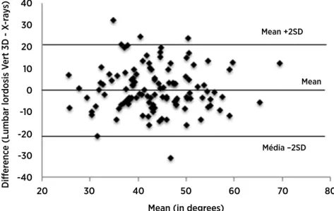
inter-evaluator reproducibility (ICC=0.84; p<0.001), moderate concurrent validity (r=0.43; p<0.001), with no signiicant diference between Vert and Cobb angles (p=0.51). he graphical analyses of Bland and Altman21 were performed only with the results from the evaluation of lumbar lordosis, since only this evaluation presented adequate levels of concurrent validity (Figure 2).
To evaluate the inluence of BMI on the Vert 3D system measurement, we evaluated the system’s repeatability (Table 1), inter-evaluator reproducibility
(Table 2), and concurrent validity (Table 3). Vert system 3D presented excellent repeatability and inter-evaluator reproducibility of thoracic kyphosis for subjects with normal BMI or overweight, and moderate levels in obese subjects. However, the validity maintained weak levels in the normal and overweight nutritional proile, and moderate level in the obese nutritional proile. For the evaluation of lumbar lordosis, we obtained excellent levels of repeatability and inter-evaluator reproducibility, in addition to moderate level of validity for all nutritional proiles.
Mean (in degrees) -40
-30 -20 -10 0 10 20
20 30 40 50 60 70 80
Média –2SD Mean Mean +2SD
30 40
Diff
er
enc
e (Lumbar lor
dosis V
ert 3D - X
-r
ay
s)
Figure 2. Graphical method of Bland and Altman between measures for lumbar lordosis obtained with Vert 3D system and x-ray exam, corresponding to the irst measurement of evaluator A
Table 1. Vert 3D System results obtained in measurements of evaluators A and B, corresponding to repeatability of Vert angles for thoracic kyphosis and lumbar lordosis in the diferent nutritional proiles
Vertebral level
Evaluator A Evaluator B
1st evaluation Mean±SD
2nd evaluation
Mean±SD p
(t Test) ICC (95%CI) p
1st evaluation Mean±SD
2nd evaluation Mean±SD
p
(t Test) ICC (95%CI) p
Normal BMI (n=69)
Kyphosis (º) 44.3±8.3 45.0±9.0 0.239 0.848
(0.766−0.867) <0.001* 44.6±9.0 45.4±9.2 0.195
0.877
(0.808−0.922) <0.001* Lordosis (º) 41.±8.7 42.8±10.2 0.331 0.795
(0.689−0.868) <0.001* 41.6±9.7 40.8±9.4 0.279
0.806
(0.704−0.875) <0.001*
Overweight BMI (n=32)
Kyphosis (º) 45.5±10.2 45.1±8.8 0.608 0.915
(0.833−0.957) <0.001* 45.7±9.7 45.0±8.7 0.390
0.880
(0.768−0.939) <0.001* Lordosis (º) 46.9±9.7 47.2±10.1 0.535 0.947
(0.894−0.974) <0.001* 47.5±9.8 47.2±9.1 0.772
0.862
(0.736−0.930) <0.001*
Obese BMI (n=14)
Kyphosis (º) 50.8±9.1 49.3±10.4 0.510 0.658
(0.217−0.876) 0.004* 48.2±6.9 50.1±11.6 0.357
0.705
(0.299−0.895) 0.002* Lordosis (º) 48.2±11.5 47.8±12.9 0.645 0.962
(0.886−0.988) <0.001* 46.1±11.3 46.9±12.8 0.376
0.959
(0.878−0.987) <0.001*
Table 3. Correlations between Cobb angles and Vert angles in measurements of Evaluator A, for thoracic kyphosis and lumbar lordosis in the diferent nutritional proiles
X-rays BMI Classiication
Vert 3D Kyphosis p
(t Test) r p
Cobb Thoracic kyphosis
Normal (n=69) <0.001** 0.25 0.036* Overweight (n=32) 0.136 0.38 0.032* Obese (n=14) 0.542 0.57 0.034*
Vert 3D Lordosis p
(t Test) r p
Cobb Lumbar lordosis
Normal (n=69) 0.232 0.40 0.001* Overweight (n=32) 0.053 0.55 0.001* Obese (n=14) 0.064 0.60 0.024*
*Signiicant correlation; **Signiicant diference.
DISCUSSION
he main indings demonstrate that: 1) Vert 3D presented excellent repeatability and inter-evaluator reproducibility levels for evaluation of thoracic kyphosis and lumbar lordosis; 2) weak concurrent validity for thoracic kyphosis and moderate for lumbar lordosis; and 3) there was signiicant diference between the angles for thoracic kyphosis obtained with the Vert 3D system and with the radiographs.
Regarding the evaluation of curvatures in sagittal plane, the literature is scarce as to validation procedures for topography instruments, as many studies focus on evaluating the frontal plane. However, the studies that present these data corroborate the results of this study. Mohokum et al.24, when using the Jenoptick Formetric
system, also obtained excellent repeatability and inter-evaluator reproducibility for the angle of thoracic kyphosis and lumbar lordosis. Goh et al.25, using the same system, reported ICC ranging from 0.98 to 0.99, for all thoracic kyphosis parameters measured.
he Milwaukee topography instrument presented excellent inter-evaluator reproducibility for thoracic kyphosis and weak inter-evaluator reproducibility for lumbar lordosis. However, in determining repeatability, we found lower levels for thoracic kyphosis, with correlation ranging from moderate to excellent and excellent for lumbar lordosis26.
he Quantec topography system was also evaluated as for repeatability; however, only the standard deviation values of these measures were presented, and we observed standard deviation of ±4.2° for lumbar lordosis and standard deviation of ±3.6° for thoracic kyphosis16. However, these data are insuicient to demonstrate that this instrument presents repeatability. Furthermore, important questions concerning validation of the Quantec system have yet to be studied, since this technology presents no results as for its intra- and inter-evaluator reproducibility27.
Even considering the clinical importance of the variables involved in a new instrument’s validation process, such as repeatability and inter-evaluator reproducibility, we found no studies reporting these evaluations in sagittal plane for the other topography systems; therefore, a gap remains in the literature regarding these aspects of validity.
In analyzing the concurrent validity of Vert 3D system, we observed that there is correlation with x-rays; however,
Table 2. Results from Vert 3D system obtained in measurements of three evaluators, as for inter-evaluator reproducibility of Vert angles for thoracic kyphosis and lumbar lordosis in the diferent nutritional proiles
Vertebral level Evaluator A
Mean±SD
Evaluator B Mean±SD
Evaluator C
Mean±SD p (Anova) ICC (CI 95%) p
Normal (n=69)
Kyphosis (º) 44.3±8.4 44.6±9.0 43.8±9.2 0.846 0.821
(0.747–0.878) <0.001* Lordosis (º) 41.7±8.7 41.7±9.7 41.1±9.8 0.913 0.786
(0.702–0.854) <0.001*
Overweight (n=32)
Kyphosis (º) 45.5±10.2 45.7±9.7 45.2±10.1 0.977 0.872
(0.785–0.931) <0.001* Lordosis (º) 46.9±9.8 47.5±9.8 47.1±9.2 0.971 0.875
(0.789–0.932) <0.001*
Obese (n=14)
Kyphosis (º) 50.8±9.1 48.2±6.9 50.5±9.8 0.704 0.723
(0.465–0.890) <0.001* Lordosis (º) 48.2±11.5 46.1±11.3 48.7±12.3 0.822 0.909
(0.795–0.967) <0.001*
this correlation presents weak level for thoracic kyphosis and moderate level for lumbar lordosis. In Figure 2, it is observed that, in spite of the mean diference of 0.63° between the two evaluation methods and of the small number of individuals out of the limits of agreement, it is not possible to airm that there is agreement between them, considering that the magnitude of the standard deviations presents a clinically important magnitude. Based on these results, it is suggested that the Vert 3D system should not be used to estimate the Cobb angle, not even in lumbar curvature.
here are few studies that investigate the concurrent validity of the topography methods in sagittal plane. Fortin et al.28 reported high correlations between the InSpeck 3D digitizer system and radiographs in evaluating thoracic kyphosis and lumbar lordosis. Kovac and Pecina29 also reported excellent correlation between the Moiré Topography system and radiological exams, in evaluating thoracic kyphosis. Importantly, in both studies the mean age of the individuals evaluated was 16.4 years (10 to 26 years) and 15.7 years (10 to 20 years), which is higher than that of those evaluated in this study, with mean age of 10.9 (6 to 13 years). Probably, physical characteristics, such as size and width of trunk, difer between the samples of the studies, which can be important factors in the evaluations, considering that the surface topography generates information based on fringe projections on the dorsum of the individual evaluated and, therefore, trunks with smaller size could hinder the system’s analysis.
According to D’Osualdo et al.30, agreement with x-rays is not the only relevant issue in surface measurements; on the contrary, reproducibility is at least as important, since only one method that allows for reproduction of its data can be used both in screening program and follow-up of changes over time and in evaluating the alterations produced by the treatment.
hus, based on the results obtained in this study, it is possible to airm that the Vert 3D topography system can be used for follow-up of thoracic and lumbar curvature, both by a single evaluator and by diferent evaluators, since it presents adequate repeatability and inter-evaluator reproducibility.
In evaluating the aspects of validation in the diferent nutritional proiles, we observed that the excellent levels of repeatability and inter-evaluator reproducibility are maintained for all proiles only for lumbar lordosis. For evaluation of thoracic kyphosis, excellent levels were obtained only for normal and
overweight subjects, whereas moderate levels were obtained for obese subjects.
he only study found in the literature that conducted this type of analysis used the Jenoptic Formetric system and found no signiicant association between BMI and levels of repeatability and inter-evaluator reproducibility, which remained excellent for the normal group and for the overweight or obese BMI group24. However, in this study24, the analyses were divided only into two groups (normal and overweight/obese), diferently from this study, in which the three groups were analyzed separately. It is suggested that the fact of collectively analyzing the overweight and obese groups may have limited the study in determining the levels of repeatability and inter-evaluator reproducibility in the obese group.
Despite this small inluence caused by obesity in the results for repeatability and inter-evaluator reproducibility, the Vert 3D system maintained levels ranging from moderate to excellent in all nutritional proiles; therefore, this instrument can be a good alternative for the evaluation of thoracic and lumbar curvatures in the cases of overweight or obese individuals, since it is automated, with no critical dependence on palpation of anatomical points, which may facilitate this type of evaluation. And, considering that currently there is an increasing number of individuals with body weight above the normal range, there is also a growing concern by health professionals, especially with regard to comorbidities associated with this condition. Within the scope of physiotherapy, taking into consideration the increase of overload on the musculoskeletal segments, the use of reproducible evaluation methods that also present conditions to analyze these individuals, as in the case of Vert 3D system, acquires importance. It is noteworthy that intra-evaluator reproducibility at diferent times was not evaluated. It is recommended that future studies organize the logistics of data collection so the participants are evaluated at diferent times with at least 24 hours of diference between each evaluation.
CONCLUSION
curvatures of children of all nutritional proiles, when utilized by diferent evaluators or by a single evaluator. However, it presented poor concurrent validity for thoracic kyphosis and moderate concurrent validity for lumbar lordosis; thus, to date, it is not possible to diagnose postural alterations using this system, not even to estimate the Cobb angle for thoracic kyphosis and lumbar lordosis. herefore, it is suggested that the Vert 3D system can be utilized as an instrument to provide additional information concerning the positioning of the dorsal surface, aiding in the clinical follow-up; however, it presents no conditions of replacing the use of radiographic examination.
REFERENCES
1. Loubresse CG, Vialle R, Wolf S. Cyphoses pathologiques. EMC-Rhumatol Orth. [Internet]. 2005 [acesso em 7 jun 2016];2(3):294-334. Disponível em: http:// w w w . s c i e n c e d i r e c t . c o m /s c i e n c e / a r t i c l e / p i i / S176242070400136X
2. Miyakoshi N, Itoi E, Kobayashi M, Kodama H. Impact of postural deformities and spinal mobility on quality of life in postmenopausal osteoporosis. Osteoporos Int. [Internet]. 2003 [acesso em 7 jun 2016];14(12):1007-12. Disponível em: http://www.ncbi.nlm.nih.gov/pubmed/14557854
3. Ensrud KE, Black DM, Harris F, Ettinger B, Cummings SR. Correlates of kyphosis in older women. The fracture intervention trial research group. J Am Geriatr Soc. [Internet]. 1997 [acesso em 7 jun 2016]; 45(6):682-7. Disponível em: http://www.ncbi.nlm.nih.gov/pubmed/9180660
4. Huang MH, Barrett-Connor E, Greendale GA, Kado DM. Hyperkyphotic posture and risk of future osteoporotic fractures: the Rancho Bernardo study. J Bone Miner Res. [Internet]. 2006 [acesso em 7 jun 2016];21(3):419-23. Disponível em: http://www.ncbi.nlm.nih.gov/ pubmed/16491290
5. Kado DM, Huang MH, Nguyen CB, Barrett-Connor E, Greendale GA. Hyperkyphotic posture and risk of injurious falls in older persons: the Rancho Bernardo Study. J Gerontol A Biol Sci Med Sci. [Internet]. 2007 [acesso em 7 jun 2016];62(6):652-7. Disponível em: http://www.ncbi.nlm. nih.gov/pubmed/17595423
6. Imagama S, Hasegawa Y, Matsuyama Y, Sakai Y, Ito Z, Hamajima N et al. Inluence of sagittal balance and physical ability associated with exercise on quality of life in middle-aged and elderly people. Arch Osteoporos. [Internet]. 2011 [acesso em 7 jun 2016];6(1-2):13-20. Disponível em: http:// www.ncbi.nlm.nih.gov/pmc/articles/PMC3235276/
7. Kado DM, Lui LY, Ensrud KE, Fink HA, Karlamangla AS, Cummings SR et al. Hyperkyphosis predicts mortality independent of vertebral osteoporosis in older women. Ann Intern Med. [Internet]. 2009 [acesso em 7 jun 2016];150(10):681-7. Disponível em: http://www.ncbi.nlm. nih.gov/pubmed/19451575
8. Chaléat-Valayer E, Mac-Thiong JM, Paquet J, Berthonnaud E, Siani F, Roussouly P. Sagittal spino-pelvic alignment in chronic low back pain. Eur Spine J. [Internet]. 2011 [acesso em 7 jun 2016];20(Suppl 5):634-40. Disponível em: http:// www.ncbi.nlm.nih.gov/pubmed/21870097
9. Ishikawa Y, Miyakoshi N, Kasukawa Y, Hongo M, Shimada Y. Spinal sagittal contour afecting falls: cut-of value of the lumbar spine for falls. Gait Posture. [Internet]. 2013 [acesso em 7 jun 2016];38(2):260-3. Disponível em: http://www. ncbi.nlm.nih.gov/pubmed/23273490
10. Carman DL, Browne RH, Birch JG. Measurement of scoliosis and kyphosis radiographs. Intraobserver and interobserver variation. J Bone Joint Surg Am. [Internet]. 1990 [acesso em 7 jun 2016];72(3):328-33. Disponível em: http://www.ncbi. nlm.nih.gov/pubmed/2312528
11. Zaina F, Donzelli S, Lusini M, Negrini S. How to measure kyphosis in everyday clinical practice: a reliability study on diferent methods. Stud Health Technol Inform. [Internet]. 2012 [acesso em 7 jun 2016];176:264-7. Disponível em: http://www.ncbi.nlm.nih.gov/pubmed/22744505
12. Miotec Biomedical Equipamentos Ltda. Vert 3D. [Internet]. [s. d.] [acesso em 7 jun 2016]. Disponível em: http://www. miotec.com.br/scanner-3d/
13. Zubović A, Davies N, Berryman F, Quraishi N, Lavy C, Gavin B et al. New method of scoliosis deformity assessment: ISIS-2 System. Spine J. ISIS-2008;8:ISIS-29S.
14. Goldberg CJ, Kaliszer M, Moore DP, Fogarty EE, Dowling FE. Surface topography, Cobb angles, and cosmetic change in scoliosis. Spine. [Internet]. 2001 [acesso em 7 jun 2016];26(4):E55-63. Disponível em: http://www.ncbi.nlm. nih.gov/pubmed/11224901
15. Drerup B, Hierholzer E. Back shape measurement using video rasterstereography and three-dimensional reconstruction of spinal shape. Clin Biomech. [Internet]. 1994 [acesso em 7 jun 2016];9(1):28-36. Disponível em: http://www.ncbi.nlm. nih.gov/pubmed/23916075
16. Thometz JG, Liu XC, Lyon R, Harris GF. Variability in three-dimensional measurements of back contour with raster stereography in normal subjects. J Pediatr Orthop. [Internet]. 2000 [acesso em 7 jun 2016];20(1):54-8. Disponível em: http://www.ncbi.nlm.nih.gov/pubmed/10641689
17. Instituto Brasileiro de Geograia e Estatística. Contagem da População. [Internet]. 2007 [acesso em 7 jun 2016]. Disponível em: http://www.ibge.gov.br/home/estatistica/ populacao/contagem2007/default.shtm
18. Cole TJ, Bellizzi MC, Flegal KM, Dietz WH. Establishing a standard deinition for child overweight and obesity worldwide: international survey. BMJ. [Internet]. 2000 [acesso em 7 jun 2016];320(7244):1240-3. Disponível em: http://www.bmj.com/content/320/7244/1240
19. Boseker EH, Moe JH, Winter RB, Koop SE. Determination of “normal” thoracic kyphosis: a roentgenographic study of 121 “normal” children. J Pediatr Orthop. [Internet]. 2000 [acesso em 7 jun 2016];20(6):796-8. Disponível em: http:// www.ncbi.nlm.nih.gov/pubmed/11097257
em 7 jun 2016];14(7):717-21. Disponível em: http://www.ncbi. nlm.nih.gov/pubmed/2772721
21. Bland JM, Altman DG. Statistical methods for assessing agreement between two methods of clinical measurement. Lancet. [Internet]. 1986 [acesso em 7 jun 2016];1(8476):307-10. Disponível em: http://www.ncbi.nlm.nih.gov/ pubmed/2868172
22. Fleiss RL. The design and analysis of clinical experiments. New York: John Wiley and Sons; 1986.
23. Gaya A. Ciências do movimento humano: introdução à metodologia da pesquisa. Porto Alegre: Artmed; 2008. 24. Mohokum M, Mendoza S, Udo W, Sitter H, Paletta JR, Skwara
A. Reproducibility of rasterstereography for kyphotic and lordotic angles, trunk length, and trunk inclination: a reliability study. Spine. [Internet]. 2010 [acesso em 7 jun 2016];35(14):1353-8. Disponível em: http://www.ncbi.nlm. nih.gov/pubmed/20505568/
25. Goh S, Price RI, Leedman P, Singer KP. Rasterstereographic analysis of the thoracic sagittal curvature: a reliability study. J Musculoskelet Res. [Internet]. 1999 [acesso em 7 jun 2016];4:137-42. Disponível em: http://cms.uwa.edu.au/ research/publications/journals/1999/goh2.shtml
26. Liu XC, Tassone JC, Thometz JG, Paulsen LC, Lyon RM, Marquez-Barrientos C et al. Development of a
3-dimensional back contour imaging system for monitoring scoliosis progression in children. Spine Deform. [Internet]. 2013 [acesso em 7 jun 2016];1(2):102-7. Disponível em: http://www.sciencedirect.com/science/article/pii/ S2212134X12000366
27. Noonan KJ. Point of view. Spine. [Internet]. 2001 [acesso em 7 jun 2016];26(11):1279. Disponível em: http://journals.lww. com/spinejournal/Citation/2001/06010/Point_of_View.21. aspx
28. Fortin C, Feldman DE, Cheriet F, Labelle H. Validity of a quantitative clinical measurement tool of trunk posture in idiopathic scoliosis. Spine. [Internet]. 2010 [acesso em 7 jun 2016];35(19):E988-94. Disponível em: http://www.ncbi.nlm. nih.gov/pubmed/20700086
29. Kovac V, Pecina M. Moiré topography in measurement of the sagittal curvatures of the spine. Coll Antropol. [Internet]. 1999 [acesso em 7 jun 2016];23(1):153-8. Disponível em: http://www.ncbi.nlm.nih.gov/pubmed/10402717