OR
IGI
N
A
L
R
E
S
E
A
R
C
H
Evaluation of longitudinal ligament of the spine of
Wistar rats in an experimental model of Suit therapy
Avaliação dos ligamentos longitudinais da coluna de ratos Wistar em modelo experimental da
terapia Suit
Evaluación de los ligamentos longitudinales de la columna vertebral de ratas Wistar desde el
modelo experimental de la terapia Suit
Marcia Cristina Dias Borges1,2, Tatiane Kamada Errero1,2, Camila Thieimi Rosa1,2, Giovanni Ribeiro
Bernardino1,2, Rose Meire Costa Brancalhão1,3, Lucinéia de Fátima Chasko Ribeiro1,3, Gladson Ricardo
Flor Bertolini2,3
Mailing address: Gladson Ricardo Flor Bertolini – State University of Western Paraná – Rua Universitária, 2069, Jd. Universitário, Caixa Postal 711 – Cascavel (PR), Brazil – CEP: 85819-110 – E-mail: [email protected] – Financing source: Nothing to declare – Conlict of interest: Nothing to declare – Presentation: Jan. 2015 – Accepted for publication: May 2016 – Approved by the Ethics Committee on the Use of Animals (CEUA) of the State University of Western Paraná (Unioeste).
1Laboratory of Cellular and Structural Biology of the State University of Western Paraná (Unioeste) – Cascavel (PR), Brazil. 2Laboratory of Study of Physiotherapeutic Injuries and Resources of Unioeste – Cascavel (PR), Brazil.
3Faculty of the Graduate Program in Biosciences and Health of Unioeste – Cascavel (PR), Brazil. ABSTRACT | Ligaments adapt according to the intensity
of physical activity and mechanical load to which they are subjected. In the last decade there have been methods and protocols in the ield of infant neurofunctional physiotherapy, which have the term “suit” in common, to characterize the existence of suits with adjustable elastic bands and the possibility of applying load on the human skeleton. Since the mechanical load can produce ibrocartilaginous changes on the ligaments and also that no studies evaluating the efect of suit therapy on ligaments of the spine were found, research with experimental methods of load are justiied. The aim of this study was to analyze thickness and morphology of longitudinal ligaments of the spine of Wistar rats when subjected to mechanical load by vertebral compression. Thirty animals were separated into ive groups (G1 – control; G2 – simulation of the use of suit; G3, G4, and G5 – maintenance of the suit). The suit experimental model, in G4 and G5, were adapted weights or elastic bands arranged in “X” for 50% of spinal overload of the weight of the animal, who remained with the suit for 40 hours over four weeks of experiment, ive days a week. There were no signiicant diferences for thickness, and morphological changes of longitudinal ligaments were also not observed. We concluded that there were no changes in longitudinal ligaments of the spine in animals subjected to the experimental model of suit therapy.
Keywords | Cerebral Palsy; Child; Physical Therapy Modalities; Spine; Rats, Wistar.
148
Descritores | Paralisia Cerebral; Criança; Modalidades de Fisioterapia; Coluna Vertebral; Ratos Wistar.
RESUMEN | Pueden adaptarse los ligamentos a la intensidad de la actividad física y a la carga mecánica sometida a ellos. En la última década, han surgido métodos y protocolos en el área de isioterapia neurofuncional infantil que tienen en común el término “suit” para caracterizar la existencia de ropas con bandas elásticas ajustables y la posibilidad de aplicación de cargas en el esqueleto humano. Debido a que la carga mecánica puede producir alteraciones ibrocartilaginosas en los ligamentos y que no han sido encontrados estudios que evaluaron el efecto de la terapia suit en los ligamentos de la columna vertebral, las investigaciones con métodos experimentales de carga son necesarias. Este estudio tiene el propósito de evaluar las espesuras y la morfología de los ligamentos longitudinales de
la columna vertebral de ratas Wistar cuando sometida a carga mecánica por compresión vertebral. Se dividieron treinta ratas en cinco grupos (G1 —grupo control; G2 —simulación de la utilización del suit; G3, G4, G5 —mantenimiento de la ropa). Desde el modelo experimental del suit, en el G4 y G5 se adaptaron pesos o elásticos puestos en “X” para la sobrecarga vertebral de 50% del peso de los animales, los cuales permanecieron con la ropa durante cuarenta horas, al largo de cuatro semanas de experimento, en cinco días semanales. No fueron observadas diferencias signiicativas para la espesura, tampoco cambios morfológicos en los ligamentos longitudinales. Se concluye que no se observaron alteraciones en los ligamentos longitudinales de la columna vertebral de los animales sometidos al modelo experimental de la terapia suit.
Palabras clave | Parálisis Cerebral; Niño; Modalidades de Fisioterapia; Columna Vertebral; Ratas Wistar.
INTRODUCTION
he joints between the vertebrae are strengthened and supported by ligaments, and among them are the anterior longitudinal ligament (ALL) and the posterior longitudinal ligament (PLL), extending from the cervical spine to the sacrum1. Ligaments adapt to the intensity
of physical activity, and may hypertrophy or atrophy, in such a way that they may change the resistance in response to exercise or immobilization2. his dynamic
behavior suggests that cells are able to detect changes in the mechanical load and to coordinate their response to change the composition of the extracellular matrix (ECM). One of the ways in which ECM can be changed is by the formation of a ibrocartilaginous matrix where ligaments are under compression3osteotendinous junctions,
osteoligamentous junctions. Individuals with vertebral slip feature ossiication of the ligamentum lavum and iniltrate with chondrocytes, suggesting the involvement of mechanical load on morphological changes4.
Ligaments have low supply of oxygen and nutrients, low cell density, and poor regenerative capacity. However, they experience some of the highest mechanical loads of the body. When these loads exceed a critical threshold, injuries may occur, resulting in morphofunctional changes and disorder of movements5. Ligaments are
functional (efective) under stress or stretch, however, without compression or functionality when shortened beyond their rest threshold2.
Mechanical stimuli, when provided in a proper manner, induce the growth and better alignment of cells, improving the quality of ECM5,6, however, the
frequency as well as the intensity of these stimuli are not yet well known2.
In the last decade, methods have emerged in the ield of neurofunctional physiotherapy, which have the term “suit” in common, in their classiications, to characterize the existence of suits that function as dynamic orthotics7,8. he basic concept is to create a support unit
to align the body as close to normal, improve and adjust the proprioceptive system through pressure exerted on joints, ligaments, and muscles, reducing pathological relexes, and restoring muscle synergies that are translated into appropriate patterns of movement7.
Elastic bands are adjustable, which means a discharge of 15 to 40 kg can be axially applied in the body8.
However, the suit sufered changes compared with its original form, to adapt the therapeutic needs within the neurological rehabilitation, and little attention has been given to the efects of this device on the musculoskeletal system, in particular spinal stabilizers ligaments.
the spine of Wistar rats submitted to mechanical load produced by an experimental model of suit therapy.
METHODOLOGY
Animals
he sample was composed of 30 Wistar rats, males, with eight weeks of age. here were no restrictions on food, being feed and natural water ad libitum, with controlled temperature (24ºC±1ºC) and light/dark cycle of 12 hours. he research was approved by the Ethics Committee on the Use of Animals (CEUA) of the State University of Western Paraná – Unioeste.
Materials and experimental protocol
An experimental model, of therapeutic suit type, was made based on the model used in suit therapy. his suit was composed of upper and lower parts, closed with Velcro, prepared with raw cotton 180-thread fabric, in which were adapted two elastic bands arranged in “X” for vertebral approximation, with 50% of the load weight of the animal, as previously described9.
For the calculation of body load, done by the displacements of the elastic bands, they were analyzed per load cell, with capacity up to 100 kgf (SB-100 of LYNX®). hrough a system of biological data collection
(BioEMG1100, LYNX®, Brazil), it was possible to
display the obtained data every two centimeters of displacement. he collected data were arranged in scatter plots (kgf versus displacement) and it was possible to ind the polynomials that have modeled the data and allowed calculating the displacements of the elastics in centimeters. he elaboration of spreadsheets, scatter graphs, and polynomial interpolation were built with the Excel program.
he sample was divided in ive groups (from G1 to G5), with six animals each:
• G1 (Group 1) – absolute control;
• G2 (Group 2) – putting the suit following removal;
• G3 (Group 3) – length of stay of 2 hours with the suit only;
• G4 (Group 4) – length of stay of 2 hours a day with the suit and weights adapted;
• G5 (Group 5) – length of stay of 2 hours a day with the suit and traction bands;
For G4 and G5, a load of 50% of the body weight of the animal was given. In G4, the load, through the pellets of ishing, was divided in two units coupled to the suit (±25% load on each bag); and for G5, two elastics pulled and arranged in “X” on the back of the animal (±25% load on each elastic). To this end, the animals previously weighted before the beginning of each week. he displacement of each elastic was measured with a digital caliper (Digimess® – São
Paulo/Brazil).
During the length of stay with the suit, the animals were kept in boxes (three animals per box). he researchers avoided the postural accommodation of the animals, in such a way that they would keep moving during the 2 hours proposed. Before the beginning of the study, the rats were adapted for three days for putting the suit.
he experiment lasted four weeks, performed in ive consecutive days, with two days of, totaling length of stay of 40 hours with the suit (from G3 to G5) and without it (G1 and G2). On the day following the last day of experiment, the animals were anesthetized with ketamine hydrochloride (50 mg/kg) and xylazine (10 mg/kg), then beheaded and the thoracic segment of the vertebral column dissected.
Histomorphometry of ligaments
Histomorphometric analysis was performed on a vertebra in the thoracic region (T3-T6), in the interscapular region, without a speciic segment of this region. he choice of the sample considered the visual and structural quality that best make the analyses feasible.
Figure 1. Sagittal section of the vertebral segment with presentation of vertebral bodies and spinal cord
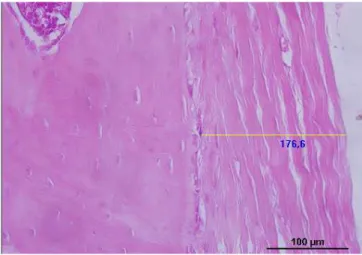
Each vertebra was analyzed in four quadrants: two quadrants for PLL (upper and lower) and two for ALL (upper and lower). he images were obtained on increase of 400× immediately below the articular cartilage and the apparent concentration of chondrocytes for upper images and immediately above the articular cartilage and the apparent concentration of chondrocytes for inferior images. he thicknesses of longitudinal ligaments were obtained by drawing a line to measure their width in micrometers (μm) and positioned in the center of the image at 50% zoom. In the presence of ligament spaces resulting from the artifacts, they were not considered in full measure. he morphological analysis was performed in each quadrant observing the presence of chondrocytes between the ibers of connective tissue and presence of neovascularization (Figure 2).
Figure 2. Morphological analysis of the anterior longitudinal ligament
Statistical analysis
he Shapiro-Wilk test was used to verify the normality of the data. Afterwards, one-way ANOVA was applied for analysis between groups, along with Tukey test. he signiicance level was 5%.
RESULTS
he indings allowed comparing the averages of the ligament thickness per group of the upper and lower regions of the vertebrae, as well as the average for each ligament, as shown in Table 1. here was no statistical diference in the comparison between groups, or in the comparison within the groups between lower and upper parts.
Table 1. Means and standard deviations of the thicknesses of longitudinal ligaments per upper and lower spine region and total means per ligament
Thicknesses G1 G2 G3 G4 G5
ALL
Upper 119.13±32.43 140.53±61.50 144.51±20.71 127.34±41.81 164.59±34.90
Lower 119.66±44.11 136.71±61.97 130.48±60.06 161.48±29.47 150.02±41.37
PLL
Upper 65.61±20.17 104.76±79.31 86.59±46.83 60.89±37.92 65.59±22.15
Lower 73.32±40.88 67.75±25.46 64.5±18.90 66.60±26.14 56.78±22.61
Total
ALL 119.39±10.71 138.62±53.21 137.49±29.12 144.40±17.90 157.30±31.73
No morphological changes were found in the ligaments, such as presence of chondrocytes or other cells between ibers of the connective tissue. Neovascularization was also not observed in such regions.
DISCUSSION
he methods that use the suit therapy, were designed in the project known as “Penguin suit”, developed by the space program of Russia that was used by astronauts in space lights to counteract the harmful efects of weightlessness and hypokinesia. he fact that it could be used for long periods of time was the basis of the creation of the intensive therapy with the use of the suit8,10, which involves a program of 80 hours of body
activity (therapeutic exercise), performed four hours a day, on ive consecutive days, for a period of four weeks and the patient staying up to three hours with the suit. Some of the concepts advocate the use of these suits with traction bands outside the clinical environment by encouraging its handling by direct caregivers, favoring their use for longer periods of time7. However, when
searching in the literature, we observed that it is still a technique lacking scientiic depth, considering the diiculty in inding research regarding its use, both clinical and experimental.
he experimental method of this research aimed to approach the most of the intensive therapy protocol, however, the animals remained only supported by their members with volunteer movements without remaining lay down. We did not determine a protocol of exercises as a way to increase body activity. hus, there was no musculoskeletal requirement beyond that imposed by the suit and the elastics bands adapted to the load of 50% of the weight of the animal, which may have contributed to have no deformation of the longitudinal ligaments by mechanical stress.
In bipedal posture, the distribution of gravity on the spine works in equilibrium with muscular forces, which is directly proportional to vertebral direction and pelvic obliquity10. In the four supports
posture, there is no one-way gravitational acting with muscle ibers of the torso, and muscle activity might have supplied the load imposed by traction bands, without enough load in the longitudinal ligament to generate morphological changes in these structures. Solomonow2 describes that muscles associated with
joints have an important role as movement limiters and, therefore, as joint stabilizers. In some joints, such as in intervertebral ones, the muscle function as stabilizer is ampliied. Since the region of collection was interscapular, in addition to the direct efect on the spine, promoted by the suit, a possible altered muscle action of those caused (or inserted) on the scapula, could have produced inluences on the ligaments.
he musculoskeletal system is able to respond to changes in the conditions of mechanical load2,3,11,12, and
this functional and structural adaptation is already well understood to muscle and bone. However, little is known about the efects of exercise on ligaments13. Tendons
and ligaments have very similar characteristics, both are recognized for presenting great tensile strength and are composed of collagen ibers. herefore, their cells behave dynamically with the ability of reorganizing before mechanical stimuli, generating changes in ECM14.
he morphology of the connective tissue can be inluenced especially during motor growth and development. A study by Kasashima et al.15 with ponies
demonstrated that there was an increase in the thickness of the supericial digital lexor tendon in groups that exercised daily with load for short periods, however long lasting. Previously, Fujie et al.16 had already observed
in rabbits aging one to three months that the changes in mechanical dimensions and properties of patellar tendons, induced by mechanical stress, were higher in younger animals. Wearing et al.13, in a review on the
impact of children’s obesity on the musculoskeletal apparatus, comment that many orthopedic conditions that manifest in obese adults may be a consequence of excessive and prolonged load imposed on young tissues. hus, it was believed that the mechanical load imposed by the suit associated with overload (of the weight or elastic) could produce some type of change to the ibrocartilaginous tissue, a fact that did not occur, as pointed to bone tissue9 and intervertebral disc17.
Waugh et al.18 evaluated the calcaneal tendon of
tissue due to lack of eicient muscular stability, would be a reasonable assumption, especially if analyzed that such loads are imposed without a validated quantitative control by methods using suit therapy.
Prolonged exposure to mechanical load can lead to chronic inlammation and degeneration of tissues of the spinal ligaments as a result of a cumulative disorder19.
Hypertrophy and failure of longitudinal ligaments – especially the PLL – results in spinal cord compression with strong potential for the development of myelopathy20,21, causing ossiication of this ligament22,23.
Such thickening may be the result of a process of ligament degeneration caused by metaplasia of collagen ibers giving rise to iniltrators of chondrocytes, proliferation of fusiform cells similar to ibroblasts, iniltration of vessels and small ossiications23. In the
research by Kasashima et al.15, the results support the
hypothesis that the imposition of additional physical activity, even after a long period of rest with little activity, did not result in compensatory activity of tendon tissue of ponies, but cumulative instead. his allows the reasoning of the stay of individuals, with little musculoskeletal capacity, before loads and in long periods, also considering suit therapy in home environment. It is worth mentioning that research on tendons previously cited had the infant population as a target, and this study was composed of young adult animals, which may have inluenced the results.
King et al.24 and Pinski et al.19 conirmed in their
investigations the presence of inlammatory cytokines in lumbar ligaments of cats, after cyclic movements of high frequency associated with the load. hese indings may not exclude the hypothesis of tissue microinjury in the ligaments investigated in this study, although they have not presented indicative of thickening and morphological changes, however, it must be considered that the sample did not carry out any repeated and long lasting spinal movement.
It is important to note that the emphasis on research on the spine has been focused on disc changes, degenerations of joint cartilage, and myelopathies associated with PLL ossiication25,27. We did not ind,
both for the experimental demarcation and for this discussion, research covering issues related to suit therapy or mechanical loads as a way of vertebral joint approach on the anterior and posterior longitudinal ligaments.
Suit therapy has as its main focus neuropediatric physiotherapy, which in turn focuses on the
treatment of motor disorders, such as non-progressive encephalopathy cases (Cerebral Palsy). In such cases, there are changes in muscle tone, as well as inadequate recruitment of motor units to generate reaction force suited to the loads imposed on the skeleton28. hus,
there is a limitation of this study, which was the use of healthy animals, not subjected to brain injury models.
his study was conducted to analyze the efects of suit therapy with traction bands on the PLL and ALL of the spine, using as samples Wistar rats. Considering this, there were other limitations to this study, such as the animals being on four supports with the torso with diferent gravitational load from the human bipedal posture and the sample being composed of young adult rats making the comparison with the population with the highest indication to suit therapy (children) diicult, yet, in addition to not performing sample size calculation for greater certainty of the exact size of the groups, cytokines that could give indications of reshaping process have not been evaluated either. Another signiicant fact was the scarce research on the subject, having no guiding methodologies to the experimental process, thus requiring the elaboration of the experimental model of the suit and methods of adapted histological analysis.
herefore, there is strong evidence of the need for quantitative and qualitative research in the ield of suit therapy, with broader investigations that may include other structures of the musculoskeletal apparatus. It is important to note that the discussion here proposed does not aim to induce, neither to conclude, that suit therapy is not a safe method to be used in the ield of children’s neurological rehabilitation. What we propose is the beginning of a discussion, based on scientiic evidence directing physical interventions, since they are techniques routinely used in clinics and rehabilitation centers and that must be supported on scientiic bases.
REFERENCES
1. Mizuno J, Nakagawa H, Song J. Symptomatic ossiication of the anterior longitudinal ligament with stenosis of the cervical spine: a report of seven cases. J Bone Joint Surg Br. 2005;87(10):1375-9.
2. Solomonow M. Ligaments: a source of musculoskeletal disorders. J Bodyw Mov Ther. 2009;13(2):136-54.
3. Benjamin M, Toumi H, Ralphs JR, Bydder G, Best TM, Milz S. Where tendons and ligaments meet bone: attachment sites (‘entheses’) in relation to exercise and/or mechanical load. J Anat. 2006;208(4):471-90.
4. Okuda T, Baba I, Fujimoto Y, Tanaka N, Sumida T, Manabe H, et al. The pathology of ligamentum lavum in degenerative lumbar disease. Spine (Phila Pa 1976). 2004;29(15):1689-97. 5. Kuo CK, Marturano JE, Tuan RS. Novel strategies in tendon
and ligament tissue engineering: Advanced biomaterials and regeneration motifs. Sports Med Arthrosc Rehabil Ther Technol. 2010;2(20).
6. Benhardt HA, Cosgrif-Hernandez EM. The role of mechanical loading in ligament tissue engineering. Tissue Eng Part B Rev. 2009;15(4):467-75.
7. Therasuit. About TheraSuit [Internet]. TheraSuit Method. 2014. Available from: www.suittherapy.com/TheraSuit%20 Information.htm
8. ADELI. Rehabilitation and training center [Internet]. 2014. Available from: http://www.adeli.gr
9. Borges MCD, Ribeiro LFC, Brancalhão RMC, Bertolini GRF. Experimental model of suit therapy with traction bands in vertebral bone remodeling in Wistar rats. J Nov Physiother. 2015;5(4).
10. Roussouly P, Pinheiro-Franco JL. Biomechanical analysis of the spino-pelvic organization and adaptation in pathology. Eur Spine J. 2011;20(Suppl 5):609-18.
11. Solomonow M. Ligaments: a source of work-related musculoskeletal disorders. J Electromyogr Kinesiol. 2004;14(1):49-60.
12. Solomonow D, Davidson B, Zhou BH, Lu Y, Patel V, Solomonow M. Neuromuscular neutral zones response to cyclic lumbar lexion. J Biomech. 2008;41(13):2821-8.
13. Wearing SC, Hennig EM, Byrne NM, Steele JR, Hills AP. The impact of childhood obesity on musculoskeletal form. Obes Rev. 2006;7(2):209-18.
14. Benjamin M, Ralphs JR. Fibrocartilage in tendons and ligaments – an adaptation to compressive load. J Anat. 1998;193(4):481-94.
15. Kasashima Y, Smith RKW, Birch HL, Takahashi T, Kusano K, Goodship AE. Exercise-induced tendon hypertrophy: cross-sectional area changes during growth are inluenced by exercise. Equine Vet J. 2002;34(Suppl 34):264-8.
16. Fujie H, Yamamoto N, Murakami T, Hayashi K. Efects of growth on the response of the rabbit patellar tendon to
stress shielding: a biomechanical study. Clin Biomech (Bristol, Avon). 2000;15(5):370-8.
17. Mattjie TF, Rosa CT, Borges MCD, Brancalhão RMC, Ribeiro LFC, Bertolini GRF. Avaliação do disco intervertebral de ratos Wistar após uso de experimental suit therapy. Fisioter Pesqui. 2015;5(3):202-9.
18. Waugh CM, Korf T, Fath F, Blazevich AJ. Efects of resistance training on tendon mechanical properties and rapid force production in prepubertal children. J Appl Physiol (1985). 2014;117(3):257-66.
19. Pinski SE, King KB, Davidson BS, Zhou BH, Lu Y, Solomonow M. High-frequency loading of lumbar ligaments increases proinlammatory cytokines expression in a feline model of repetitive musculoskeletal disorder. Spine J. 2010;10(12):1078-85.
20. Okamoto K, Kobashi G, Washio M, Sasaki S, Yokoyama T, Miyake Y, et al. Dietary habits and risk of ossiication of the posterior longitudinal ligaments of the spine (OPLL); indings from a case-control study in Japan. J Bone Miner Metab. 2004;22(6):612-7.
21. Sugita D, Yayama T, Uchida K, Kokubo Y, Nakajima H, Yamagishi A, et al. Indian hedgehog signaling promotes chondrocyte diferentiation in enchondral ossiication in human cervical ossiication of the posterior longitudinal ligament. Spine (Phila Pa 1976). 2013;38(22):E1388-96. 22. Ikuta K, Arima J, Sasaki K, Oga M, Nakano S, Tanaka T, et al.
Hypertrophy of the posterior longitudinal ligament in the thoracic spine. Spinal Cord. 2006;44(3):200-2.
23. Saetia K, Cho D, Lee S, Kim DH, Kim SD. Ossiication of the posterior longitudinal ligament: a review. Neurosurg Focus. 2011;30(3):E1.
24. King K, Davidson B, Zhou BH, Lu Y, Solomonow M. High magnitude cyclic load triggers inlammatory response in lumbar ligaments. Clin Biomech (Bristol, Avon). 2009;24(10):792-8.
25. Fu Z, Shi J, Jia Jr. L, Yuan Jr. W, Guan Z. Intervertebral thoracic disc calciication associated with ossiication of posterior longitudinal ligament in an eleven-year-old child. Spine (Phila Pa 1976). 2011;36(12):E808-10.
26. Uchida K, Yayama T, Sugita D, Nakajima H, Guerrero AR, Watanabe S, et al. Initiation and progression of ossiication of the posterior longitudinal ligament of the cervical spine in the hereditary spinal hyperostotic mouse (twy/twy). Eur Spine J. 2012;21(1):149-55.
27. Nishida N, Kanchiku T, Kato Y, Imajo Y, Yoshida Y, Kawano S, et al. Biomechanical analysis of cervical myelopathy due to ossiication of the posterior longitudinal ligament: Efects of posterior decompression and kyphosis following decompression. Exp Ther Med. 2014;7(5):1095-9.