ORIGIN
AL RESEAR
CH
Long-term aquatic training triggers positive
electrical alterations and other parameters
in adult female rats
Treinamento aquático em longo prazo desencadeia alterações
elétricas positivas e outros parâmetros em ratas adultas
El entrenamiento acuático en largo plazo produce alteraciones
eléctricas positivas y otros parámetros en ratas adultas
Ana Claudia Petrini1, Vitor Alexandre Pezolato1, Douglas Massoni Ramos2,
Carlos Alberto da Silva1,2, Adriana Pertille1,2
Mailing address: Profa. Dra. Adriana Pertille – Rodovia do Açúcar, 7000 – CEP 13423-170 – Piracicaba (SP), Brasil – E-mail: [email protected] – Presentation: Sept. 2014 – Accepted for publication: Aug. 2015 – Funding: National Council for Scientiic and Technological Development (CNPq - process No. 477686/2011-7) – Approval by CEUA UNIMEP No. 09/13.
Research conducted at the Universidade Metodista de Piracicapa (UNIMEP) – Piracicaba (SP), Brazil.
1Physical therapist with a master’s degree in Ciências da Reabilitação from Universidade Norte do Paraná (UNOPAR) – Londrina (PR),
Brazil.
2Postgraduate Program in Human Movement Sciences, School of Health Sciences, Universidade Metodista de Piracicaba (UNIMEP) –
Piracicaba (SP), Brazil.
ABSTRACT | The purpose of this study was to assess cardiac electrical alterations and efects on body weight, water and food consumption, and relative weight of organs of adult female rats submitted to long-term swimming training. We used adult Wistar female rats, healthy, divided into n=10 groups, groups S (sedentary) and TR (trained), which had aquatic training for sixty minutes, three times a week, for 16 weeks. We evaluated body weight (W), water and food consumption, heart rate (HR), myocardial angle (SAQRS), intervals QRS, QTc, and PR, and relative weight of adrenal glands, heart, spleen, and kidneys. For statistical analysis, we used the statistical package “SPSS version 17.0”; data distribution was veriied by the Kolmogorov-Smirnov normality test. For data with parametric distribution, we used student’s t test for independent samples, while for data with nonparametric distribution we applied Mann-Whitney test (p<0.05). Rats from group TR had higher water and food consumption; however, body weight was similar between groups TR and S. Group TR had electrical alterations in HR, myocardial angle (SAQRS), S and T wave, demonstrating signiicant bradycardia at rest and possible cardiac hypertrophy, in addition to the diference found for relative weight of spleen and kidney. Training protocol used for adult female rats favored positive electrical changes in the heart, resulting in bradycardia during rest and improvement in cardiovascular itness.
370
Keywords | Exercise; Swimming/statistics & numerical data; Heart Rate; Rats, Wistar; Adaptation.
do baço e dos rins. O protocolo de treinamento utilizado para as ratas adultas favoreceu as alterações elétricas positivas do coração, resultando em bradicardia durante repouso e melhora no condicionamento cardiovascular.
Descritores | Exercício; Natação/estatística & dados numéricos; Frequência Cardíaca; Ratos Wistar; Adaptação.
RESUMEN | Este estudio tuvo el propósito de evaluar las alteraciones eléctricas cardíacas y los resultados sobre la masa corporal, consumo de agua y alimentos, además del peso relacionado a los órganos de las ratas adultas sometidas a un entrenamiento de natación en largo plazo. Se utilizó de ratas Wistar, adultas, saludables, divididas en grupos de n=10, siendo S (sedentarias) y E (entrenadas), las que realizaron el entrenamiento acuático de 60 minutos, tres veces por semana, durante 16 semanas. Se evaluaron el peso corporal (P), el consumo de agua y alimentos, la frecuencia cardiaca (FC), el ángulo del miocardio (SAQRS), los intervalos QRS, QTc y PR y el peso relacionado a las glándulas adrenales, al corazón,
al bazo y a los riñones. En el análisis estadístico se empleó el sistema estadístico SPSS versión 17.0; se evaluó la distribución de los datos a través del test de normalidad Kolmogorov-Smirnov. En los datos con distribución paramétrica se empleó el test t Student, en las muestras independientes y en los datos con distribución no paramétrica se aplicó el test Mann-Whitney (p<0,05). Las ratas del grupo E presentaron un mayor consumo de agua y de alimentos, no obstante, la masa corporal fue semejante entre los grupos E y S. El grupo E presentó alteraciones eléctricas en la FC, en el ángulo del miocardio (SAQRS), ondas S y T, lo que muestra una bradicardia en reposo y una posible hipertroia cardiaca, además de la diferencia encontrada en el peso del bazo y de los riñones. El protocolo de entrenamiento empleado en las ratas adultas les favoreció las alteraciones eléctricas positivas del corazón, resultando en bradicardia durante reposo y en una mejora de su condición cardiovascular.
Palabras clave | Ejercicio; Natación/estadística y datos numéricos; Frecuencia Cardiaca; Ratas Wistar; Adaptación.
INTRODUCTION
Systemic changes in the body are observed which result from diferent stimuli, such as in response to physical activity or to natural conditions such as those that arise during the aging process1.
Physical exercise before and/or during the aging process leads to changes in metabolic capacities and general physical mobility. herefore, it is presumable that regular physical activity can mitigate the degenerative process of aging2, reducing and/or preventing a number of functional declines3,4.
In recent years, there has been a considerable increase in the incidence of heart failure (HF) in the population. he electrocardiogram (ECG), a simple and reproducible test, contributes to the efective diagnosis of heart disease, thus contributing to increased overall life expectancy5.
It is known that, by combating any risk factor for cardiovascular disease, the possibility of developing heart problems is decreased; however, adequate myocardial vascularization is also necessary6. hus, regular physical exercise has a favorable impact on almost all risk factors, in addition to maintaining or increasing the supply of blood to the heart7.
However, the efectiveness of exercising in relation to an individual’s life will depend on the type of exercise, its intensity, and the duration of the activity performed8.
Regular aerobic exercises are the main origin of changes in the cardiovascular system. Reductions in heart rate and blood pressure are the main parameters for which such changes are evident9,10.
Low-intensity exercise has a signiicant efect in reducing peripheral vascular resistance, marked by attenuation of vasoconstriction, expansion of endothelial function and structural changes in microcirculation11.
Systemic adaptations stimulated by regular physical training also involve various organs, such as the heart, kidneys, spleen, and adrenal glands, which may change in volume as indication of efectiveness of exercise12.
Most studies found in the literature directed to analyze the efectiveness of prior physical training in relation to diferent variables in animals utilized the physical exercise protocol of swimming, sixty minutes in length or more and ive or six times a week13-16. his study aimed to analyze the cardiovascular efects of a long-term swimming training, with no load and performed only three times a week in healthy female adult rats, as well as to evaluate the efects of such training on body mass, water and food consumption, in addition to relative weight of adrenal glands, heart, spleen, and kidneys.
World Health Organization (WHO) for adults and seniors (18–64 years), which corresponds to aerobic physical activity, of moderate intensity, for at least 150 minutes during per week4, the hypothesis of this study is that the type of training proposed may present positive changes in cardiac variables, identiiable through electrocardiographic examination.
METHODOLOGY
Experimental study with 20 adult female Wistar rats, healthy, eight months old, from the Vivarium of Methodist University of Piracicaba and kept in the Vivarium of the School of Health Sciences (FACIS-UNIMEP) under ambient temperature of 23°C ± 2°C, subjected to light/ dark cycle of 12 h, with water and food ad libitum. he study was approved by the UNIMEP Ethics Committee on Animal Use under protocol No. 09/13.
Animal were randomly divided into two experimental groups named sedentary group (S, n=10 animals) and trained group (TR, n=10 animals).
Animals in group S were kept in large polyethylene cages, without performing any kind of activity, for a sixteen week period. Animals in group TR were submitted to physical swimming exercise three times a week, for sixty minutes and without additional load, during the same time period. At the end of the experiment, the adult female rats completed twelve months of age.
Animal training protocol
Based on recommendations from the WHO4, seeking a close similarity in relation to humans, the training protocol for adult female rats in this study had, altogether, 180 minutes per week of physical activity (swimming) divided into three interspersed days of the week and two days of rest, each session with sixty minutes of duration15.
Initially, in order to adapt to the aquatic environment, animals performed ten minutes of activity, with ive extra minutes per session, up to a total of sixty minutes15,17.
Training was conducted in a rectangular aquarium, 1 meter long and 45 cm wide, with thermal heating system and water drainage, so water could be replaced as needed, which had capacity for 5 animals per session. Water temperature was monitored by a thermometer in
order to remain at 30°C ± 2°C, to minimize the stress associated with exposure to cold or hot water18.
All training sessions were held in the afternoon and, after the exercise, animals were dried with hot air blast and then returned to the boxes17. here was no sample loss.
Electrocardiographic analysis
After the training period, animals were anesthetized with sodium pentobarbital (40mg/kg, ip) for electrocardiographic analysis. he choice of anesthetic agent was based on the context that the barbiturates do not alter the cardiovascular electric proile19.
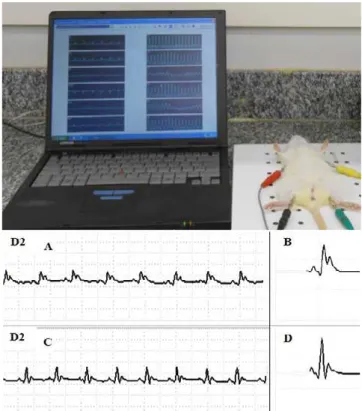
Electrodes were connected to the channels of the electrocardiography device (HeartWare® System) and recorded three bipolar derivations (DI, DII, and DIII) and three ampliied derivations (aVR, aVL, and aVF), with sensibility N and speed of 50 mm/second (Figure 1).
QT interval was measured in ten consecutive beats, from the beginning of the QRS complex to the point of return of the isoelectric T wave deined as TP segment. QT interval was corrected by heart rate using the Bazett formula (QTc = QT/ √RR).
Figure 1. Representation of the HeartWare® system to obtain the
Monitoring of body mass and water and food consumption
he animals’ body mass weight was monitored through fortnightly assessments, using a digital scale (GEHAKA, BG 1000). Group TR was irst weighed before beginning the irst session of swimming.
Every three days, each box with four rats received 450 grams of food and 700 ml of water. At the end of the third day, the remaining food was weighed (digital scale, GEHAKA, BG 1000), calculating the proportional food consumption per animal, while water consumption was calculated by means of a test tube, in order to determine the volume consumed in each box.
Euthanasia of animals
In the following day after electrocardiographic analysis, animals were anesthetized with an intraperitoneal injection of a mixture of Ketalar® (50mg/mL) and Rompun® (2g/100mL), proportion of 1:1, dosage of 0.3mL/100g of body mass. After signals of general anesthesia, the following organs were removed and weighed: adrenal glands, heart, spleen, and kidneys.
To calculate relative weight of organs of animals, we followed the protocol described by Lana, Paulino and Gonçalves13, which divided the weight of each organ (in grams) by the body weight of each animal on the collection day, and multiplied the value obtained by 100. hus, the result was expressed in grams/100 grams of live weight (g/100g LW)
STATISTICAL ANALYSIS
he data collected were tabulated and subsequently analyzed using the statistical package “SPSS version 17.0.” Statistical analysis was preceded by application of the Kolmogorov-Smirnov (KS) test to verify the normality of the data. hus, for comparisons between groups the Student t test for independent samples was applied to data with parametric distribution, while the Mann-Whitney test applied to data with nonparametric distribution. In all cases, p<0.05 was adopted for statistical signiicance.
RESULTS
In comparing the average daily food and water consumption per animal, through the nonparametric statistical test Mann-Whitney, we observed that the value was signiicantly higher (p=0.002 and p=0,045) for group TR (21±2.9g / 45.3±6.5mL) compared to group S (17.3±2.1 g / 40.2±8mL).
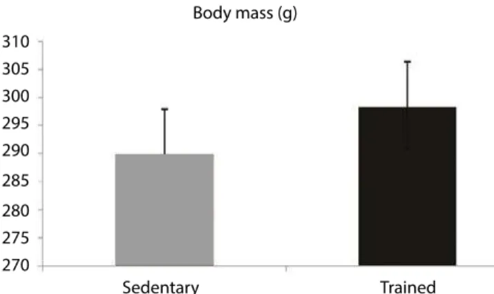
However, as observed in Figure 2, the inal body mass of the animals was similar between groups (Student t parametric test; p>0.05).
Body mass (g)
Sedentary Trained
310 305 300 295 290 285 280 275 270
Figure 2. Body mass (g) of rats in the groups studied. The values correspond to mean ± SD, n=10
Concerning the relative weight of organs, comparison between groups using parametric Student’s t test showed statistically signiicant increase (p<0.05) for kidneys and spleen of female rats submitted to training compared to the sedentary group. Weight of adrenal glands and heart was similar between groups (Figure 3).
0.45 0.4 0.35 0.3 0.25
0.2 0.15 0.1
0.05 0
Relative weight of organs
Sedentary
Trained
Kidneys Spleen Adrenal
glands
Heart
g/100 g L
W
Figure 3. Relative weight of organs of female rats in the groups studied. Values correspond to mean ± SD, n=10. *p<0.05 difers from group S
peak of two consecutive R waves. Parametric Student’s t test was applied, and statistically signiicant mean values that were 15% lower were observed for the TR group (Table 1).
Concerning results obtained for heart angle (SAQRS), we found that female rats from the TR group had a greater statistically signiicant angle (parametric Student’s t test, p<0.05), with approximately 50.6° of diference in relation to rats in the S group, this being considered a more vertical heart (Table 1).
For QTc interval values, which corresponds to the time required to achieve myocardial repolarization, statistically signiicant diferences were not observed (parametric Student’s t test), although, in this case, the TR group had values 22.37% lower compared to the S group, as shown in Table 1.
For data with nonparametric distribution, IPR interval (ms) and SPR segment (ms), the Mann-Whitney test showed no statistically signiicant diferences between groups.
Table 1. Heart rate (HR, beats/min), myocardium angle (SAQRS) and parameters of intervals and segments of electrocardiographic waves (ms) of sedentary (S) and trained (TR) female rats. Values are expressed in mean ± SD, n=10. *p<0.05 difers from group S
HR (beats/min) SAQRS QTc (ms) QRS (ms) IPR (ms) SPR ( ms)
S 324.50±7.4 23.2±6.4 143.25±9.8 45.7±0.7 37.7±1.1 11.1±0.5 TR 275.58±9.0* 73.8±5.7* 111.20±8.6 47.1±1.1 38.3±1.5 12.4±1.4
Furthermore, the parametric Student’s t test showed statistically signiicant diferences in amplitude for the R wave, which corresponds to ventricular activation, and for S wave, which corresponds to the irst negative delection during ventricular depolarization; it was found that the voltage of the R wave was 94% higher, while the S wave was increased by 15% as a result of training (Figure 4).
Sedentary Trained
S Wave R Wave
R and S Wave Amplitude
0.6 0.5 0.4 0.3 0.2 0.1 0
W
a
ve A
mplitude (mV
)
Figure 4. R and S wave amplitude (mV) for the groups studied. Values correspond to mean ± SD, n=10. *p<0.05 difers from group S
DISCUSSION
Long-term swimming training, with no load, three times a week, favored bradycardia at rest, heart verticalization, and increase in amplitude for the R and S waves, thus resulting in cardiovascular itness.
Moreover, we observed that trained female rats consumed a greater amount of food and water; however, body mass was not increased. Training three times a week also promoted increase in mass for kidneys and spleen.
Regarding the animals’ body mass weight, the fact that there were no statistically signiicant diferences between the groups corroborates other studies12–16, which observed no diferences among the body mass weight of animals trained by swimming ive times a week.
Reduction in heart rate at rest is among the main cardiovascular parameters subject to changes due to aerobic exercise, as evidenced by the hypertrophied heart of trained rats10,13.
he expected heart rate of sedentary rats is approximately 344 bpm20. Results of this study show that the S group follows the expected pattern, while the TR group showed an average decrease of 15%, or 48 bpm.
hus, beneicial cardiovascular anatomical adaptations were observed, causing the female rats submitted to training to have bradycardia at rest, corroborating the results of the study of Cardinot et al.21, who evaluated the behavior of blood pressure and heart rate in mice submitted to preventive aerobic training.
cardiovascular system, considering the physical training in rats a good experimental model to study these adaptations.
In order to observe if left ventricular hypertrophy occurred, data of the present study are in accordance with the research described in the literature21, reporting that hypertrophied left ventricles show an increase in amplitude for R wave, which evidences ventricular depolarization.
Although, indications of cardiac hypertrophy were observed in the ECG results from the present study, there was no statistically signiicant diference for relative weight of the heart.
he raise in the R wave voltage can be justiied by the angle of the heart, since it is more vertical in the TR group, thus increasing the R wave amplitude in the D2 variation22. It is assumed that this verticalization is due to left ventricle hypertrophy, so the heart adapts anatomically to surrounding structures. Since the left ventricle internal dimensions uniformly increase due to the overload from exercise, there is an enlargement of the cavity which, subsequently, is followed by hypertrophy in the left ventricle myocardium23.
he S wave is scarcely described in experimental studies; however, in humans, increase in voltage may be a signal of left ventricle hypertrophy23. hus, its increased amplitude in long-term trained female rats suggests a high demand for electricity, in order depolarize a hypertrophied ventricle.
A smaller QTc interval indicates a short or early repolarization24. hus, the reduction of QTc interval (QT interval corrected by Bazett formula) for the TR group, although not statistically signiicant, is considered an important inding in this study. Since the QRS interval between the groups is similar, it is possible to infer that there was a power adjustment in order to support the exercise load, starting the myocardial repolarization at an early stage.
Regarding the efect of regular exercise for prevention of cardiovascular diseases, such as acute myocardial infarction, there are diferences among studies in the literature.
he study conducted by Veiga et al.16 evaluated the efects of myocardial infarction (MI) in rats submitted to prior physical exercise for eight weeks, indicating that exercise by swimming did not attenuate the changes induced by MI in female rats.
In contrast, the study by Brown et al.25 observed reduction in infarction area and preservation of coronary artery low in female rats previously trained for 20 weeks through a running protocol. hese data corroborate the
study by Freimann et al.14, who conducted physical training of swimming followed by induction of MI in male rats, observing considerable reduction in MI area, scar reduction, and increased density of arterioles.
While the objective of this study was not to induce MI, the electrical changes demonstrated by ECG in trained animals result in cardiovascular itness and suggest the efectiveness of this type of training for the prevention of cardiovascular events.
With respect to the weight of organs, the literature shows increase in the relative weight of kidneys of animals only when they are submitted to high-intensity exercise, for an eleven-week period12. hus, the increase in the relative weight of kidneys during physical exercise, even with no load, can be justiied considering the longer training. According to Shizuru et al.26, renal responses resulting from physical exercises are, in fact, related to their time and intensity.
he relative weight of spleen found in this study difers from results in the literature, since because of physical exercise the weight of the organ can be reduced as a result of increased expulsion of blood stored in the structure, when trained and immobilized animals are compared. However, rats had higher immune and endocrine activity, which can be induced by stress27. Aging associated with long-term exercise may have contributed to the relative increase in spleen size.
With regard to the weight of the adrenal gland, we observed no statistically signiicant diference, probably due to the type of training to which the animals were submitted, with no load, since data in the literature indicate that changes in the weight of adrenal glands occur more frequently in response to training with load and high intensity12.
his research enabled better understanding of the aspects related to structural changes in the body of adult female rats, with emphasis on electrical variables of the myocardium, when submitted to physical training, with no load, performed only three times a week for a long period of time. A limitation of the experiment is that no data was collected concerning the blood pressure of animals in both groups.
CONCLUSION
characterizing an efective and preventive cardiovascular physical training.
Acknowledgements
he authors gratefully acknowledge the National Council for Scientiic and Technological Development (CNPq) for the inancial support.
REFERENCES
1. Sá A, Porto A. Envelhecimento e saúde ocupacional. Rev Geriatr. 1990; 3(28):16-24.
2. Degens, H. Age-related skeletal muscle dysfunction: causes and mechanisms. J Musculoskelet Neuronal Interact. 2007;7(3):246-52.
3. Nelson ME, Rejeski WJ, Blair SN, Duncan PW, Judge JO, King AC, et al. Physical activity and public health in older adults: recommendation from the American College of Sports Medicine and the American Heart Association. Med Sci Sports Exerc. 2007;39(8):1435-45.
4. Organização Mundial de Saúde (OMS). O papel da atividade física no Envelhecimento Saudável. Florianópolis: 2006. 5. Barreto ACP, Drumond Neto C, Mady C, Albuquerque DC,
Brindélio Filho DF, Braile DM, et al. Revisão das II diretrizes da Sociedade Brasileira de Cardiologia para o diagnóstico e tratamento da insuiciência cardíaca. Arq Bras Cardiol. 2002;79 (Suppl 4):1-30.
6. Libby P, Theroux P. Pathophysiology of coronary artery disease. Circulation. 2005;111:3481-8.
7. Duncker DJ, Bache RJ. Regulation of coronary blood low during exercise. Physiol Rev. 2008;88(3):1009-86.
8. Negrão CE, Rondon MUPB. Exercício físico, hipertensão e controle barorrelexo da pressão arterial. Rev Bras Hipertens. 2001;8(1):89-95.
9. Brum PC, Forjaz CLM, Tinucci T, Negrão CE. Adaptações agudas e crônicas do exercício físico no sistema cardiovascular. Rev Paul Educ Fís. 2004;18:21-31.
10. Barros JGD, Redondo FR, Zarno FS, Mattos KC, De Angelis K, Irogoyen MC, et al. Treinamento físico de natação promove remodelamento cardíaco e melhora a perfusão sanguínea no músculo cardíaco de SHR via mecanismo dependente de adenosina. Rev Bras Med Esporte. 2011;17(3):193-7.
11. Negrão CE, Moreira ED, Santos MCLM, Farah VMA, Krieger EM. Vagal function impairment after exercise training. J Appl Physiol. 1992;72:1749-53.
12. Lana AC, Paulino CA, Gonçalves DI. Inluência dos exercícios físicos de baixa e alta intensidade sobre o limiar de hipernocicepção e outros parâmetros em ratos. Rev Bras Med Esporte. 2006;15(5):248-54.
13. Medeiros A, Gianolla RM, Kalil LMP, Bacurau RFP, Rosa LFBC, Negrão CE, et al. Efeito do treinamento físico com natação sobre o sistema cardiovascular de ratos normotensos. Rev Paul Educ Fís. 2000;14(1):7-15.
14. Freimann S, Scheinowitz M, Yekutieli D, Feinberg MS, Eldar M, Kessler-Icekson G. Prior exercise training improves the outcome of acute myocardial infarction in the rat. J Am Coll Cardiol. 2005;45(6):931-8.
15. Nascimento CCF, Padula N, Milani JGPO, Shimano AC, Martinez EZ, Mattiello-Sverzut AC. Histomorphometric analysis of the response of rat skeletal muscle to swimming, immobilization and rehabilitation. Braz J Med Biol Res. 2008; 41(9):818-24.
16. Veiga ECA, Portes LA, Bocalini DS, Antonio EL, Santos AA, Santos MH, et al. Repercussões cardíacas após exercício e infarto do miocárdio em ratas submetidas previamente a exercício físico. Arq Bras Cardiol. 2013;100(1):37-43.
17. Oliveira LS, Sobral LL, Takeda SYM, Betini J, Guirro RRJ, Somazz MC, et al. Estimulación eléctrica y natación en la fase aguda de la axonotmesis: inluencia sobre la regeneración nerviosa y la recuperación funcional. Rev Neurol. 2008;47:11-5.
18. Manuel Rosety-Rodríguez, Alejandra Camacho, Miguel Ángel Rosety, Gabriel Fornieles, Antonio Jesús Diaz, Ignacio Rosety, et al. A 6-week training program increase muscle antioxidant system in eldery diabetic fatty rats. Med Sci Monit. 2012;18(9):346-50.
19. Kumar S, Kela AK, Mehta VL, Shukla AK. Preferred anesthetic agents in experimental cardiology: A study on rat electrocardiogram. Indian J Pharmacol. 1995;27:127-9. 20. Santos MRV, Souza VH, Menezes IAC, Bitencurt JL,
Rezende-Neto JM, Barreto AS, et al. Parâmetros bioquímicos, isiológicos e morfológicos de ratos (Rattus novergicus linhagem Wistar) produzidos pelo Biotério Central da Universidade de Sergipe. Scientia Plena. 2010;6(10):1-6.
21. Cardinot T, Moretti A, Mattos K, Brum P, Souza H. Comportamento da pressão arterial e frequência cardíaca em camundongos LDLr-/- submetidos a programa preventivo de treinamento físico aeróbico. Col Pesq Educ Fís. 2013; 12(1):71-8.
22. Póvoa R, Souza D. Análise crítica do eletrocardiograma e do ecocardiograma na detecção da hipertroia ventricular esquerda. Rev Bras Hipertens. 2008; 15(2):81-9.
23. Camarozano A, Rabischofsky A, Maciel BC, Brindeiro Filho D, Horowitz ES, Pena JLB, et al. Diretrizes das indicações da ecocardiograia. Soc Bras Cardiol. 2009;93(6):e265-e302. 24. Corrado D, Basso C, Pavei A, Michieli P, Schiavon M, Thiene G.
Trends in sudden cardiovascular death in young competitive athletes after implementation of a preparticipation screenning program. JAMA. 2006;296:1593-601.
infarct size after ischemia-reperfusion in rat heart. J Appl Physiol. 2003;95:2510-8.
26. Shizuru EM, Freud BJ, Hashiru GM, Clay-Baugh JR. Hormonal electrolyte and renal responses to exercise are intensity dependent. J Appl Physiol. 1991;70: 900-6.