Copyright © 2007 by Sociedade Brasileira de Pediatria
O
RIGINALA
RTICLECoagulation disorder in children and adolescents
with moderate to severe traumatic brain injury
Carolina A. Affonseca,1 Luís Fernando A. Carvalho,2 Sérgio D. Guerra,3
Alexandre R. Ferreira,4 Eugênio M. A. Goulart4
Abstract
Objectives: To describe the epidemiological profile of children and adolescents with moderate to severe traumatic brain injury admitted to an intensive care unit; to describe the frequency of coagulation disorders in these patients; to determine the relationship between coagulopathy and trauma severity; to assess the factors associated with coagulopathy; and to assess the effect of coagulopathy on the mortality of these patients.
Methods:Cross-sectional study with 301 patients aged up to 16 years admitted to an intensive care unit due to moderate to severe traumatic brain injury, carried out over a 5-year period. The coagulation profile was associated with clinical, epidemiological and CT findings. Univariate and multivariate analyses were used to check the association between coagulopathy and mortality.
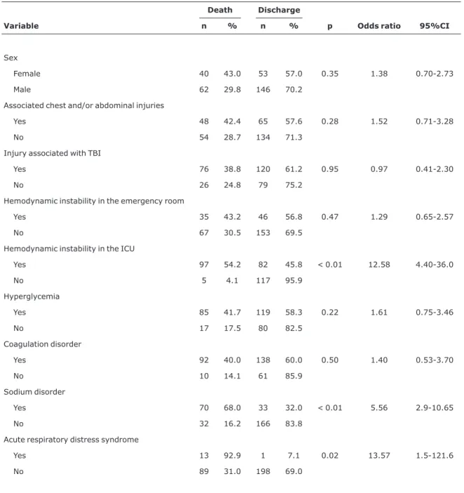
Results:Minimum age was 23 days, and maximum age was 16 years (mean of 7.9 years). About 77% of patients had coagulopathy, whose occurrence was directly associated with the severity of the trauma, but not with the rise in mortality. The factors associated with the presence of coagulopathy were the following: severity of the traumatic brain injury (OR = 2.83; 95%CI 1.58-5.07), diagnosis of brain swelling on cranial computed tomography (OR = 2.11; 95%CI 1.13-4.07) and occurrence of chest and/or abdominal injury (OR = 2.07; 95%CI 1.11-4.00). Approximately 35% of patients died. The multivariate analysis showed that the factors associated with an increased risk of death were presence of sodium disorders (OR = 5.56; 95%CI 2.90-10.65), hypotension in the intensive care unit (OR = 12.58; 95%CI 4.40-35.00) and acute respiratory distress syndrome (OR = 13.57; 95%CI 1.51-121.66).
Conclusion:The development of coagulopathy is a frequent complication in patients with moderate to severe traumatic brain injury. Even though it is not closely associated with death in this study, it may be regarded as a marker of injury severity.
J Pediatr (Rio J). 2007;83(3):274-282:Traumatic brain injury, blood coagulation, child, adolescent.
1. Mestre, Faculdade de Medicina, Universidade Federal de Minas Gerais (UFMG), Belo Horizonte, MG, Brasil. Intensivista pediátrica.
2. Mestre, Faculdade de Medicina, UFMG, Belo Horizonte, MG, Brasil. Intensivista pediátrico. Diarista, UTI Pediátrica, Hospital João XXIII, Belo Horizonte, MG, Brasil.
3. Mestre, Faculdade de Medicina, UFMG, Belo Horizonte, MG, Brasil. Intensivista pediátrico. Coordenador, UTI Pediátrica, Hospital João XXIII, Belo Horizonte, MG, Brasil.
4. Doutor, Faculdade de Medicina, UFMG, Belo Horizonte, MG, Brasil. Professor adjunto, Departamento de Pediatria, Faculdade de Medicina, UFMG, Belo Horizonte, MG, Brasil.
Manuscript received Oct 30 2006, accepted for publication Feb 28 2007.
Suggested citation:Affonseca CA, Carvalho LF, Guerra SD, Ferreira AR, Goulart EM. Coagulation disorder in children and adolescents with moderate to severe traumatic brain injury. J Pediatr (Rio J). 2007;83(3):274-282.
doi 10.2223/JPED.1639
Introduction
Traumatic injuries are one of the major preventable health problems among children. In Brazil, they are the major cause of death among individuals aged 10 to 29 years and account for approximately 40% of deaths among children aged 5 to 9 years and 18% among those aged 1 to 4 years,1
and brain injury is the main cause of death and of sequelae in these age groups.2
Primary brain injury is that which results from the mechanical injury that occurs at the time of the trauma, as a result of the blow to the head. Secondary brain injury is not closely related to the mechanism of injury. It occurs after the initial trauma and is defined as the neuronal injury originating from local or systemic response to the initial injury. Several factors, such as hypotension and hypoxia, have been correlated with the development of secondary injuries.1-8
Other factors, such as the presence of coagulation disorders, are currently being investigated, so as to define their importance among the complex factors that influence the prognosis of patients with traumatic brain injury (TBI).
Since 1974, when Goodnight et al.9 first ascribed
secondary systemic hemostatic failure to acute defibrillation associated with traumatic brain injury that destroyed the brain tissue, several other authors have described the occurrence of coagulopathy in adults and children with TBI,10-19as well as the association between the intensity of
the coagulation disorder and the severity of TBI, which reflects the magnitude of the brain injury.12,16-18
Albeit widely described, the incidence of post-TBI coagulopathy has not been accurately determined among children.16 In 1982, Miner et al.12 reported that 71% of
patients aged 2 to 18 years admitted to the intensive care unit (ICU) due to TBI had abnormal results in at least one coagulation test. Fifteen years later, May et al.,14in a study
with adults, found out that 81% of patients with an ECG score
≤6 on the Glasgow coma scale and that 100% of those with an ECG score < 5 showed laboratory evidence of coagulopathy on admission.
The association between the presence of coagulopathy and a worse prognosis with higher mortality among TBI patients was described by several authors.8,12,13,15-18,20
Since the type and extension of the brain injury determine the prognosis and as such injury is subject to deterioration due to impaired hemostasis, the importance and efficacy of treatment of coagulopathy have been widely discussed.21-23
Goodnight et al.9 suggest that the early treatment of
coagulopathy may have contributed to a better prognosis of adult patients, which was described by other authors some years later.6,14 A study with similar results carried out in
children younger than 16 years was published only in 2001.17
In that same year, however, Vavilala et al.18could not prove
there was a relationship between the treatment of the coagulation disorder and a more favorable prognosis in children.
The aims of the present study are to describe the epidemiological profile of children and adolescents with moderate to severe traumatic brain injury admitted to an ICU; to describe the frequency of coagulation disorders in these patients; to determine the relationship between coagulopathy and trauma severity; to assess the factors associated with coagulopathy; and to assess the effect of coagulopathy on the mortality of these patients.
Methods
This cross-sectional study consisted of a two-step analysis of the medical records of 318 children and adolescents diagnosed with moderate TBI (ECG score between 9 and 13) or severe TBI (ECG score ≤ 8) and admitted to the pediatric ICU of Hospital João XXIII between 09/01/1998 and 08/31/2003.
Those patients whose medical records did not provide any information on the ECG score at hospital admission or on the results of coagulation tests performed in the first 24 hours of admission were excluded; and so were those patients whose injury had been caused by penetrating trauma and also those aged over 16 years who had been occasionally admitted to a pediatric ICU. None of the patients had a past history of coagulation disorders or vitamin K deficiency.
João XXIII is a public hospital that belongs to the Hospital Foundation of the State of Minas Gerais, Brazil. It is a referral center for the emergency treatment of adults and children with trauma, severe exogenous poisonings, burns, and venomous animal bites or stings, and it attends to approximately 150,000 patients a year. About 250 children and adolescents are admitted to the ICU every year; of these, 80 are brain injury victims.
During their stay in the ICU, patients with TBI are treated according to the guidelines published in consensus agreements that are based on the current literature and revised regularly. The last revision was made in 2003.2,24
The blood sample is handled using an automated system. Quality control is performed on a daily basis, and the equipment is calibrated every time reagents are changed, in case of coagulation tests, or every week in case of platelet count.
Coagulation disorder is diagnosed when PA is lower than 70% and/or the PT is greater than 16 seconds and/or the aPTT is greater than 10 seconds when compared to controls and/or the total platelet count is less than 150,000/mm3.
A coagulation test is therefore performed every 12 hours until test results are normal.
The data were collected in a two-step procedure. Initially, the data about patients admitted to Hospital João XXIII between September 1998 and December 2001 were collected from medical records stored at the Division of Medical and Statistical Files (Serviço de Arquivo Médico e Estatístico, SAME), between August 2001 and September 2002. Further data were collected between January 2002 and August 2003 by the daily analysis of medical records of ICU patients. The study protocol was approved by the Center for Teaching and Research of Hospital João XXIII, by the Research Ethics Committee of the Hospital Foundation of the State of Minas Gerais, by the Department Board of the Department of Pediatrics of the School of Medicine of Universidade Federal de Minas Gerais (UFMG) and by the Research Ethics Committee of UFMG.
A written informed consent was obtained from parents or surrogates, whereby they authorized the participation of their children in the study.
Since this study includes all patients who met the inclusion criteria (convenience sample), the sample size was not calculated.
The statistical analysis was made using Epi-Info. The chi-square test was used for comparison of ratios, and Fisher’s exact test was used whenever necessary. The chi-square test for linear trend was used in cases of progression. The analysis of variance was used to compare the means of normally distributed data, with the use of non-parametric tests when variances were not homoge-neous. Variables with p < 0.25 in the univariate analysis were simultaneously reassessed in the logistic regression used for the multivariate analysis through the MULTLR program. In the final model, the variables with a p value < 0.05 were considered to be statistically significant.
Results
A total of 318 patients with moderate to severe TBI were admitted to the ICU during the study period. Of these patients, 17 were excluded from the analysis: four whose ECG score on admission was unknown; eight who had penetrating brain injury; three whose coagulation test results
were not retrieved; and two whose ages were greater than 16 years. The study group therefore included 301 patients.
Minimum age was 23 days, and maximum age was 16 years, with mean age of 7.9±4.3 years and median of 7.8 years. Of the analyzed patients, 69.1% were male. The length of hospital stay averaged 10.9 days for those who had been discharged from the ICU, and 5.5 days for those who eventually died. The major mechanisms of injury associated with moderate to severe TBI were: motor vehicle-pedestrian collisions (45.8%), falls (20.3%), car accidents (15.6%) and bicycle accidents (9.3%). In 2.7% of cases, there was intentional injury, i.e., physical violence.
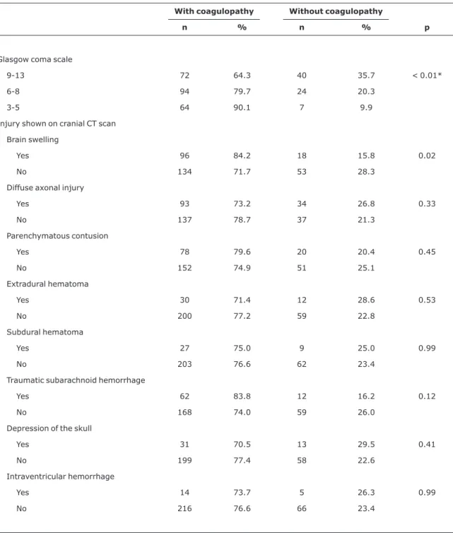
As to the severity of TBI, 112 patients (37.2%) showed moderate injury and 189 (62.8%) had severe injury. Among those patients with severe TBI, 71 (23.6%) showed abnormal motor behavior regarding flexion or extension or lack of motor response (ECG score 3 to 5) on hospital admission. Among those patients with moderate TBI, 21 patients (18.7%) died, whereas the mortality rate among patients with severe TBI amounted to 43.9% (83 patients). The frequency of coagulopathy corresponded to 76.4%, and was closely related to the severity of TBI (Table 1).
The main injuries observed on cranial CT were: diffuse axonal injury (42.2%); brain swelling (37.9%); and intraparenchymatous contusions (32.6%), but the same patient may have one or more findings on the CT scan. The distribution of coagulopathy according to intracranial injuries is shown in Table 1.
Most patients had injuries associated with TBI. Sixty-five percent of patients were diagnosed with injuries in other parts of the body, such as chest, abdomen, extremities, and spinal column (spinal cord injury).
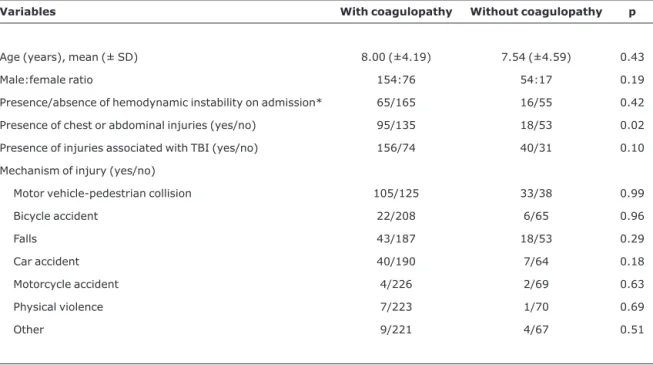
The presence of coagulopathy was not statistically different between the patients with other injuries associated with TBI and those with TBI only (p = 0.10). By separately analyzing patients with chest and abdominal injuries, one notes that the frequency of coagulation disorders is significantly higher in this group than in patients who were not diagnosed with these injuries (p = 0.02).
By separately analyzing the groups with and without coagulation disorders, one perceives that they share similarities in terms of mean age, distribution by sex, presence of hemodynamic instability on admission, presence of injuries associated with TBI and mechanism of injury (Table 2).
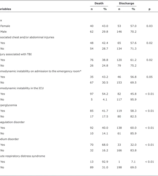
The univariate analysis showed that female patients were statistically more likely to die than their male counterparts (p = 0.03). Likewise, according to this same analysis, the
following factors played a role in the rise in mortality: presence of injuries associated with TBI; presence of hemodynamic instability on hospital admission and during
Table 1- Frequency of coagulation disorders according to the Glasgow coma scale on hospital admission and based on the main CT scan findings of patients with moderate to severe traumatic brain injury
With coagulopathy Without coagulopathy
n % n % p
Glasgow coma scale
9-13 72 64.3 40 35.7 < 0.01*
6-8 94 79.7 24 20.3
3-5 64 90.1 7 9.9
Injury shown on cranial CT scan
Brain swelling
Yes 96 84.2 18 15.8 0.02
No 134 71.7 53 28.3
Diffuse axonal injury
Yes 93 73.2 34 26.8 0.33
No 137 78.7 37 21.3
Parenchymatous contusion
Yes 78 79.6 20 20.4 0.45
No 152 74.9 51 25.1
Extradural hematoma
Yes 30 71.4 12 28.6 0.53
No 200 77.2 59 22.8
Subdural hematoma
Yes 27 75.0 9 25.0 0.99
No 203 76.6 62 23.4
Traumatic subarachnoid hemorrhage
Yes 62 83.8 12 16.2 0.12
No 168 74.0 59 26.0
Depression of the skull
Yes 31 70.5 13 29.5 0.41
No 199 77.4 58 22.6
Intraventricular hemorrhage
Yes 14 73.7 5 26.3 0.99
No 216 76.6 66 23.4
the ICU stay; presence of metabolic disorders, such as hyperglycemia or sodium disorders; and development of acute respiratory distress syndrome (Table 3).
However, according to the multivariate analysis of all these factors, only sodium disorders, hypotension during ICU stay and acute respiratory distress syndrome were significantly associated with a rise in mortality (Table 4).
Discussion
Brain injury is highly prevalent among children who suffer head trauma, and its incidence has increased year after year. When not fatal, it causes injuries that result in severe or incapacitating sequelae.
Despite continuous improvement in resuscitation techniques, in intensive care, and in neurological monitoring, the prognosis of children with severe TBI has been dismal. This fact is corroborated by the high mortality rate (around 34%) observed, which is quite similar to that described in the literature.7,20
When conjointly analyzed, motor vehicle-pedestrian collisions account for over 70% of cases of moderate to severe TBI in children and adolescents. Such association has also been demonstrated by other authors,7,8,16,20 who
regard falls as another cause of this type of injury.
Coagulation disorders, the major object of investigation in this study, were observed in 77% of patients and their prevalence, as previously pointed out by other authors,12,17
was closely related to the severity of trauma, whose classification is based on an ECG score that shows the intensity of the brain injury.
Patients with moderate or severe TBI with other associated traumas did not usually show a prevalence of coagulopathy greater than the one presented by patients with TBI only, except when the group of patients with TBI associated with chest and/or abdominal injuries, in whom the occurrence of coagulation disorders was twice as high, was compared to the group of patients with TBI, but with no associated injuries. This fact, already described by other authors,12can be explained by analyzing the mechanism of
Table 2- Comparison of general characteristics between patients with moderate to severe traumatic brain injury, based on the presence or absence of coagulation disorders
Variables With coagulopathy Without coagulopathy p
Age (years), mean (± SD) 8.00 (±4.19) 7.54 (±4.59) 0.43
Male:female ratio 154:76 54:17 0.19
Presence/absence of hemodynamic instability on admission* 65/165 16/55 0.42
Presence of chest or abdominal injuries (yes/no) 95/135 18/53 0.02
Presence of injuries associated with TBI (yes/no) 156/74 40/31 0.10
Mechanism of injury (yes/no)
Motor vehicle-pedestrian collision 105/125 33/38 0.99
Bicycle accident 22/208 6/65 0.96
Falls 43/187 18/53 0.29
Car accident 40/190 7/64 0.18
Motorcycle accident 4/226 2/69 0.63
Physical violence 7/223 1/70 0.69
Other 9/221 4/67 0.51
injury that most likely causes coagulopathy in TBI: tissue injury with release of thromboplastin, which activates the coagulation cascade. Not only the brain, but also the lungs and liver contain a large amount of thromboplastin in their tissues.11 The lack of statistical significance between the
occurrence of injuries associated with TBI and the presence of
coagulopathy is actually due to the fact that the skeletal muscles are predominantly affected, whose injury does not directly cause coagulation disorders.
The role of coagulopathy in the prognosis of these patients has not been appropriately clarified in the literature.
Table 3- Univariate analysis of factors associated with the mortality of patients with moderate to severe traumatic brain injuries admitted to the intensive care unit
Death Discharge
Variables n % n % p
Sex
Female 40 43.0 53 57.0 0.03
Male 62 29.8 146 70.2
Associated chest and/or abdominal injuries
Yes 48 42.4 65 57.6 0.02
No 54 28.7 134 71.3
Injury associated with TBI
Yes 76 38.8 120 61.2 0.02
No 26 24.8 79 75.2
Hemodynamic instability on admission to the emergency room*
Yes 35 43.2 46 56.8 0.05
No 67 30.5 153 69.5
Hemodynamic instability in the ICU
Yes 97 54.2 82 45.8 < 0.01
No 5 4.1 117 95.9
Hyperglycemia
Yes 85 41.7 119 58.3 < 0.01
No 17 17.5 80 82.5
Coagulation disorder
Yes 92 40.0 138 60.0 < 0.01
No 10 14.1 61 85.9
Sodium disorder
Yes 70 68.0 33 32.0 < 0.01
No 32 16.2 166 83.8
Acute respiratory distress syndrome
Yes 13 92.9 1 7.1 < 0.01
No 89 31.0 198 69.0
In the present study, after the multivariate analysis of the factors associated with the mortality of patients with moderate to severe TBI, the presence of coagulopathy was not considered to play an important role in the increase in
mortality. During the review of the literature, such association was found and described by several authors. Some ascribed the rise in mortality to the presence of coagulopathy, even if mild.6,11,13,14,17,20
Table 4- Multivariate analysis of factors associated with the mortality of patients with moderate to severe traumatic brain injuries admitted to the intensive care unit
Death Discharge
Variable n % n % p Odds ratio 95%CI
Sex
Female 40 43.0 53 57.0 0.35 1.38 0.70-2.73
Male 62 29.8 146 70.2
Associated chest and/or abdominal injuries
Yes 48 42.4 65 57.6 0.28 1.52 0.71-3.28
No 54 28.7 134 71.3
Injury associated with TBI
Yes 76 38.8 120 61.2 0.95 0.97 0.41-2.30
No 26 24.8 79 75.2
Hemodynamic instability in the emergency room
Yes 35 43.2 46 56.8 0.47 1.29 0.65-2.57
No 67 30.5 153 69.5
Hemodynamic instability in the ICU
Yes 97 54.2 82 45.8 < 0.01 12.58 4.40-36.0
No 5 4.1 117 95.9
Hyperglycemia
Yes 85 41.7 119 58.3 0.22 1.61 0.75-3.46
No 17 17.5 80 82.5
Coagulation disorder
Yes 92 40.0 138 60.0 0.50 1.40 0.53-3.70
No 10 14.1 61 85.9
Sodium disorder
Yes 70 68.0 33 32.0 < 0.01 5.56 2.9-10.65
No 32 16.2 166 83.8
Acute respiratory distress syndrome
Yes 13 92.9 1 7.1 0.02 13.57 1.5-121.6
No 89 31.0 198 69.0
When the studies published by the authors who found this association are analyzed in more detail, one observes that the factors that could have contributed to a larger number of deaths among TBI patients were not assessed.
Hulka et al.,11who analyzed 159 adult patients in 1996,
stated that TBI patients who developed coagulopathy were nine times more likely to die than those who did not develop it. However, only one univariate analysis was performed, and other factors associated with the prognosis, such as trauma associated with TBI, hypotension, and metabolic disorders, were not assessed. In 2001, Keller et al.17assessed children
with TBI and noticed that those with coagulopathy were at a higher risk of death. Nevertheless, as they used a small sample (n = 53) and performed a descriptive analysis, their data were not statistically significant. Quite recently, two studies with pediatric patients were published, in which an attempt was made at correlating the presence of coagulation disorder with prognosis. The first study, which assessed a small number of patients (n = 51), shows a statistically significant correlation between coagulopathy and prognosis (p < 0.001). However, once again, other important prognostic variables were not assessed, and only a univariate analysis was carried out.20The first study that included a
larger number of variables and used a multivariate analysis was the one conducted by Chiaretti et al.,8published in 2002.
These authors found a significant correlation between the development of disseminated intravascular coagulation and a worse prognosis (p = 0.03). Thus, there is a paucity of methodologically well-designed studies in the literature investigating the effect of coagulation disorder on mortality of TBI patients and including the multiple factors associated with it.
Therefore, the question about the role of coagulopathy in the increase in the mortality of TBI patients remains unanswered. According to the findings of the present study, coagulation disorder is not a determining factor of mortality, but a marker of the severity of brain injury, which means that patients with such a disorder should be more closely and intensively monitored.
The literature describes that the appropriate initial treatment of patients with head trauma seems to play a crucial role in the final prognosis.8,14Thus, the follow-up of
patients with moderate to severe TBI should include the analysis of their coagulation profile, in addition to maintaining homeostasis and appropriate brain perfusion, so that coagulopathy, which is highly prevalent in this group, can be treated properly and timely. Besides contributing to the judicious use of hospital resources, this also avoids delayed treatment, which may positively influence the neurological prognosis of these patients.
A limitation of this study is that some of the data were obtained retrospectively. Despite the careful collection of
data and filling out of forms, important information about some patients could not be retrieved.
References
1. Guerra SD, Jannuzzi MA, Moura AD. Traumatismo
cranioencefálico em pediatria. J Pediatr (Rio J). 1999;75 Supl 2:S279-93.
2. Marik PE, Varon J, Trask T.Management of head trauma. Chest. 2002;122:699-711.
3. Mazzola CA, Adelson PD.Critical care management of head trauma in children. Crit Care Med. 2002;30(11 Suppl):S393-401.
4. Colégio Americano de Cirurgiões. Suporte avançado de vida no trauma: programa para médicos. São Paulo: Colégio Americano de Cirurgiões; 1997.
5. American College of Surgeons. Basic and advanced prehospital life support. 4th ed. Chicago: American College of Surgeons; 1999.
6. Piek J, Chesnut RM, Marshall LF, van Berkum-Clark M, Klauber MR, Blunt BA, et al.Extracranial complications of severe head injury. J Neurosurg. 1992;77:901-7.
7. Kokoska ER, Smith GS, Pittman T, Weber TR.Early hypotension worsens neurological outcome in pediatric patients with moderately severe head trauma. J Pediatr Surg. 1998;33:333-8.
8. Chiaretti A, Piastra M, Pulitano S, Pietrini D, De Rosa G, Barbaro R, et al.Prognostic factors and outcome of children with severe head injury: an 8-year experience. Childs Nerv Syst. 2002;18:129-36.
9. Goodnight SH, Kenoyer G, Rapaport SI, Patch MJ, Lee JA, Kurze T. Defibrination after brain-tissue destruction: a serious complication of head injury. N Engl J Med. 1974;290:1043-7.
10. Jacoby RC, Owings JT, Holmes J, Battistella FD, Gosselin RC, Paglieroni TG.Platelet activation and function after trauma. J Trauma. 2001;51:639-47.
11. Hulka F, Mullins RJ, Frank EH.Blunt brain injury activates the coagulation process. Arch Surg. 1996;131:923-7; discussion 927-8.
12. Miner ME, Kaufman HH, Graham SH, Haar FH, Gildenberg PL. Disseminated intravascular coagulation fibrinolytic syndrome following head injury in children: frequency and prognostic implications. J Pediatr. 1982;100:687-91.
13. Hymel KP, Abshire TC, Luckey DW, Jenny C.Coagulopathy in pediatric abusive head trauma. Pediatrics. 1997;99:371-5.
14. May AK, Young JS, Butler K, Bassam D, Brady W.Coagulopathy in severe closed head injury: is empiric therapy warranted?Am Surg. 1997;63:233-6; discussion 236-7.
15. Becker S, Schneider W, Kreuz W, Jacobi G, Scharrer I, Nowak-Gottl U.Post-trauma coagulation and fibrinolysis in children suf-fering from severe cerebro-cranial trauma. Eur J Pediatr. 1999;158 Suppl 3:S197-202.
16. Chiaretti A, Pezzotti P, Mestrovic J, Piastra M, Polidori G, Storti S, et al. The influence of hemocoagulative disorders on the outcome of children with head injury. Pediatr Neurosurg. 2001;34:131-7.
18. Vavilala MS, Dunbar PJ, Rivara FP, Lam AM. Coagulopathy predicts poor outcome following head injury in children less than 16 years of age. J Neurosurg Anesthesiol. 2001;13:13-8.
19. Sorensen JV, Jensen HP, Rahr HB, Borris LC, Lassen MR, Fedders O, et al.Haemostatic activation in patients with head injury with and without simultaneous multiple trauma. Scand J Clin Lab Invest. 1993;53:659-65.
20. Pfenninger J, Santi A.Severe traumatic brain injury in children -are the results improving?Swiss Med Wkly. 2002;132:116-20.
21. Stein SC, Young GS, Talucci RC, Greenbaum BH, Ross SE. Delayed brain injury after head trauma: significance of coagulopathy. Neurosurgery. 1992;30:160-5.
22. Stein SC, Spettell CM.Delayed and progressive brain injury in children and adolescents with head trauma. Pediatr Neurosurg. 1995;23:299-304.
23. Oertel M, Kelly DF, McArthur D, Boscardin WJ, Glenn TC, Lee JH, et al.Progressive hemorrhage after head trauma: predictors and consequences of the evolving injury. J Neurosurg. 2002;96:109-16.
24. Carney NA, Chesnut RM, Kochanek PM; American Association for Surgery of Trauma; Child Neurology Society; International Society for Pediatric Neurosurgery; et al. Guidelines for the acute medical management of severe traumatic brain injury in infants, children, and adolescents. Pediatr Crit Care Med. 2003;4(3 Suppl):S1.
Correspondence:
Carolina de Araújo Affonseca