Assessment of cardiac influence on peripheral
vascular Doppler in the elderly
Avaliação da influência de alterações cardíacas na ultrassonografia
vascular periférica de idosos
Alcides José Araújo Ribeiro1
*
, Andréa Campos de Oliveira Ribeiro2, Márcia Marisia Maciel Rodrigues3, Sandra de Barros Cobra Negreiros3, Ana Cláudia Cavalcante Nogueira3, Osório Luís Rangel Almeida3,
José Carlos Quináglia e Silva3, Ana Patrícia de Paula4
Abstract
Background: Heart diseases can cause changes to vascular ultrasonography (VUS) waveforms in peripheral vessels. hese changes are typically bilateral and systemic, they have been little studied, and little is known about them.
Objective: To assess peripheral VUS waveforms in elderly patients in order to identify changes caused by heart diseases. Methods: During 2014, a total of 183 elderly patients were examined with peripheral VUS and the results were analyzed. Results: he sample comprised 102 women (55.7%) and 81 men (44.3%) with ages ranging from 60 to 91 years (mean of 70.4±7.2 years). Abnormalities were identiied in VUS waveforms in 84 patients (45.9%). A total of 138 abnormalities were identiied and classiied into eight of the 13 categories described in the literature, as follows: arrhythmia, systolic pulsus bisferiens, low peak systolic velocity, pulsatile low in femoral veins, bradycardia, tachycardia, pulsus tardus et parvus and pulsus alternans. here was low agreement between presence/absence of VUS abnormalities and cardiological assessments. Analysis of speciic abnormalities revealed variable levels of agreement between VUS and cardiological assessments, ranging from good for tachycardia, moderate for arrhythmia, to low for bradycardia. here was no agreement between VUS and cardiological examinations for the remaining categories of abnormalities. Conclusions: Certain cardiac abnormalities can be identiied in elderly patients by analysis of peripheral VUS waveforms. It is important to recognize and report the presence of these abnormalities because there is a possibility that they may serve to signal hitherto unidentiied diagnoses in these patients. However, further studies are needed to determine the importance of changes to peripheral Doppler waveforms to recognition of heart diseases.
Keywords: Doppler ultrasonography; heart diseases; diagnosis; aortic valve insuiciency; vein femoral; pulsating low.
Resumo
Contexto: As cardiopatias podem causar alterações no formato das ondas da ultrassonograia vascular (UV) em vasos periféricos. Essas alterações, tipicamente bilaterais e sistêmicas, são pouco conhecidas e estudadas. Objetivo: Avaliar as ondas periféricas da UV de pacientes idosos para identiicar alterações decorrentes de cardiopatias. Métodos: Foram estudados 183 pacientes idosos submetidos a UV periférica no ano de 2014. Resultados: Foram avaliados 102 mulheres (55,7%) e 81 homens (44,3%) com idade entre 60 e 91 anos (média de 70,4±7,2 anos). Encontraram-se alterações pela UV em 84 pacientes (45,9%). Foram identiicadas 138 alterações de oito dos 13 tipos descritos na literatura: arritmia, onda bisferiens de pico sistólico, baixa velocidade de pico sistólico, pulsatilidade em veias femorais, bradicardia, taquicardia, onda de pulso parvus tardus e onda de pulso alternans. Houve baixa concordância entre a presença e a não presença de alterações na UV e na avaliação cardiológica. Na análise especíica das alterações, os exames tiveram uma concordância variável, que foi boa para o achado de taquicardia, moderada para arritmia e baixa para bradicardia. Não houve concordância entre a UV e os exames cardiológicos para as demais alterações. Conclusões: É possível identiicar determinadas alterações cardíacas em idosos por meio da análise do formato das ondas periféricas da UV. É importante reconhecer e relatar a presença dessas alterações, pela possibilidade de alertar para um diagnóstico ainda não identiicado nesses pacientes. Entretanto, mais estudos são necessários para que seja deinida a importância das alterações no formato das ondas Doppler periféricas no reconhecimento de cardiopatias.
Palavras-chave: ultrassonograia Doppler; cardiopatias; diagnóstico; insuiciência da valva aórtica; veia femoral; luxo pulsátil.
1 Hospital de Base do Distrito Federal – HBDF, Unidade de Cirurgia Vascular e Angiologia, Brasília, DF, Brazil. 2 Clínica Villas Boas, Brasília, DF, Brazil.
3 Hospital de Base do Distrito Federal – HBDF, Unidade de Cardiologia, Brasília, DF, Brazil. 4 Hospital de Base do Distrito Federal – HBDF, Brasília, DF, Brazil.
Financial support: None.
Conlicts of interest: No conlicts of interest declared concerning the publication of this article. Submitted: April 23, 2015. Accepted: June 20, 2016.
INTRODUCTION
According to the federal government of Brazil, cardiovascular diseases are responsible for 29.4% of all deaths recorded each year, which places Brazil among the countries with the highest mortality rates due to these diseases.1
Heart diseases are common among elderly patients who undergo VUS. As such, when interpreting peripheral Doppler waveforms from these patients, the fact that cardiac function could be abnormal should be taken into account, since this could cause abnormalities in the spectral waves of peripheral examinations.2
Vascular ultrasound low assessments should take
into account elements of cardiovascular physiology, including cardiac rhythm and function and parameters associated with preload and afterload. Changes to cardiac rhythm and systolic and/or diastolic function, presence of valve disease, and the hemodynamic conditions under which VUS was conducted should
also be considered when interpreting low patterns.3,4
Standards for normal peripheral VUS waves were published in 1985. Later, other patterns related to vascular diseases were recognized.5
By deinition, the possible changes to peripheral
VUS examination waveforms resulting from cardiac effects tend to be systemic and bilateral, but they are neither well publicized nor well known. In the great majority of cases these signs go unnoticed during examinations and when examiners do recognize them they very often ignore them and do not include them in their examination reports.
The following cardiac-induced changes to peripheral VUS waveforms are described in the literature: arrhythmia2,6,7 (Figures 1, 2 and 3), pulsatile waves in
the common femoral and popliteal veins2,8 (Figure 4),
systolic pulsus bisferiens6,9-11 (Figures 3 and 5), low
peak systolic velocity,1 bradycardia2 (Figure 5),
tachycardia,2 pulsus alternans,12 pulsus tardus et
parvus,11-13 water hammer pulse,9 systolic spike and
dome,13 elevated peak systolic velocity caused by
high cardiac output,10 pulsus paradoxus,14 and waves
caused by cardiac assistance devices.11,12,15
The objectives of this study were to evaluate the presence of these spectral changes in VUS waveforms from elderly patients who underwent vascular arterial and/or venous echography and to analyze the degree
of agreement between these indings and cardiological
examinations and diagnoses.
METHODS
The study was conducted at the Hospital de Base do Distrito Federal (HBDF), in Brasília, DF, Brazil, from December 2013 to December 2014, after
prior approval from the Ethics Committee. It is a cross-sectional and analytical study of a sample comprising 183 elderly patients.
Inclusion criteria: patients aged over 60 years, who underwent peripheral VUS, who were capable of providing the information needed, who were able to attend for cardiological assessments when requested, and who signed a free and informed consent form. Exclusion criteria: patients with hemodynamic instability, and/or who were unable to provide the information
Figure 5. Pulsus bisferiens and bradycardia.
Figure 1. Atrial ibrillation.
Figure 2. Atrial ibrillation.
Figure 3. Pulsus bisferiens and extrasystole.
needed and attend for cardiological assessments when requested. These patients were subdivided into two subsets. Group I comprised 133 patients without prior diagnoses of heart disease, 72 (54.13%) of whom had already been scheduled for VUS and were invited to attend for examinations and 61 (45.87%) of whom who had not been scheduled for VUS, but were invited to take part in the study during assessments at HBDF internal medicine clinics, when they were referred for carotid screening tests. Group II comprised 50 patients with prior diagnoses of heart disease made by a cardiologist. For both groups, the examiner who performed the ultrasound examinations was unaware of the patients’ prior medical histories. Patients in group I were referred for cardiological assessment after their VUS examinations unless they provided evidence of a cardiological consultation during the preceding 30 days. The cardiology consultant was unaware of the VUS results and when necessary requested routine supplementary cardiological tests, conducted at the HBDF cardiology unit. Electrocardiograms were conducted for all patients in the study.
Ultrasonography protocol
The following devices were used for VUS: a Toshiba Aplio 50 (Toshiba, Japan), a Sonosite M-Turbo (Sonosite, United States), and an Aloka SSD-1700 DYNAVIEW II (Aloka, Japan), with a linear 4 to 7 MHz transducer and unheated gel. All examinations were conducted by a specialist in
angiology and vascular surgery, certiied to practice
Doppler vascular echography. Examinations were conducted with patients in decubitus dorsal, the leg
in a passive position at neutral rotation, slight lexion
of the knee and rotation of the head contralateral to the side being studied. The spectral patterns of the Doppler waves were analyzed for six arteries (right and left common carotid arteries, right and left brachial arteries, and right and left common femoral arteries) in order to enable assessment of bilateralism and presence of systemic arterial involvement, and the right and left common femoral veins to enable assessment of cardiac repercussions.
Statistical analysis
The sample was described statistically in terms of mean and standard deviation for age and in terms of frequencies for all other data. Agreement between the results of VUS and of cardiological tests was assessed using the Kappa index of agreement. This was categorized according to the Landis & Kopp
classiication, in which agreement ranging from 0 to 0.2 is considered insigniicant; from 0.21 to 0.40 is low;
from 0.41 to 0.60 is moderate; from 0.61 to 0.80 is good; and agreement indices in the range 0.81 to 1.00
are excellent.16,17
The statistical analyses in this study were conducted using IBM SPSS 20 (Statistical Package for the Social Sciences, Chicago, United States).
RESULTS
A total of 183 patients (102 women and 81 men) aged from 60 to 91 years, with a mean age of 70.4 (±7.2) years, were assessed. The majority of these patients (57.4%) did not have a prior history of heart
disease. Eight of the 13 abnormal VUS indings
described in the literature were detected.2-15 Abnormal
VUS waveforms were found in 84 patients (45.9%), 54 (40.6%) of whom were in group I and 30 (60%) of whom were in group II. Within group I, there was
a larger number of abnormal indings among patients
for whom tests for a range of different pathologies had already been requested (63.6%) than among those patients who had been invited to take part at the clinic
(36.36%). The 138 abnormal indings are listed in
Table 1. Just one abnormality of the Doppler waveforms was observed in 62 of the patients (72.94%), while 13 (15.29%) patients had two abnormalities, seven (8.23%) patients had three, and two (2.35%) patients
had four abnormal indings. Just 24 of the patients who
underwent the VUS examination did not complete their cardiological assessments and were therefore excluded from the analysis of agreement. There was a low level of agreement between presence/absence of
abnormal VUS indings and the results of cardiological
assessment (Kappa = 0.251). Agreement for group II
was insigniicant (Kappa = 0.109), because almost all
of the patients exhibited some type of abnormality on cardiological assessment, whereas VUS only detected abnormalities in half of the patients. Sixty percent of the patients who exhibited Doppler waveform abnormalities had a prior history of heart disease. When agreement for each type of abnormality was analyzed separately the level of agreement varied,
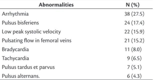
Table 1. Abnormalities of Doppler waveforms.
Abnormalities N (%)
Arrhythmia 38 (27.5)
Pulsus bisferiens 24 (17.4)
Low peak systolic velocity 22 (15.9)
Pulsating low in femoral veins 21 (15.2)
Bradycardia 11 (8.0)
Tachycardia 9 (6.5)
Pulsus tardus et parvus 7 (5.1)
with good agreement for tachycardia (Kappa = 0.66), moderate for arrhythmia (Kappa = 0.494), and low for bradycardia (Kappa = 0.264). There was no agreement between the other VUS waveform abnormalities and cardiological test results (Table 2), but when agreement between VUS abnormalities and other cardiological
indings and diagnoses related to the abnormality in
question, there was a greater level of agreement for pulsus tardus et parvus, pulsus bisferiens and pulsus alternans waves (Table 3). The levels of agreement
between cardiac indings and the pulsus bisferiens
and pulsus tardus et parvus waves were considered
insigniicant (Kappa = 0.135 and 0.104, respectively),
but the correlations between pulsus bisferiens and aortic
insuficiency, and between pulsus tardus et parvus
and aortic valve sclerosis were higher, increasing
from insigniicant to low (Kappa = 0.224 and 0.265,
respectively). There was no agreement between VUS
indings of pulsus alternans and cardiological assessment, but when correlated with other cardiological indings and diagnoses, agreement increased to insigniicant
(Kappa = 0.003). The following abnormalities were not detected: water hammer pulse, systolic spike and dome, elevated peak systolic velocity due to high cardiac output, pulsus paradoxus, and waves caused by cardiac assistance devices.
DISCUSSION
This study demonstrates that analysis of peripheral Doppler waveforms from elderly patients can suggest diagnoses of cardiac diseases or abnormalities. As far as we are aware, there are no similar studies in the
literature, comparing Doppler waveform indings
with cardiological assessment and this study is the
irst to investigate all 13 of the abnormalities that
have been described to date. The overall agreement between presence/absence of VUS abnormalities and cardiological assessment results was low. The number of abnormalities found was higher in the subset of cardiac disease patients than in the subset of patients with no previous diagnosis of heart disease (60% vs. 41.35%), as was expected. There were higher numbers of both
abnormal VUS indings and of abnormal cardiological assessment indings among the patients with prior
history of heart diseases. Agreement between each
speciic type of VUS abnormality and cardiological
assessment results was variable, with higher levels of agreement for tachycardia, arrhythmia, and bradycardia. There was initially no agreement between VUS and the cardiological tests for the other types of VUS
abnormality, but when other cardiological indings and diagnoses that had a relationship with the speciic
abnormalities in question were taken into account, the levels of agreement for pulsus tardus et parvus,
Table 3. Agreement between tests and groups of additional cardiological indings.
Symptom
Doppler ultrasonography
(n = 159)
Cardiological tests (n = 159)
Kappa
Arrhythmia/Chagas heart disease/cardiac thrombus 21.4% 20.1% 0.464
Pulsating low in femoral vein/tricuspid insuiciency 11.3% 25.8% 0.175
Pulsus bisferiens/aortic insuiciency 12.6% 23.3% 0.224
Low peak systolic velocity/myocardial ischemia/low ejection fraction/hypo-contractility/coronary stenosis/ myocardial hypoperfusion
5.7% 24.5% -0.010
Bradycardia 6.9% 1.9% 0.264
Tachycardia 1.9% 1.9% 0.66
Pulsus alternans/myocardial ischemia/low ejection fraction/coronary steno-sis/hypoperfusion
2.5% 23.3% 0.003
Pulsus tardus et parvus/aortic valve sclerosis 3.8% 12.6% 0.265
Water hammer low 0.0% 23.3%
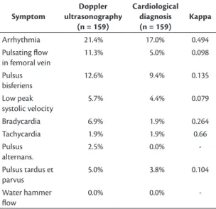
-Table 2. Agreement between Doppler ultrasonography indings
and cardiological diagnoses.
Symptom
Doppler ultrasonography
(n = 159)
Cardiological diagnosis (n = 159)
Kappa
Arrhythmia 21.4% 17.0% 0.494
Pulsating low in femoral vein
11.3% 5.0% 0.098
Pulsus bisferiens
12.6% 9.4% 0.135
Low peak systolic velocity
5.7% 4.4% 0.079
Bradycardia 6.9% 1.9% 0.264
Tachycardia 1.9% 1.9% 0.66
Pulsus alternans.
2.5% 0.0%
-Pulsus tardus et parvus
5.0% 3.8% 0.104
Water hammer low
-pulsus bisferiens and -pulsus alternans waves increased.
The non-signiicant results between groups may be
because of the small sample size. The possibility of identifying abnormalities in Doppler waveforms during VUS examinations may offer one more mechanism for investigating the cardiovascular health of elderly patients and also offer the opportunity of identifying relevant cardiac abnormalities, which are a major cause of death in this age group.
CONCLUSIONS
Peripheral Doppler waveforms can enable detection
of indings indicative of diagnoses or useful for cardiac
workup. The correlations between abnormal peripheral Doppler waveforms and cardiological diagnosis vary. The best level of agreement was observed for tachycardia, followed by arrhythmia and bradycardia.
Other correlations were not signiicant.
These abnormalities should be detailed on VUS reports, which will help the treating physician to arrive at a diagnosis. However, more studies are needed to determine the importance of abnormalities of peripheral Doppler waveforms to recognition of heart diseases.
REFERENCES
1. Portal Brasil [site na Internet]. Brasília [atualizada 2013 fev 01; citado 2013 fev 01]. http://www.brasil.gov.br/sobre/saude/ saudedohomem/doencas-cardiovasculares
2. Bendick PJ. Cardiac effects on peripheral vascular Doppler waveforms. JVU. 2011;35(4):237-43.
3. Rohren EM, Kliewer MA, Carroll BA, Hertzberg BS. A spectrum of Doppler waveforms in the carotid and vertebral arteries. AJR Am J Roentgenol. 2003;181(6):1695-704. PMid:14627599. http:// dx.doi.org/10.2214/ajr.181.6.1811695.
4. Romualdo AP. Hemodinâmica aplicada ao estudo Doppler. In: Romualdo AP. Doppler sem segredos. Rio de Janeiro: Elsevier; 2015. p. 45-64.
5. O’Boyle MK, Vibhakar NI, Chung J, Keen WD, Gosink BB. Duplex sonography of the carotid arteries in patients with isolated aortic stenosis: imaging findings and relation to severity of stenosis. AJR. 1996;166(1):197-202. PMid:8571875. http://dx.doi.org/10.2214/ ajr.166.1.8571875.
6. Necas M. Arterial spectral Doppler waveforms: hemodynamic principles and clinical observations. ASUM Ultrasound Bulletin. 2006;9(1):13-22.
7. Needham T. Cardiovascular influences on vascular testing: how does it affect the waveform? In: Congresso da Sociedade de Ultrassom Vascular; 2009; Chattanooga, TN, EUA.
8. Abu-Yousef MM, Mufid M, Woods KT, Brown BP, Barloon TJ. Normal lower limb venous Doppler flow phasicity: is it cardiac or respiratory? AJR. 1997;169(6):1721-5. PMid:9393197. http:// dx.doi.org/10.2214/ajr.169.6.9393197.
9. Kervancioglu S, Davutoglu V, Ozkur A, et al. Duplex sonography of the carotid arteries in patients with pure aortic regurgitation:
pulse waveform and hemodynamic changes and a new indicator of the severity of aortic regurgitation. Acta Radiol. 2004;45(4):411-6. PMid:15323393. http://dx.doi.org/10.1080/02841850410005381.
10. Malaterre HR, Kallee K, Giusiano B, Letallec L, Djiane P. Holodiastolic reverse flow in the common carotid: another indicator of the severity of aortic regurgitation. Int J Cardiovasc Imaging. 2001;17(5):333-7. PMid:12025946. http://dx.doi.org/10.1023/A:1011921501967. 11. Scoutt LM, Lin FL, Kliewer M. Waveform analysis of the carotid
arteries. Ultrasound Clin. 2006;1(1):133-59. http://dx.doi. org/10.1016/j.cult.2005.09.012.
12. Wood MM, Romine LE, Lee YK, et al. Spectral Doppler signature waveforms in ultrasonography. A review of normal and abnormal waveforms. Ultrasound Q. 2010;26(2):83-99. PMid:20498564. http://dx.doi.org/10.1097/RUQ.0b013e3181dcbf67.
13. Kim ESH. Carotid duplex sonography: getting to the heart of the matter and beyond. In: SDMS Annual Conference; 2013 Out 10; Las Vegas, EUA. Dallas: SDMS.
14. Size GP, Losansky L, Russo T. Cardiac effects on Spectral Doppler. In: Size GP. Vascular reference guide. Pearce: Insideultrasound; 2013. p. 336-344.
15. Ginat DT, Bhatt S, Sidhu R, Dogra V. Carotid and vertebral artery Doppler ultrasound waveforms. A pictorial review. Ultrasound Q. 2011;27(2):81-5. PMid:21606790. http://dx.doi.org/10.1097/ RUQ.0b013e31821c7f6a.
16. Siegel S, Castellan N. Nonparametric statistics for the behavior sciences. 2. ed. New York: McGraw-Hill; 1988. p. 284-285.
17. Landis JR, Koch GG. The measurement of observer agreement for categorical data. Biometrics. 1977;33(1):159-74. PMid:843571. http://dx.doi.org/10.2307/2529310.
*
Correspondence
Alcides José Araújo Ribeiro Clínica de Veias SEPS 715/915, conjunto A, bloco D, sala 317 - Asa Sul CEP 70390-155 - Brasília (DF), Brazil Tel.: +55 (61) 3202-4332 E-mail: [email protected]
Author information
AJAR - Angiologist, vascular surgeon and vascular sonographer, Clínica de Veias and Hospital de Base do Distrito Federal (HBDF); MSc in Health Sciences, Escola Superior de Ciências da Saúde (ESCS), Fundação de Ensino e Pesquisa em Ciências da Saúde (FEPECS). ACOR - Radiologist, Clínica Villas Boas. MMMR, SBCN, ACCN, OLRA and JCQS - Cardiologists, Hospital de Base do Distrito Federal (HBDF). APP - Clinical Medicine, Rheumatology, Hospital de Base do Distrito Federal (HBDF); director at HBDF; professor and advisor of the MSc program at Fundação de Ensino e Pesquisa em Ciências da Saúde (FEPECS)/Secretaria de Saúde do Distrito Federal (SES-DF).
Author contributions
Conception and design: AJAR, APP Analysis and interpretation: AJAR, APP Data collection: AJAR, JCQS, MMMR, SBCN, ACCN, OLRA Writing the article: AJAR, ACOR, APP Critical revision of the article: AJAR, APP, ACOR, JCQS Final approval of the article*: AJAR, APP, ACOR Statistical analysis: N/A. Overall responsibility: AJAR